Application
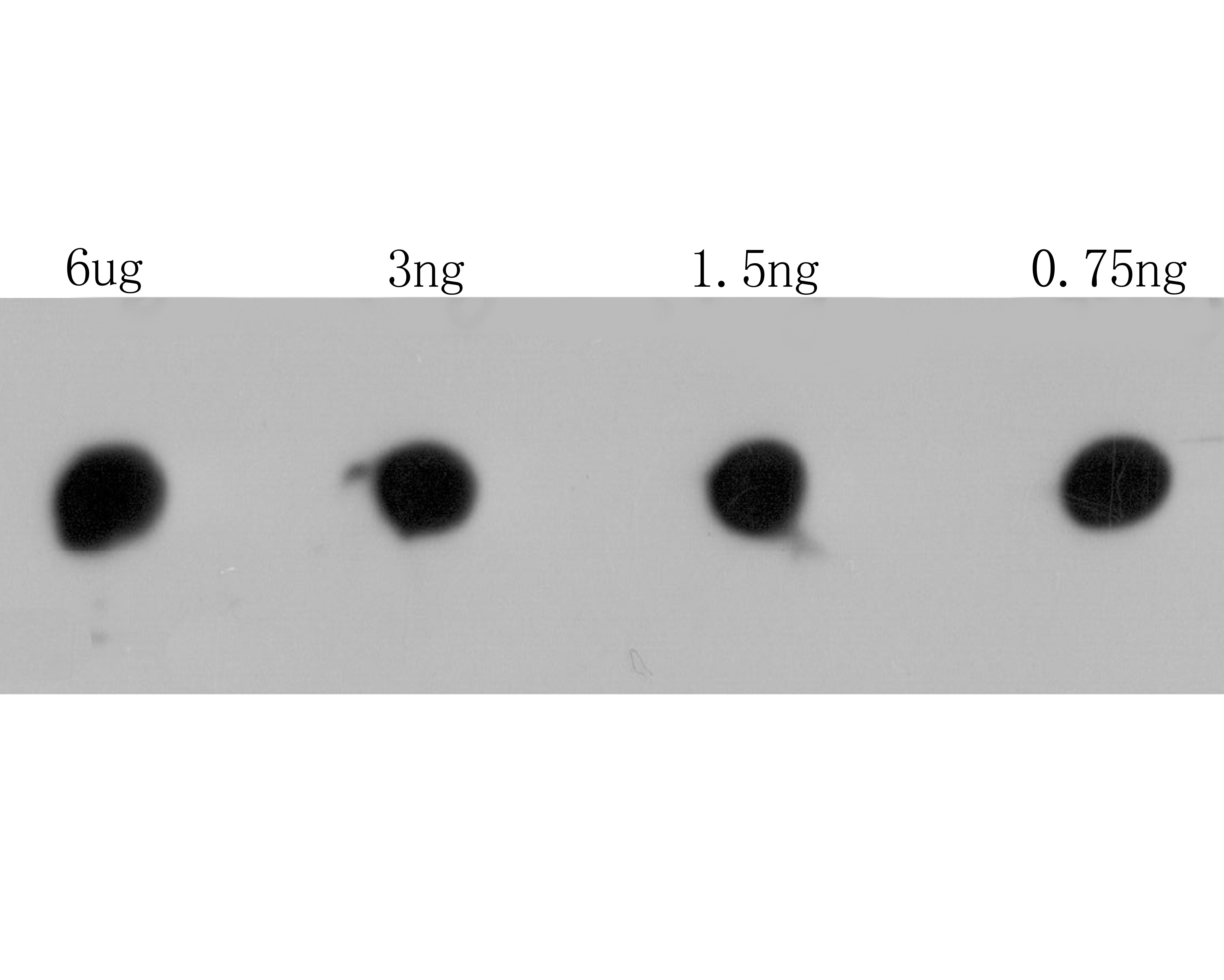
Fig1: Dot blot analysis of RYR3 on RYR3 recombinant protein. The primary antibody was used at a 1:2,000 dilution in 5% BSA at room temperature for 2 hours. Goat Anti-Rabbit IgG - HRP Secondary Antibody (HA1001) at 1:5,000 dilution was used for 1 hour at room temperature.Application
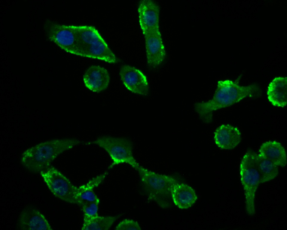
Fig2: ICC staining RYR3 in Lovo cells (green). Formalin fixed cells were permeabilized with 0.1% Triton X-100 in TBS for 10 minutes at room temperature and blocked with 1% Blocker BSA for 15 minutes at room temperature. Cells were probed with the antibody (ER1803-92) at a dilution of 1:200 for 1 hour at room temperature, washed with PBS. Alexa Fluorc™ 488 Goat anti-Rabbit IgG was used as the secondary antibody at 1/100 dilution. The nuclear counter stain is DAPI (blue).Application
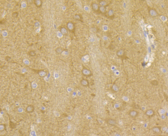
Fig3: Immunohistochemical analysis of paraffin-embedded rat brain tissue using anti-RYR3 antibody. The section was pre-treated using heat mediated antigen retrieval with Tris-EDTA buffer (pH 8.0-8.4) for 20 minutes.The tissues were blocked in 5% BSA for 30 minutes at room temperature, washed with ddH2O and PBS, and then probed with the antibody (ER1803-92) at 1/50 dilution, for 30 minutes at room temperature and detected using an HRP conjugated compact polymer system. DAB was used as the chrogen. Counter stained with hematoxylin and mounted with DPX.Application
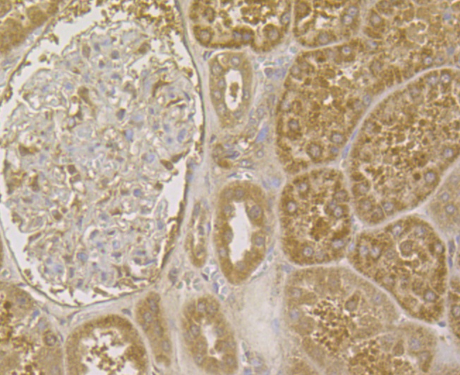
Fig4: Immunohistochemical analysis of paraffin-embedded human kidney tissue using anti-RYR3 antibody. The section was pre-treated using heat mediated antigen retrieval with Tris-EDTA buffer (pH 8.0-8.4) for 20 minutes.The tissues were blocked in 5% BSA for 30 minutes at room temperature, washed with ddH2O and PBS, and then probed with the antibody (ER1803-92) at 1/200 dilution, for 30 minutes at room temperature and detected using an HRP conjugated compact polymer system. DAB was used as the chrogen. Counter stained with hematoxylin and mounted with DPX.Application
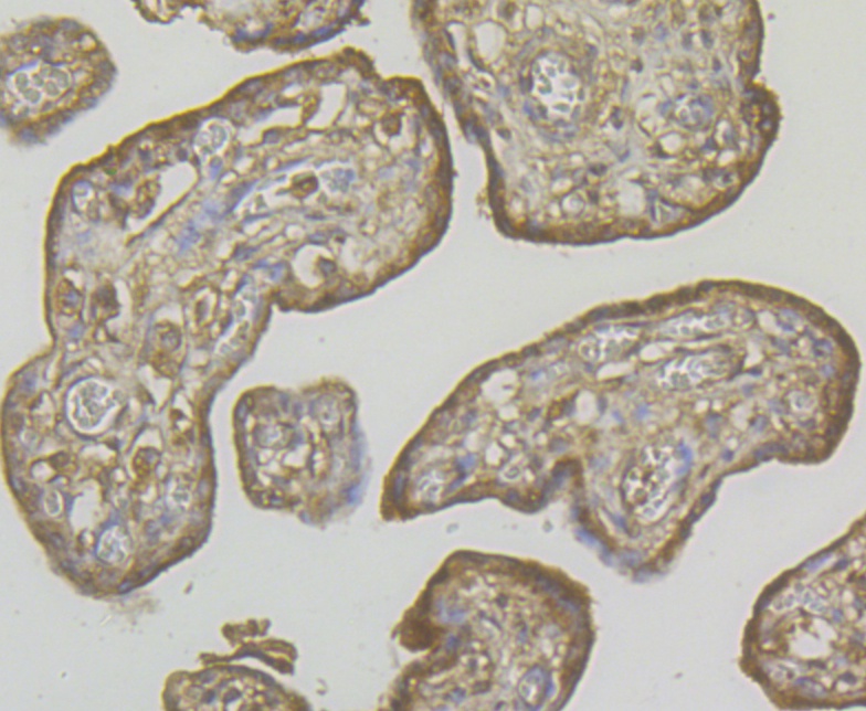
Fig5: Immunohistochemical analysis of paraffin-embedded human placenta tissue using anti-RYR3 antibody. The section was pre-treated using heat mediated antigen retrieval with Tris-EDTA buffer (pH 8.0-8.4) for 20 minutes.The tissues were blocked in 5% BSA for 30 minutes at room temperature, washed with ddH2O and PBS, and then probed with the antibody (ER1803-92) at 1/200 dilution, for 30 minutes at room temperature and detected using an HRP conjugated compact polymer system. DAB was used as the chrogen. Counter stained with hematoxylin and mounted with DPX.Application
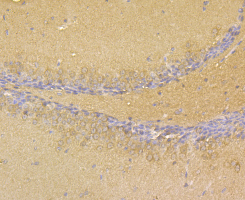
Fig6: Immunohistochemical analysis of paraffin-embedded mouse brain tissue using anti-RYR3 antibody. The section was pre-treated using heat mediated antigen retrieval with Tris-EDTA buffer (pH 8.0-8.4) for 20 minutes.The tissues were blocked in 5% BSA for 30 minutes at room temperature, washed with ddH2O and PBS, and then probed with the antibody (ER1803-92) at 1/50 dilution, for 30 minutes at room temperature and detected using an HRP conjugated compact polymer system. DAB was used as the chrogen. Counter stained with hematoxylin and mounted with DPX.| Product Name | RYR3 Rabbit Polyclonal Antibody |
|---|---|
| Antibody Type | Primary Antibodies |
| Product description | The protein encoded by this gene is a ryanodine receptor, which functions to release calcium from intracellular storage for use in many cellular processes. For example, the encoded protein is involved in skeletal muscle contraction by releasing calcium from the sarcoplasmic reticulum followed by depolarization of T-tubules. Two transcript variants encoding different isoforms have been found for this gene. Calcium channel that mediates the release of Ca2+ from the sarcoplasmic reticulum into the cytoplasm in muscle and thereby plays a role in triggering muscle contraction. May regulate Ca2+ release by other calcium channels. Calcium channel that mediates Ca2+-induced Ca2+ release from the endoplasmic reticulum in non-muscle cells. Contributes to cellular calcium ion homeostasis (By similarity). Plays a role in cellular calcium signaling. |
| Immunogen | Recombinant protein within Human RYR3 aa 1-120. |
| Clonality | Polyclonal |
|---|---|
| Isotype | IgG |
| Host Species | Rabbit |
| Tested Applications | DotICCICC/IFIHC |
| ELISA:1:5,000-1:10,000: Dot Blot:1:2000-1:20,000: ICC:1:100: IHC:1:50-1:200: |
|
| Species Reactivity | HumanMouseRat |
| Concentration | 1mg/ml |
| Purification | Affinity purified |
| Alternative Names | Brain ryanodine receptor-calcium release channel Brain-type ryanodine receptor Type 3 ryanodine receptor Ryanodine receptor 3 Brain ryanodine receptor calcium release channel antibody Brain type ryanodine receptor antibody Cardiac muscle ryanodine receptor calcium release channel antibody Cardiac muscle type ryanodine receptor antibody CCO antibody Central core disease of muscle antibody HBRR antibody MHS antibody Ryanodine receptor 3 antibody Ryanodine receptor type1 antibody RYDR antibody RYR 3 antibody RYR antibody RYR3 antibody Sarcoplasmic reticulum calcium release channel antibody Skeletal muscle calcium release channel antibody Skeletal muscle ryanodine receptor antibody Skeletal muscle type ryanodine receptor antibody Skeletal muscle-type ryanodine receptor antibody SKRR antibody VTSIP antibody |
|---|---|
| Molecular Weight(MW) | 552 kDa |
| Cellular Localization | Endoplasmic reticulum, Membrane, Sarcoplasmic reticulum. |
Application
Fig1: Dot blot analysis of RYR3 on RYR3 recombinant protein. The primary antibody was used at a 1:2,000 dilution in 5% BSA at room temperature for 2 hours. Goat Anti-Rabbit IgG - HRP Secondary Antibody (HA1001) at 1:5,000 dilution was used for 1 hour at room temperature.
Application
Fig2: ICC staining RYR3 in Lovo cells (green). Formalin fixed cells were permeabilized with 0.1% Triton X-100 in TBS for 10 minutes at room temperature and blocked with 1% Blocker BSA for 15 minutes at room temperature. Cells were probed with the antibody (ER1803-92) at a dilution of 1:200 for 1 hour at room temperature, washed with PBS. Alexa Fluorc™ 488 Goat anti-Rabbit IgG was used as the secondary antibody at 1/100 dilution. The nuclear counter stain is DAPI (blue).
Application
Fig3: Immunohistochemical analysis of paraffin-embedded rat brain tissue using anti-RYR3 antibody. The section was pre-treated using heat mediated antigen retrieval with Tris-EDTA buffer (pH 8.0-8.4) for 20 minutes.The tissues were blocked in 5% BSA for 30 minutes at room temperature, washed with ddH2O and PBS, and then probed with the antibody (ER1803-92) at 1/50 dilution, for 30 minutes at room temperature and detected using an HRP conjugated compact polymer system. DAB was used as the chrogen. Counter stained with hematoxylin and mounted with DPX.
Application
Fig4: Immunohistochemical analysis of paraffin-embedded human kidney tissue using anti-RYR3 antibody. The section was pre-treated using heat mediated antigen retrieval with Tris-EDTA buffer (pH 8.0-8.4) for 20 minutes.The tissues were blocked in 5% BSA for 30 minutes at room temperature, washed with ddH2O and PBS, and then probed with the antibody (ER1803-92) at 1/200 dilution, for 30 minutes at room temperature and detected using an HRP conjugated compact polymer system. DAB was used as the chrogen. Counter stained with hematoxylin and mounted with DPX.
Application
Fig5: Immunohistochemical analysis of paraffin-embedded human placenta tissue using anti-RYR3 antibody. The section was pre-treated using heat mediated antigen retrieval with Tris-EDTA buffer (pH 8.0-8.4) for 20 minutes.The tissues were blocked in 5% BSA for 30 minutes at room temperature, washed with ddH2O and PBS, and then probed with the antibody (ER1803-92) at 1/200 dilution, for 30 minutes at room temperature and detected using an HRP conjugated compact polymer system. DAB was used as the chrogen. Counter stained with hematoxylin and mounted with DPX.
Application
Fig6: Immunohistochemical analysis of paraffin-embedded mouse brain tissue using anti-RYR3 antibody. The section was pre-treated using heat mediated antigen retrieval with Tris-EDTA buffer (pH 8.0-8.4) for 20 minutes.The tissues were blocked in 5% BSA for 30 minutes at room temperature, washed with ddH2O and PBS, and then probed with the antibody (ER1803-92) at 1/50 dilution, for 30 minutes at room temperature and detected using an HRP conjugated compact polymer system. DAB was used as the chrogen. Counter stained with hematoxylin and mounted with DPX.| Application Notes | ELISA:1:5,000-1:10,000: Dot Blot:1:2000-1:20,000: ICC:1:100: IHC:1:50-1:200: |
|---|
| Form | Liquid |
|---|---|
| Storage Instructions | Store at +4℃ after thawing. Aliquot store at -20℃. Avoid repeated freeze / thaw cycles. |
| Storage Buffer | 1*TBS (pH7.4), 1%BSA, 50%Glycerol. Preservative: 0.05% Sodium Azide. |


