Application
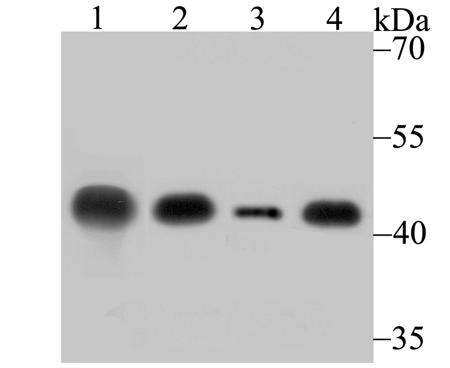
Fig1: Western blot analysis of Creatine kinase B type on different lysates using anti-Creatine kinase B type antibody at 1/500 dilution. Positive control: Lane 1: Mouse brain Lane 2: Mouse brain Lane 3: Rat brain Lane 4: 293Application
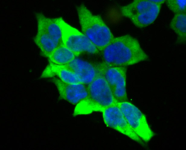
Fig2: ICC staining Creatine kinase B type in 293T cells (green). The nuclear counter stain is DAPI (blue). Cells were fixed in paraformaldehyde, permeabilised with 0.25% Triton X100/PBS.Application
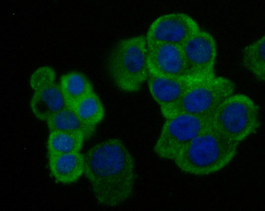
Fig3: ICC staining Creatine kinase B type in LOVO cells (green). The nuclear counter stain is DAPI (blue). Cells were fixed in paraformaldehyde, permeabilised with 0.25% Triton X100/PBS.Application
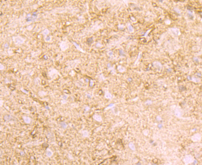
Fig4: Immunohistochemical analysis of paraffin-embedded rat brain tissue using anti-Creatine kinase B type antibody. Counter stained with hematoxylin.Application
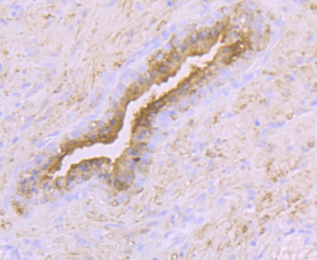
Fig5: Immunohistochemical analysis of paraffin-embedded human prostate tissue using anti-Creatine kinase B type antibody. Counter stained with hematoxylin.Application
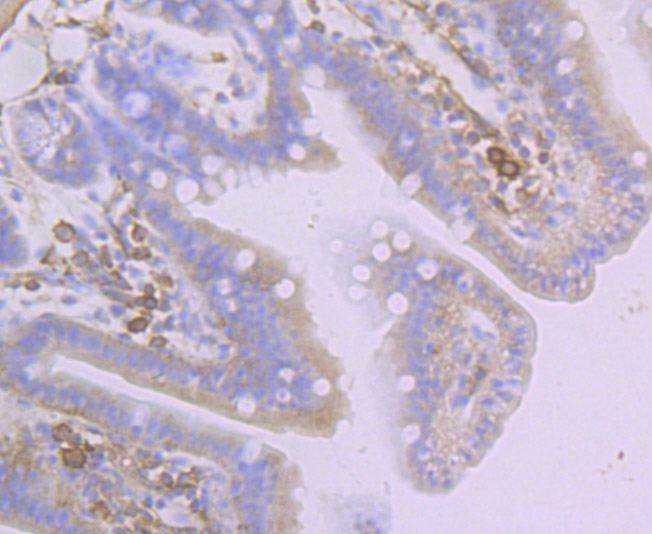
Fig6: Immunohistochemical analysis of paraffin-embedded mouse colon tissue using anti-Creatine kinase B type antibody. Counter stained with hematoxylin.Application
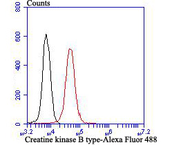
Fig7: Flow cytometric analysis of 293T cells with Creatine kinase B type antibody at 1/100 dilution (red) compared with an unlabelled control (cells without incubation with primary antibody; black). Alexa Fluor 488-conjugated goat anti rabbit IgG was used as the secondary antibody.| Product Name | Creatine kinase B type [JB78-34] |
|---|---|
| Antibody Type | Primary Antibodies |
| Antigen Alias | B CK antibody B-CK antibody BB-CK antibody BCK antibody Brain creatine kinase antibody Ckb antibody CKBB antibody Creatine kinase B antibody Creatine kinase B chain antibody Creatine kinase B type antibody Creatine kinase B-type antibody Creatine Kinase BB Isoenzyme antibody Creatine kinase brain antibody Creatine kinase brain type antibody Creatine phosphokinase BB antibody Epididymis luminal protein 211 antibody Epididymis secretory protein Li 29 antibody HEL 211 antibody HEL S 29 antibody KCRB_HUMAN antibody |
| Product description | Creatine kinases (CKs) are a large family of isoenzymes that regulate levels of ATP in subcellular compartments, where they provide ATP at sites of fluctuating energy demand by the transfer of phosphates between creatine and adenine nucleotides. Creatine kinases provide the energy of phosphate hydrolysis necessary to drive the normal function of many cellular systems including muscle, electrocytes, retina photoreceptor cells, brain cells, kidney, salt glands, myometrium, placenta, pancreas, thymus, thyroid, intestinal epithelial cells, endothelial cells, cartilage and bone cells, macrophages, blood platelets, and tumor and cancer cells. Human cytoplasmic creatine kinase-B, also designated CK-B and BCK, is a 381 amino acid, brain tissue specific isoform of creatine kinase. Human cytoplasmic creatine kinase-muscle (CK-M, MCK) is a muscle tissue-specific isoform of creatine kinase. Human cytoplasmic creatine kinase-Mi (Mi-CK, MtCK) is a 416 amino acid mitochondrial-specific isoform of creatine kinase. Cytosolic creatine kinases are important in the energetic regulation of Ca2+-pumps and in the maintenance of Ca2+-homeostasis. |
| Immunogen | Recombinant protein |
| Clonality | Monoclonal |
|---|---|
| Isotype | IgG |
| Host Species | Recombinant rabbit |
| Tested Applications | WBICCIFIHCFC |
| WB:1:500-1:2,000 ICC:1:50-1:200 IHC:1:50-1:200 FC:1:50-1:100 |
|
| Species Reactivity | HumanMouseRat |
| Concentration | 1mg/ml |
| Alternative Names | B CK antibody B-CK antibody BB-CK antibody BCK antibody Brain creatine kinase antibody Ckb antibody CKBB antibody Creatine kinase B antibody Creatine kinase B chain antibody Creatine kinase B type antibody Creatine kinase B-type antibody Creatine Kinase BB Isoenzyme antibody Creatine kinase brain antibody Creatine kinase brain type antibody Creatine phosphokinase BB antibody Epididymis luminal protein 211 antibody Epididymis secretory protein Li 29 antibody HEL 211 antibody HEL S 29 antibody KCRB_HUMAN antibody |
|---|---|
| Molecular Weight(MW) | 43 kDa |
| Cellular Localization | Cytoplasm. |
Application
Fig1: Western blot analysis of Creatine kinase B type on different lysates using anti-Creatine kinase B type antibody at 1/500 dilution. Positive control: Lane 1: Mouse brain Lane 2: Mouse brain Lane 3: Rat brain Lane 4: 293
Application
Fig2: ICC staining Creatine kinase B type in 293T cells (green). The nuclear counter stain is DAPI (blue). Cells were fixed in paraformaldehyde, permeabilised with 0.25% Triton X100/PBS.
Application
Fig3: ICC staining Creatine kinase B type in LOVO cells (green). The nuclear counter stain is DAPI (blue). Cells were fixed in paraformaldehyde, permeabilised with 0.25% Triton X100/PBS.
Application
Fig4: Immunohistochemical analysis of paraffin-embedded rat brain tissue using anti-Creatine kinase B type antibody. Counter stained with hematoxylin.
Application
Fig5: Immunohistochemical analysis of paraffin-embedded human prostate tissue using anti-Creatine kinase B type antibody. Counter stained with hematoxylin.
Application
Fig6: Immunohistochemical analysis of paraffin-embedded mouse colon tissue using anti-Creatine kinase B type antibody. Counter stained with hematoxylin.
Application
Fig7: Flow cytometric analysis of 293T cells with Creatine kinase B type antibody at 1/100 dilution (red) compared with an unlabelled control (cells without incubation with primary antibody; black). Alexa Fluor 488-conjugated goat anti rabbit IgG was used as the secondary antibody.| Positive Control | Rat brain tissue, human prostate tissue, mouse colon tissue, mouse brain tissue lysate, 293, 293T, LOVO. |
|---|---|
| Application Notes | WB:1:500-1:2,000 ICC:1:50-1:200 IHC:1:50-1:200 FC:1:50-1:100 |
| Form | Liquid |
|---|---|
| Storage Instructions | Store at +4℃ after thawing. Aliquot store at -20℃ or -80℃. Avoid repeated freeze / thaw cycles. |
| Storage Buffer | 1*TBS (pH7.4), 1%BSA, 40%Glycerol. Preservative: 0.05% Sodium Azide. |


