Application
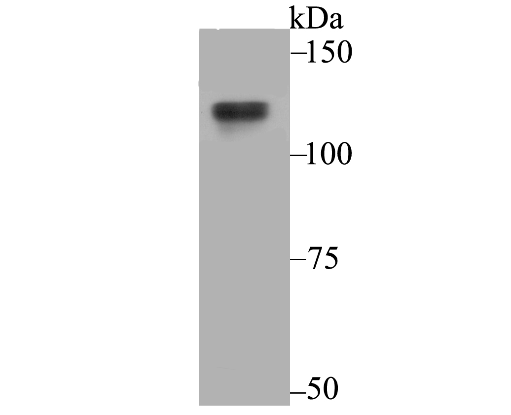
Fig1: Western blot analysis of USP7 on 293T cell using anti-USP7 antibody at 1/500 dilution.Application
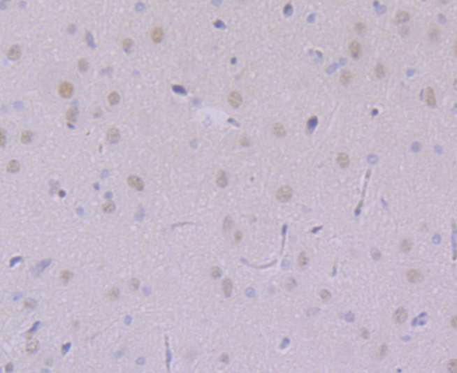
Fig2: Immunohistochemical analysis of paraffin-embedded rat brain tissue using anti-USP7 antibody. Counter stained with hematoxylin.Application
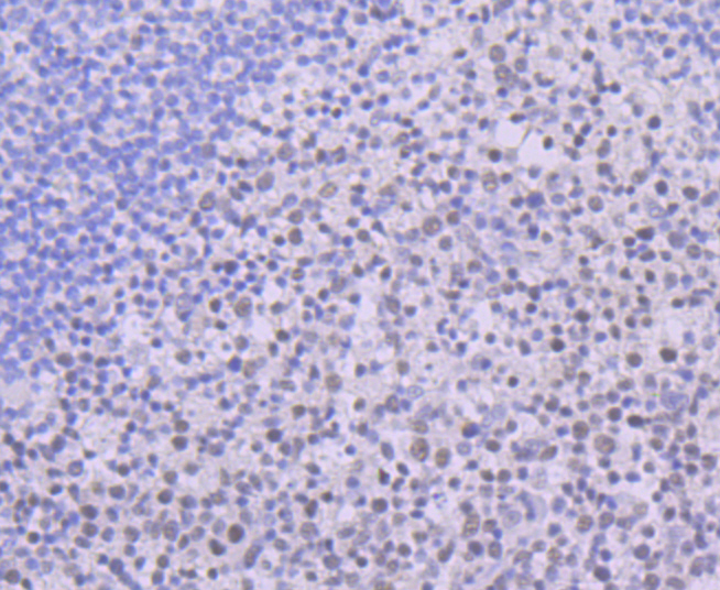
Fig3: Immunohistochemical analysis of paraffin-embedded human tonsil tissue using anti-USP7 antibody. Counter stained with hematoxylin.Application
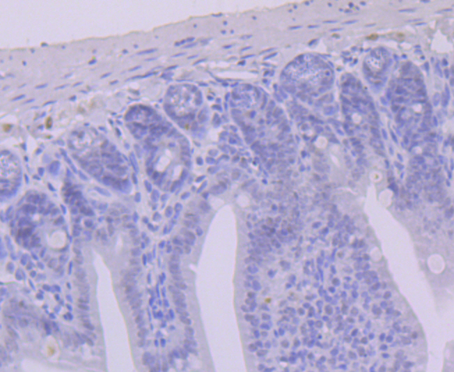
Fig4: Immunohistochemical analysis of paraffin-embedded mouse colon tissue using anti-USP7 antibody. Counter stained with hematoxylin.Application
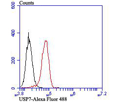
Fig5: Flow cytometric analysis of PC-3M cells with USP7 antibody at 1/100 dilution (red) compared with an unlabelled control (cells without incubation with primary antibody; black). Alexa Fluor 488-conjugated goat anti-rabbit IgG was used as the secondary antibody.| Product Name | USP7 [JB80-36] |
|---|---|
| Antibody Type | Primary Antibodies |
| Antigen Alias | Deubiquitinating enzyme 7 antibody HAUSP antibody Herpes virus associated ubiquitin specific protease antibody Herpesvirus-associated ubiquitin-specific protease antibody TEF 1 antibody tef-1 antibody TEF1 antibody Ubiquitin carboxyl terminal hydrolase 7 antibody Ubiquitin carboxyl-terminal hydrolase 7 antibody Ubiquitin specific peptidase 7 (herpes virus associated) antibody Ubiquitin specific peptidase 7 antibody Ubiquitin specific peptidase 7 herpes virus associated antibody Ubiquitin specific processing protease 7 antibody Ubiquitin specific protease 7 (herpes virus associated) antibody Ubiquitin specific protease 7 antibody Ubiquitin specific protease 7 herpes virus associated antibody Ubiquitin thioesterase 7 antibody Ubiquitin thiolesterase 7 antibody Ubiquitin-specific-processing protease 7 antibody UBP 7 antibody UBP-7 antibody UBP7 antibody UBP7_HUMAN antibody USP 7 antibody usp-7 antibody Usp7 antibody VMW110-ASSOCIATED PROTEIN, 135-KD antibody |
| Product description | HAUSP (herpesvirus-associated ubiquitin-specific protease, USP7) is a ubiquitin-specific protease. HAUSP localizes predominantly to the nucleus, in a TD-dependent manner, where it associates with ND10. ND10 are small nuclear structures implicated in a variety of cellular processes including response to stress and interferons, oncogenesis, and viral infection. HAUSP binds strongly to Vmw110, a herpesvirus regulatory protein which has the ability to disrupt ND10. HAUSP, a novel p53-interacting protein, functions to deubiquitinize p53 in an important pathway for p53 stabilization. HAUSP strongly stabilizes p53 even in the presence of excess Mdm2, and also induces p53-dependent cell growth repression and apoptosis. The HAUSP protein is distributed in the nucleus in a micropunctate pattern with a limited number of larger discrete foci, some of which co-localize with PML in ND10. The gene encoding HAUSP maps to human chromosome band 16p13.2. |
| Immunogen | Recombinant protein within human USP7 aa 100-200. |
| Clonality | Monoclonal |
|---|---|
| Isotype | IgG |
| Host Species | Recombinant rabbit |
| Tested Applications | WBIHCFC |
| WB:1:500-1:1,000 IHC:1:50 FC:1:50-1:100 |
|
| Species Reactivity | HumanMouseRat |
| Concentration | 1mg/ml |
| Alternative Names | Deubiquitinating enzyme 7 antibody
HAUSP antibody
Herpes virus associated ubiquitin specific protease antibody
Herpesvirus-associated ubiquitin-specific protease antibody
TEF 1 antibody
tef-1 antibody
TEF1 antibody
Ubiquitin carboxyl terminal hydrolase 7 antibody
Ubiquitin carboxyl-terminal hydrolase 7 antibody
Ubiquitin specific peptidase 7 (herpes virus associated) antibody
Ubiquitin specific peptidase 7 antibody
Ubiquitin specific peptidase 7 herpes virus associated antibody
Ubiquitin specific processing protease 7 antibody
Ubiquitin specific protease 7 (herpes virus associated) antibody
Ubiquitin specific protease 7 antibody
Ubiquitin specific protease 7 herpes virus associated antibody
Ubiquitin thioesterase 7 antibody
Ubiquitin thiolesterase 7 antibody
Ubiquitin-specific-processing protease 7 antibody
UBP 7 antibody
UBP-7 antibody
UBP7 antibody
UBP7_HUMAN antibody
USP 7 antibody
usp-7 antibody
Usp7 antibody
VMW110-ASSOCIATED PROTEIN 135-KD antibody |
|---|---|
| Molecular Weight(MW) | 128 kDa |
| Cellular Localization | Nucleus. Cytoplasm. |
Application
Fig1: Western blot analysis of USP7 on 293T cell using anti-USP7 antibody at 1/500 dilution.
Application
Fig2: Immunohistochemical analysis of paraffin-embedded rat brain tissue using anti-USP7 antibody. Counter stained with hematoxylin.
Application
Fig3: Immunohistochemical analysis of paraffin-embedded human tonsil tissue using anti-USP7 antibody. Counter stained with hematoxylin.
Application
Fig4: Immunohistochemical analysis of paraffin-embedded mouse colon tissue using anti-USP7 antibody. Counter stained with hematoxylin.
Application
Fig5: Flow cytometric analysis of PC-3M cells with USP7 antibody at 1/100 dilution (red) compared with an unlabelled control (cells without incubation with primary antibody; black). Alexa Fluor 488-conjugated goat anti-rabbit IgG was used as the secondary antibody.| Positive Control | 293T, rat brain tissue, human tonsil tissue, mouse colon tissue, PC-3M. |
|---|---|
| Application Notes | WB:1:500-1:1,000 IHC:1:50 FC:1:50-1:100 |
| Form | Liquid |
|---|---|
| Storage Instructions | Store at +4℃ after thawing. Aliquot store at -20℃ or -80℃. Avoid repeated freeze / thaw cycles. |
| Storage Buffer | 1*TBS (pH7.4), 1%BSA, 40%Glycerol. Preservative: 0.05% Sodium Azide. |


