Application
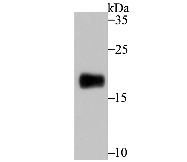
Fig1: Western blot analysis of CD3G on mouse thymus tissue lysate using anti-CD3G antibody at 1/500 dilution.Application
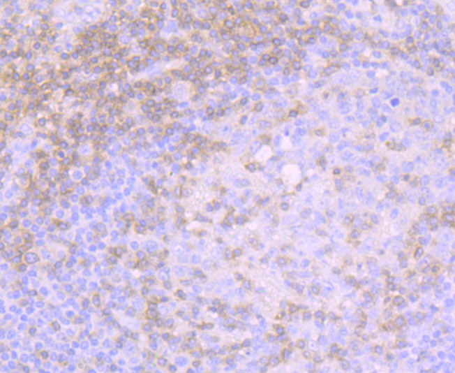
Fig2: Immunohistochemical analysis of paraffin-embedded human tonsil tissue using anti-CD3G antibody. Counter stained with hematoxylin.Application
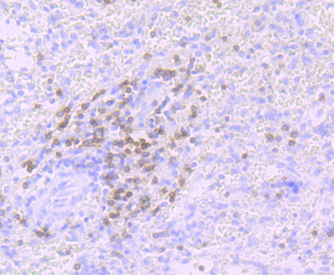
Fig3: Immunohistochemical analysis of paraffin-embedded human spleen tissue using anti-CD3G antibody. Counter stained with hematoxylin.Application
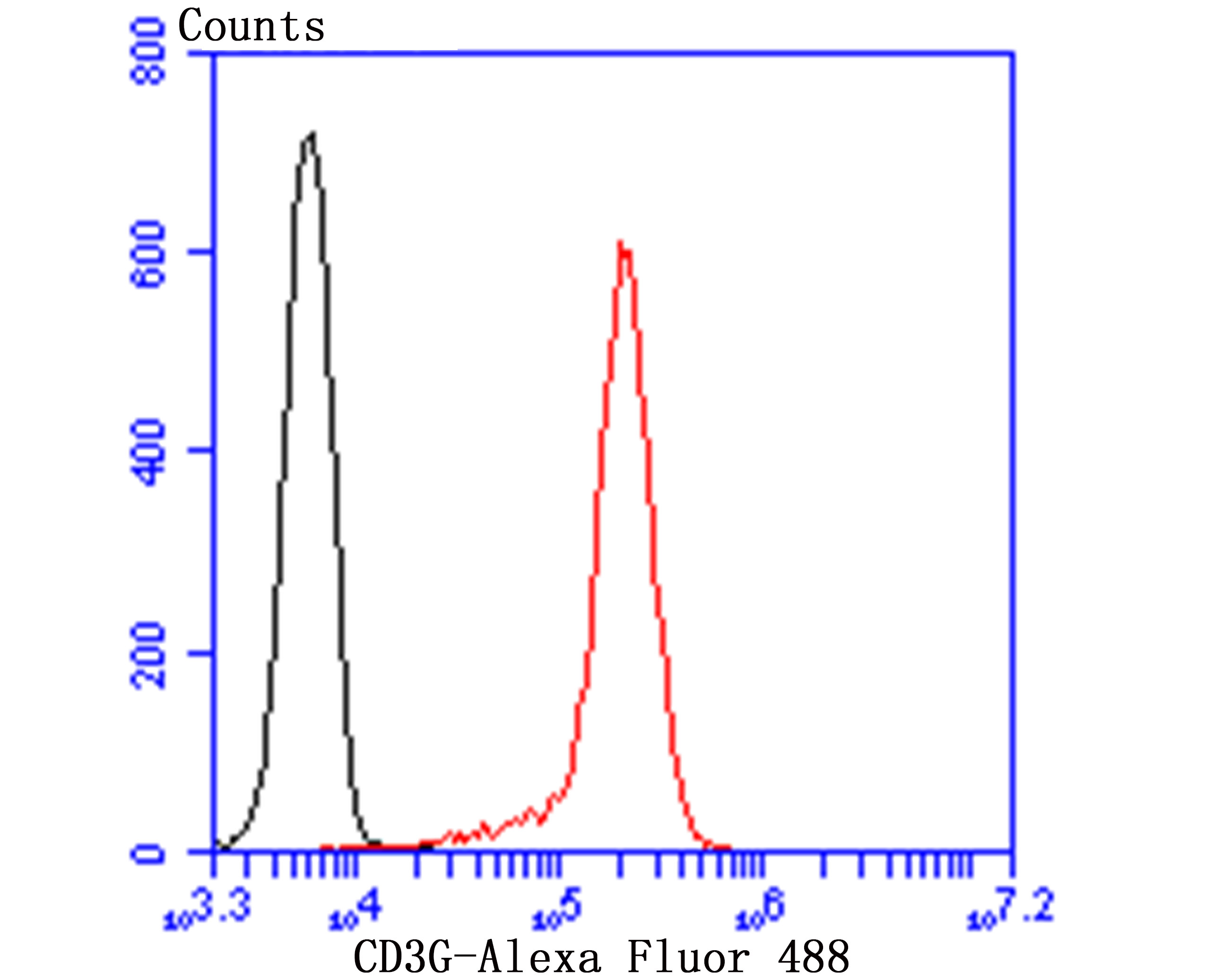
Fig4: Flow cytometric analysis of Jurkat cells with CD3G antibody at 1/100 dilution (red) compared with an unlabelled control (cells without incubation with primary antibody; black). Alexa Fluor 488-conjugated goat anti-rabbit IgG was used as the secondary antibody.| Product Name | CD3G [JB38-29] |
|---|---|
| Antibody Type | Primary Antibodies |
| Antigen Alias | CD3 gamma antibody CD3-GAMMA antibody CD3g antibody CD3g antigen gamma polypeptide (TiT3 complex) antibody CD3g antigen gamma polypeptide antibody CD3g molecule gamma (CD3 TCR complex) antibody CD3g molecule gamma antibody CD3G_HUMAN antibody FLJ17620 antibody FLJ17664 antibody FLJ79544 antibody FLJ94613 antibody MGC138597 antibody T cell antigen receptor complex gamma subunit of T3 antibody T-cell receptor T3 gamma chain antibody T-cell surface glycoprotein CD3 gamma chain antibody T3G antibody |
| Product description | The T cell antigen receptor (TCR) recognizes foreign antigens and translates such recognition events into intracellular signals that elicit a change in the cell from a dormant to an activated state. Much of this signaling process can be attributed to a multisubunit complex of proteins that associates directly with the TCR. This complex has been designated CD3 (cluster of differentiation 3). It is composed of five invariant polypeptide chains that associate to form three dimers: a heterodimer of gamma and epsilon chains (γε), a heterodimer of delta and epsilon chains (δε) and a homodimer of two zeta chains (ζζ) or a heterodimer of zeta and eta chains (ζη). The ζ and η chains are encoded by the same gene but differ in their carboxyl-terminal ends due to an alternative splicing event. The γ, ε and δ chains each contain a single copy of a conserved immunoreceptor tyrosine-based activation motif (ITAM). |
| Immunogen | Recombinant protein |
| Clonality | Monoclonal |
|---|---|
| Isotype | IgG |
| Host Species | Recombinant rabbit |
| Tested Applications | WBIHCFCIP |
| WB:1:500-1:1,000 IHC:1:50-1:200 FC:1:50-1:100 IP:1:10-1:50 |
|
| Species Reactivity | HumanMouse |
| Concentration | 1mg/ml |
| Alternative Names | CD3 gamma antibody CD3-GAMMA antibody CD3g antibody CD3g antigen gamma polypeptide (TiT3 complex) antibody CD3g antigen gamma polypeptide antibody CD3g molecule gamma (CD3 TCR complex) antibody CD3g molecule gamma antibody CD3G_HUMAN antibody FLJ17620 antibody FLJ17664 antibody FLJ79544 antibody FLJ94613 antibody MGC138597 antibody T cell antigen receptor complex gamma subunit of T3 antibody T-cell receptor T3 gamma chain antibody T-cell surface glycoprotein CD3 gamma chain antibody T3G antibody |
|---|---|
| Molecular Weight(MW) | 20 kDa |
| Cellular Localization | Cell membrane. Membrane. |
Application
Fig1: Western blot analysis of CD3G on mouse thymus tissue lysate using anti-CD3G antibody at 1/500 dilution.
Application
Fig2: Immunohistochemical analysis of paraffin-embedded human tonsil tissue using anti-CD3G antibody. Counter stained with hematoxylin.
Application
Fig3: Immunohistochemical analysis of paraffin-embedded human spleen tissue using anti-CD3G antibody. Counter stained with hematoxylin.
Application
Fig4: Flow cytometric analysis of Jurkat cells with CD3G antibody at 1/100 dilution (red) compared with an unlabelled control (cells without incubation with primary antibody; black). Alexa Fluor 488-conjugated goat anti-rabbit IgG was used as the secondary antibody.| Positive Control | Human tonsil tissue, human spleen tissue, mouse thymus tissue lysate, Jurkat. |
|---|---|
| Application Notes | WB:1:500-1:1,000 IHC:1:50-1:200 FC:1:50-1:100 IP:1:10-1:50 |
| Form | Liquid |
|---|---|
| Storage Instructions | Store at +4℃ after thawing. Aliquot store at -20℃ or -80℃. Avoid repeated freeze / thaw cycles. |
| Storage Buffer | 1*TBS (pH7.4), 1%BSA, 40%Glycerol. Preservative: 0.05% Sodium Azide. |


