Application
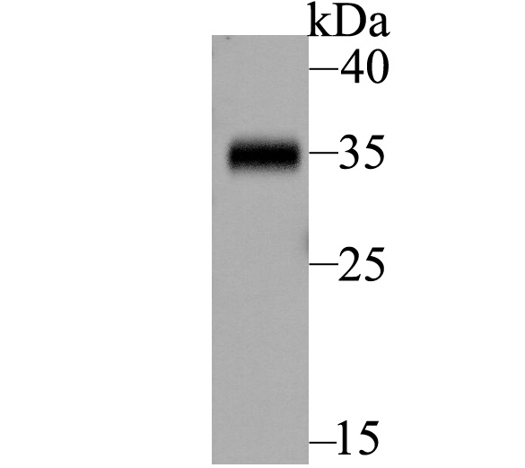
Fig1: Western blot analysis of BOB1 on Daudi lysate. Proteins were transferred to a PVDF membrane and blocked with 5% BSA in PBS for 1 hour at room temperature. The primary antibody was used at a 1:500 dilution in 5% BSA at room temperature for 2 hours. Goat Anti-Rabbit IgG - HRP Secondary Antibody (HA1001) at 1:5,000 dilution was used for 1 hour at room temperature.Application
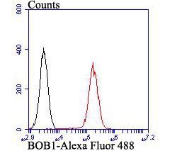
Fig2: Flow cytometric analysis of BOB1 was done on Daudi cells. The cells were fixed, permeabilized and stained with BOB1 antibody at 1/100 dilution (red) compared with an unlabelled control (cells without incubation with primary antibody; black). After incubation of the primary antibody on room temperature for an hour, the cells was stained with a Alexa Fluor™ 488-conjugated goat anti-rabbit IgG Secondary antibody at 1/500 dilution for 30 minutes.| Product Name | BOB1 |
|---|---|
| Antibody Type | Primary Antibodies |
| Antigen Alias | B cell Oct binding protein 1 antibody B cell specific coactivator OBF 1 antibody B cell specific coactivator OBF1 antibody B-cell-specific coactivator OBF-1 antibody BOB 1 antibody BOB-1 antibody OBF 1 antibody OBF1 antibody OBF1_HUMAN antibody OCA B antibody OCA-B antibody OCAB antibody OCT binding factor 1 antibody OCT-binding factor 1 antibody POU class 2 associating factor 1 antibody POU domain class 2 associating factor 1 antibody POU domain class 2-associating factor 1 antibody Pou2af1 antibody |
| Product description | Transcriptional coactivator that specifically associates with either OCT1 or OCT2. It boosts the OCT1 mediated promoter activity and to a lesser extent, that of OCT2. It has no intrinsic DNA-binding activity. It recognizes the POU domains of OCT1 and OCT2. It is essential for the response of B-cells to antigens and required for the formation of germinal centers. Reduced activation of Bob1-deficient B cells following antigenic stimulation is often reported. The action of Bob1 on B cell maturation and tolerance could be mediated by effects on B cell activation, in part by regulating the expression of immunosuppressive miRNAs. Bob1 has long been considered a B cell‐specific factor that interacts with the transcription factors Oct1 and Oct2 to enhance octamer‐dependent transcription. Bob1 can directly bind to and transactivate the promoters of Bcl6 and Btla. |
| Immunogen | Recombinant protein within Human BOB1 aa 10-180. |
| Clonality | Polyclonal |
|---|---|
| Isotype | IgG |
| Host Species | Rabbit |
| Tested Applications | WBFC |
| WB:1:500: FC:1:50-1:100: |
|
| Species Reactivity | HumanMouseRat |
| Concentration | 1mg/ml |
| Alternative Names | B cell Oct binding protein 1 antibody B cell specific coactivator OBF 1 antibody B cell specific coactivator OBF1 antibody B-cell-specific coactivator OBF-1 antibody BOB 1 antibody BOB-1 antibody OBF 1 antibody OBF1 antibody OBF1_HUMAN antibody OCA B antibody OCA-B antibody OCAB antibody OCT binding factor 1 antibody OCT-binding factor 1 antibody POU class 2 associating factor 1 antibody POU domain class 2 associating factor 1 antibody POU domain class 2-associating factor 1 antibody Pou2af1 antibody |
|---|---|
| Molecular Weight(MW) | 35 kDa (predicted molecular weight: 27 kDa) |
| Cellular Localization | Nucleus. |
Application
Fig1: Western blot analysis of BOB1 on Daudi lysate. Proteins were transferred to a PVDF membrane and blocked with 5% BSA in PBS for 1 hour at room temperature. The primary antibody was used at a 1:500 dilution in 5% BSA at room temperature for 2 hours. Goat Anti-Rabbit IgG - HRP Secondary Antibody (HA1001) at 1:5,000 dilution was used for 1 hour at room temperature.
Application
Fig2: Flow cytometric analysis of BOB1 was done on Daudi cells. The cells were fixed, permeabilized and stained with BOB1 antibody at 1/100 dilution (red) compared with an unlabelled control (cells without incubation with primary antibody; black). After incubation of the primary antibody on room temperature for an hour, the cells was stained with a Alexa Fluor™ 488-conjugated goat anti-rabbit IgG Secondary antibody at 1/500 dilution for 30 minutes.| Positive Control | Daudi. |
|---|---|
| Application Notes | WB:1:500: FC:1:50-1:100: |
| Form | Liquid |
|---|---|
| Storage Instructions | Store at +4℃ after thawing. Aliquot store at -20℃. Avoid repeated freeze / thaw cycles. |
| Storage Buffer | 1*TBS (pH7.4), 1%BSA, 50%Glycerol. Preservative: 0.05% Sodium Azide. |


