Application
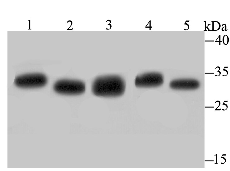
Fig1: Western blot analysis of Proteasome 20S C2 on different lysates using anti-Proteasome 20S C2 antibody at 1/500 dilution. Positive control: Lane 1: PC-3M Lane 2: K562 Lane 3: Rat testis Lane 4: Mouse testis Lane 5: Mouse colonApplication
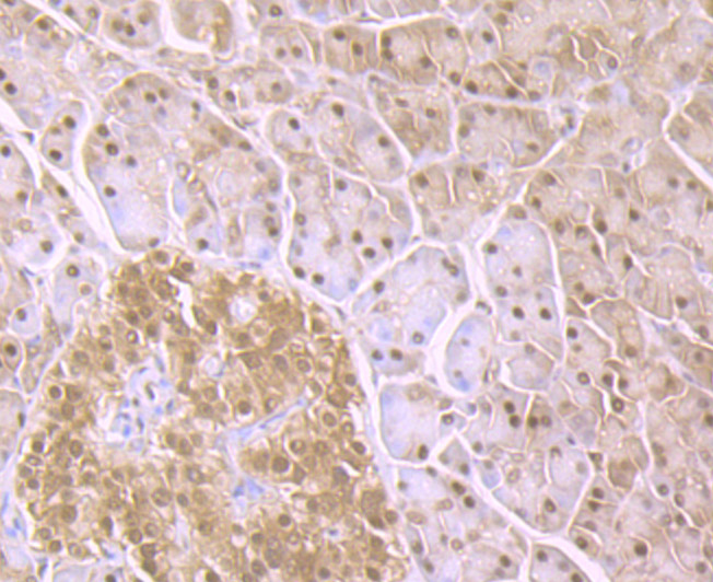
Fig2: Immunohistochemical analysis of paraffin-embedded human pancreas tissue using anti-Proteasome 20S C2 antibody. Counter stained with hematoxylin.Application
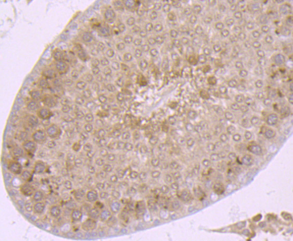
Fig3: Immunohistochemical analysis of paraffin-embedded rat testis tissue using anti-Proteasome 20S C2 antibody. Counter stained with hematoxylin.Application
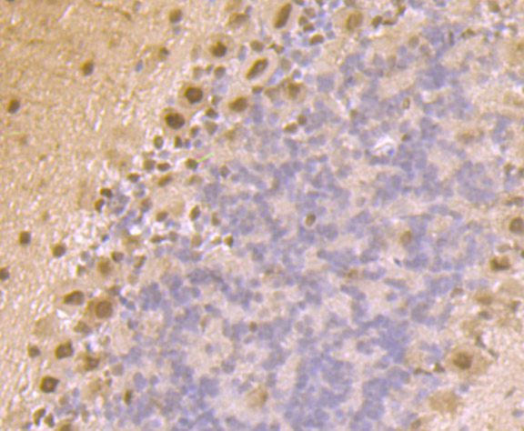
Fig4: Immunohistochemical analysis of paraffin-embedded mouse cerebellum tissue using anti-Proteasome 20S C2 antibody. Counter stained with hematoxylin.Application
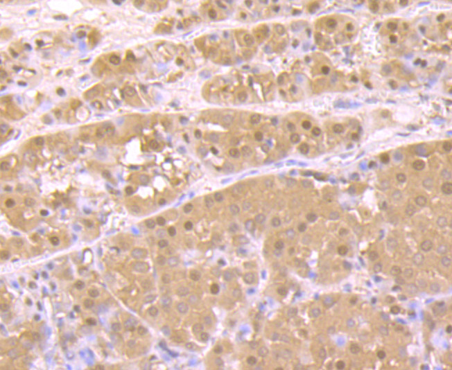
Fig5: Immunohistochemical analysis of paraffin-embedded human liver cancer tissue using anti-Proteasome 20S C2 antibody. Counter stained with hematoxylin.Application
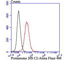
Fig6: Flow cytometric analysis of K562 cells with Proteasome 20S C2 antibody at 1/100 dilution (red) compared with an unlabelled control (cells without incubation with primary antibody; black). Alexa Fluor 488-conjugated goat anti rabbit IgG was used as the secondary antibody.| Product Name | Proteasome 20S C2 [JB89-30] |
|---|---|
| Antibody Type | Primary Antibodies |
| Antigen Alias | 30 kDa prosomal protein antibody HC 2 antibody HC2 antibody Macropain subunit C2 antibody Macropain subunit nu antibody MGC14542 antibody MGC14575 antibody MGC14751 antibody MGC1667 antibody MGC21459 antibody MGC22853 antibody MGC23915 antibody Multicatalytic endopeptidase complex subunit C2 antibody NU antibody PROS 30 antibody PROS-30 antibody PROS30 antibody Proteasome (prosome macropain) subunit alpha type 1 antibody Proteasome alpha 1 subunit antibody Proteasome component C2 antibody Proteasome nu chain antibody Proteasome subunit alpha type 1 antibody Proteasome subunit alpha type I antibody Proteasome subunit alpha type-1 antibody Proteasome subunit nu antibody Protein P30 33K antibody PSA1_HUMAN antibody PSC 2 antibody PSC2 antibody PSMA 1 antibody psmA1 antibody |
| Product description | Component of the 20S core proteasome complex involved in the proteolytic degradation of most intracellular proteins. This complex plays numerous essential roles within the cell by associating with different regulatory particles. Associated with two 19S regulatory particles, forms the 26S proteasome and thus participates in the ATP-dependent degradation of ubiquitinated proteins. The 26S proteasome plays a key role in the maintenance of protein homeostasis by removing misfolded or damaged proteins that could impair cellular functions, and by removing proteins whose functions are no longer required. Associated with the PA200 or PA28, the 20S proteasome mediates ubiquitin-independent protein degradation. This type of proteolysis is required in several pathways including spermatogenesis (20S-PA200 complex) or generation of a subset of MHC class I-presented antigenic peptides (20S-PA28 complex). |
| Immunogen | Recombinant protein |
| Clonality | Monoclonal |
|---|---|
| Isotype | IgG |
| Host Species | Recombinant rabbit |
| Tested Applications | WBIHCFCIP |
| WB:1:500-1:2,000 IHC:1:50-1:200 FC:1:50-1:100 ICC:1:50-1:200 |
|
| Species Reactivity | HumanMouseRat |
| Concentration | 1mg/ml |
| Alternative Names | 30 kDa prosomal protein antibody HC 2 antibody HC2 antibody Macropain subunit C2 antibody Macropain subunit nu antibody MGC14542 antibody MGC14575 antibody MGC14751 antibody MGC1667 antibody MGC21459 antibody MGC22853 antibody MGC23915 antibody Multicatalytic endopeptidase complex subunit C2 antibody NU antibody PROS 30 antibody PROS-30 antibody PROS30 antibody Proteasome (prosome macropain) subunit alpha type 1 antibody Proteasome alpha 1 subunit antibody Proteasome component C2 antibody Proteasome nu chain antibody Proteasome subunit alpha type 1 antibody Proteasome subunit alpha type I antibody Proteasome subunit alpha type-1 antibody Proteasome subunit nu antibody Protein P30 33K antibody PSA1_HUMAN antibody PSC 2 antibody PSC2 antibody PSMA 1 antibody psmA1 antibody |
|---|---|
| Molecular Weight(MW) | 30 kDa |
| Cellular Localization | Cytoplasm. Nucleus. |
Application
Fig1: Western blot analysis of Proteasome 20S C2 on different lysates using anti-Proteasome 20S C2 antibody at 1/500 dilution. Positive control: Lane 1: PC-3M Lane 2: K562 Lane 3: Rat testis Lane 4: Mouse testis Lane 5: Mouse colon
Application
Fig2: Immunohistochemical analysis of paraffin-embedded human pancreas tissue using anti-Proteasome 20S C2 antibody. Counter stained with hematoxylin.
Application
Fig3: Immunohistochemical analysis of paraffin-embedded rat testis tissue using anti-Proteasome 20S C2 antibody. Counter stained with hematoxylin.
Application
Fig4: Immunohistochemical analysis of paraffin-embedded mouse cerebellum tissue using anti-Proteasome 20S C2 antibody. Counter stained with hematoxylin.
Application
Fig5: Immunohistochemical analysis of paraffin-embedded human liver cancer tissue using anti-Proteasome 20S C2 antibody. Counter stained with hematoxylin.
Application
Fig6: Flow cytometric analysis of K562 cells with Proteasome 20S C2 antibody at 1/100 dilution (red) compared with an unlabelled control (cells without incubation with primary antibody; black). Alexa Fluor 488-conjugated goat anti rabbit IgG was used as the secondary antibody.| Positive Control | Rat testis tissue, human liver cancer tissue, human pancreas tissue, mouse cerebellum tissue, K562, PC-3M, mouse testis tissue lysate, mouse colon tissue lysate. |
|---|---|
| Application Notes | WB:1:500-1:2,000 IHC:1:50-1:200 FC:1:50-1:100 ICC:1:50-1:200 |
| Form | Liquid |
|---|---|
| Storage Instructions | Store at +4℃ after thawing. Aliquot store at -20℃ or -80℃. Avoid repeated freeze / thaw cycles. |
| Storage Buffer | 1*TBS (pH7.4), 1%BSA, 40%Glycerol. Preservative: 0.05% Sodium Azide. |


