Application
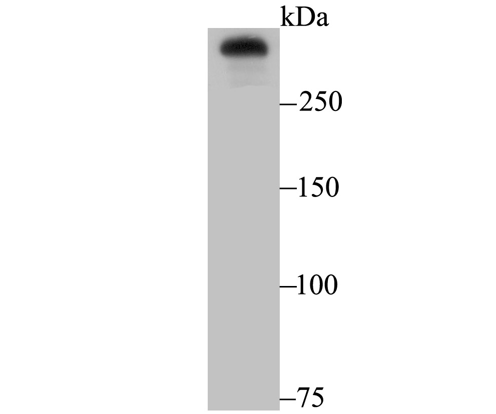
Fig1: Western blot analysis of Huntingtin on A431 cell using anti-Huntingtin antibody at 1/500 dilution.Application
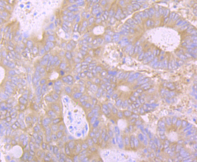
Fig2: Immunohistochemical analysis of paraffin-embedded human colon cancer tissue using anti-Huntingtin antibody. Counter stained with hematoxylin.Application
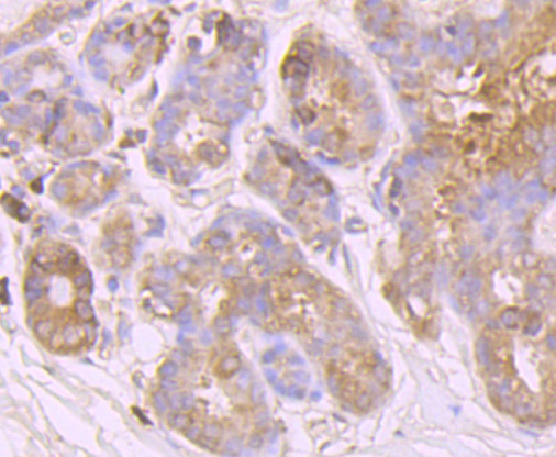
Fig3: Immunohistochemical analysis of paraffin-embedded human breast tissue using anti-Huntingtin antibody. Counter stained with hematoxylin.Application
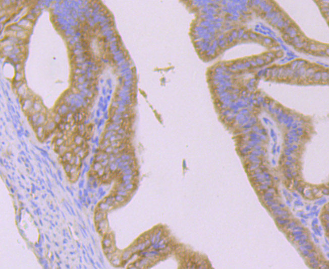
Fig4: Immunohistochemical analysis of paraffin-embedded mouse epididymis tissue using anti-Huntingtin antibody. Counter stained with hematoxylin.Application
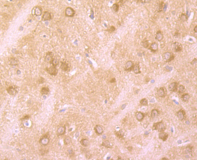
Fig5: Immunohistochemical analysis of paraffin-embedded rat brain tissue using anti-Huntingtin antibody. Counter stained with hematoxylin.Application
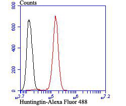
Fig6: Flow cytometric analysis of SH-SY-5Y cells with Huntingtin antibody at 1/100 dilution (red) compared with an unlabelled control (cells without incubation with primary antibody; black). Alexa Fluor 488-conjugated goat anti rabbit IgG was used as the secondary antibody.| Product Name | Huntingtin [JB89-34] |
|---|---|
| Antibody Type | Primary Antibodies |
| Antigen Alias | AI256365 antibody C430023I11Rik antibody HD antibody HD protein antibody HD_HUMAN antibody HDH antibody HTT antibody Huntingtin antibody HUNTINGTON CHOREA antibody Huntington disease protein antibody Huntington's disease protein homolog antibody IT 15 antibody IT15 antibody OTTMUSP00000026909 antibody ZHD antibody |
| Product description | The huntingtin gene, also called the HTT or HD (Huntington disease) gene, is the IT15 ("interesting transcript 15") gene, which codes for a protein called the huntingtin protein. It is variable in its structure, as the many polymorphisms of the gene can lead to variable numbers of glutamine residues present in the protein. The mass of huntingtin protein is dependent largely on the number of glutamine residues it has, the predicted mass is around 350 kDa. Normal huntingtin is generally accepted to be 3144 amino acids in size. The exact function of this protein is not known, but it plays an important role in nerve cells. Within cells, huntingtin may be involved in signaling, transporting materials, binding proteins and other structures, and protecting against programmed cell death (apoptosis). The huntingtin protein is required for normal development before birth. It is expressed in many tissues in the body, with the highest levels of expression seen in the brain. |
| Immunogen | Recombinant protein |
| Clonality | Monoclonal |
|---|---|
| Isotype | IgG |
| Host Species | Recombinant rabbit |
| Tested Applications | WBIHCFCICC |
| WB:1:500 IHC:1:50-1:200 FC:1:50-1:100 ICC:1:50 |
|
| Species Reactivity | HumanMouseRat |
| Concentration | 1mg/ml |
| Alternative Names | AI256365 antibody C430023I11Rik antibody HD antibody HD protein antibody HD_HUMAN antibody HDH antibody HTT antibody Huntingtin antibody HUNTINGTON CHOREA antibody Huntington disease protein antibody Huntington's disease protein homolog antibody IT 15 antibody IT15 antibody OTTMUSP00000026909 antibody ZHD antibody |
|---|---|
| Molecular Weight(MW) | 347 kDa |
| Cellular Localization | Cytoplasm. Nucleus. |
Application
Fig1: Western blot analysis of Huntingtin on A431 cell using anti-Huntingtin antibody at 1/500 dilution.
Application
Fig2: Immunohistochemical analysis of paraffin-embedded human colon cancer tissue using anti-Huntingtin antibody. Counter stained with hematoxylin.
Application
Fig3: Immunohistochemical analysis of paraffin-embedded human breast tissue using anti-Huntingtin antibody. Counter stained with hematoxylin.
Application
Fig4: Immunohistochemical analysis of paraffin-embedded mouse epididymis tissue using anti-Huntingtin antibody. Counter stained with hematoxylin.
Application
Fig5: Immunohistochemical analysis of paraffin-embedded rat brain tissue using anti-Huntingtin antibody. Counter stained with hematoxylin.
Application
Fig6: Flow cytometric analysis of SH-SY-5Y cells with Huntingtin antibody at 1/100 dilution (red) compared with an unlabelled control (cells without incubation with primary antibody; black). Alexa Fluor 488-conjugated goat anti rabbit IgG was used as the secondary antibody.| Positive Control | Rat brain tissue, human colon cancer tissue, human breast tissue, mouse epididymis tissue, SH-SY-5Y, A431. |
|---|---|
| Application Notes | WB:1:500 IHC:1:50-1:200 FC:1:50-1:100 ICC:1:50 |
| Form | Liquid |
|---|---|
| Storage Instructions | Store at +4℃ after thawing. Aliquot store at -20℃ or -80℃. Avoid repeated freeze / thaw cycles. |
| Storage Buffer | 1*TBS (pH7.4), 1%BSA, 40%Glycerol. Preservative: 0.05% Sodium Azide. |


