Application
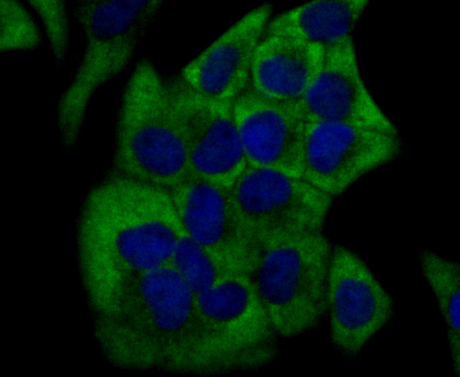
Fig1: ICC staining Cytochrome P450 17A1 in Hela cells (green). The nuclear counter stain is DAPI (blue). Cells were fixed in paraformaldehyde, permeabilised with 0.25% Triton X100/PBS.Application
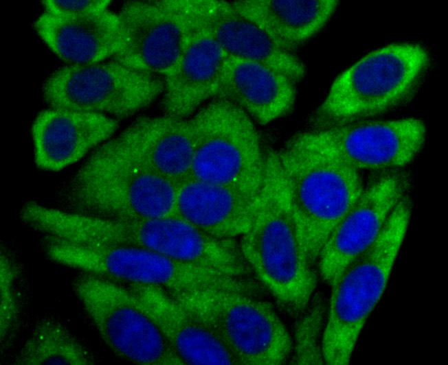
Fig2: ICC staining Cytochrome P450 17A1 in HepG2 cells (green). The nuclear counter stain is DAPI (blue). Cells were fixed in paraformaldehyde, permeabilised with 0.25% Triton X100/PBS.Application
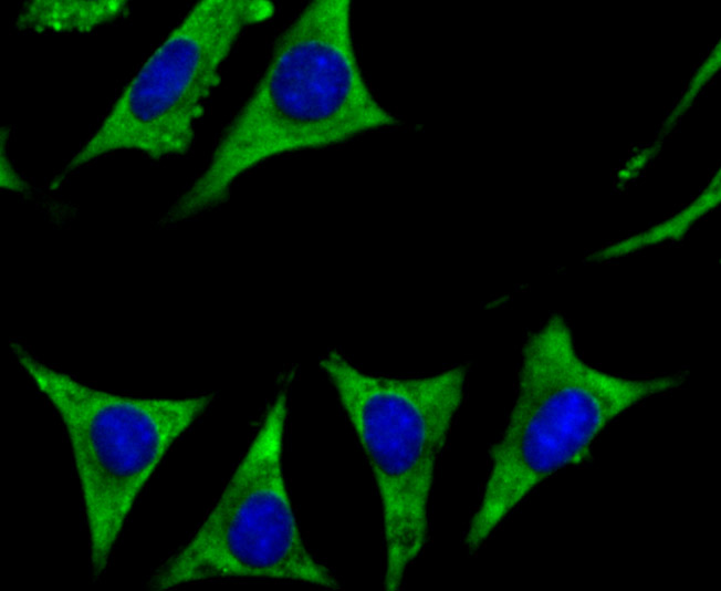
Fig3: ICC staining Cytochrome P450 17A1 in SH-SY-5Y cells (green). The nuclear counter stain is DAPI (blue). Cells were fixed in paraformaldehyde, permeabilised with 0.25% Triton X100/PBS.Application
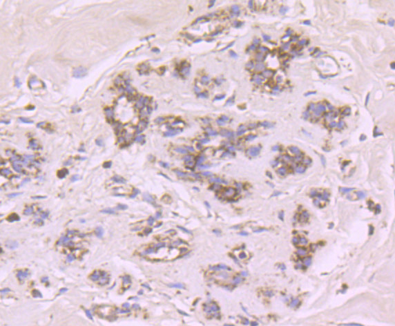
Fig4: Immunohistochemical analysis of paraffin-embedded human breast tissue using anti-Cytochrome P450 17A1 antibody. Counter stained with hematoxylin.Application
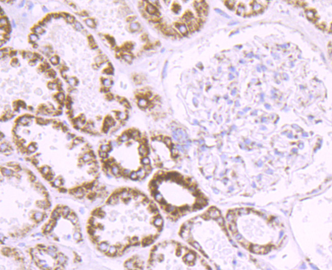
Fig5: Immunohistochemical analysis of paraffin-embedded human kidney tissue using anti-Cytochrome P450 17A1 antibody. Counter stained with hematoxylin.Application
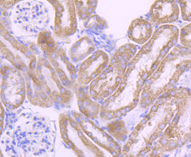
Fig6: Immunohistochemical analysis of paraffin-embedded mouse kidney tissue using anti-Cytochrome P450 17A1 antibody. Counter stained with hematoxylin.Application
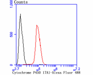
Fig7: Flow cytometric analysis of SH-SY-5Y cells with Cytochrome P450 17A1 antibody at 1/100 dilution (red) compared with an unlabelled control (cells without incubation with primary antibody; black). Alexa Fluor 488-conjugated goat anti-rabbit IgG was used as the secondary antibody.| Product Name | Cytochrome P450 17A1 [JB93-32] |
|---|---|
| Antibody Type | Primary Antibodies |
| Antigen Alias | 20 lyase antibody CP17A_HUMAN antibody CPT7 antibody CYP17 antibody CYP17A1 antibody CYPXVII antibody Cytochrome P450 17A1 antibody Cytochrome P450 family 17 antibody Cytochrome P450 family 17 subfamily A polypeptide 1 antibody Cytochrome p450 subfamily XVII (steroid 17 alpha hydroxylase) adrenal hyperplasia antibody Cytochrome p450 XVIIA1 antibody Cytochrome P450-C17 antibody Cytochrome P450c17 antibody OTTHUMP00000020382 antibody P450 C17 antibody P450c17 antibody S17AH antibody Steroid 17 alpha hydroxylase/17,20 lyase antibody Steroid 17 alpha monooxygenase antibody Steroid 17-alpha-hydroxylase/17 antibody Steroid 17-alpha-monooxygenase antibody |
| Product description | The cytochrome P450 proteins are monooxygenases that catalyze many reactions involved in drug metabolism and synthesis of cholesterol, steroids and other lipids. P450 enzymes are classified into subfamilies, such as CYP1A, CYP2A, CYP2C, CYP2D, CYP4A14, CYP7A, CYP7B, CYP8B, CYP11A, CYP17A1, CYPI9 and CYP27A, based on sequence similarities. CYP17A (17alpha-hydroxylase/17,20-lyase) is important for the conversion of pregnenolone and progesterone to dehydroepiandrosterone (DHEA) and androstenedione. In this process, it catalyzes both the 17-alpha-hydroxylation and the 17,20-lyase reaction. CYP17A1 is crucial during sexual development, both during fetal development and during puberty, and is intracellularly regulated by cAMP levels. Defects in the CYP17A1 gene, which encodes for the protein, may cause adrenal hyperplasia type V (AH-V) which is characterized by hypokalemia and hypertension. Male patients affected by AH-V do not undergo normal sexual differentiation and develop female external genitalia and do not undergo pubertal develoment. |
| Immunogen | Recombinant protein |
| Clonality | Monoclonal |
|---|---|
| Isotype | IgG |
| Host Species | Recombinant rabbit |
| Tested Applications | WBICCIFIHCFC |
| ICC:1:50-1:200 IHC:1:50-1:200 FC:1:50-1:100 WB:1:500 |
|
| Species Reactivity | HumanMouseRat |
| Concentration | 1mg/ml |
| Alternative Names | 20 lyase antibody
CP17A_HUMAN antibody
CPT7 antibody
CYP17 antibody
CYP17A1 antibody
CYPXVII antibody
Cytochrome P450 17A1 antibody
Cytochrome P450 family 17 antibody
Cytochrome P450 family 17 subfamily A polypeptide 1 antibody
Cytochrome p450 subfamily XVII (steroid 17 alpha hydroxylase) adrenal hyperplasia antibody
Cytochrome p450 XVIIA1 antibody
Cytochrome P450-C17 antibody
Cytochrome P450c17 antibody
OTTHUMP00000020382 antibody
P450 C17 antibody
P450c17 antibody
S17AH antibody
Steroid 17 alpha hydroxylase/17 20 lyase antibody Steroid 17 alpha monooxygenase antibody Steroid 17-alpha-hydroxylase/17 antibody Steroid 17-alpha-monooxygenase antibody |
|---|---|
| Molecular Weight(MW) | 57 kDa |
| Cellular Localization | Membrane. |
| SwissProt ID | P05093 |
|---|
Application
Fig1: ICC staining Cytochrome P450 17A1 in Hela cells (green). The nuclear counter stain is DAPI (blue). Cells were fixed in paraformaldehyde, permeabilised with 0.25% Triton X100/PBS.
Application
Fig2: ICC staining Cytochrome P450 17A1 in HepG2 cells (green). The nuclear counter stain is DAPI (blue). Cells were fixed in paraformaldehyde, permeabilised with 0.25% Triton X100/PBS.
Application
Fig3: ICC staining Cytochrome P450 17A1 in SH-SY-5Y cells (green). The nuclear counter stain is DAPI (blue). Cells were fixed in paraformaldehyde, permeabilised with 0.25% Triton X100/PBS.
Application
Fig4: Immunohistochemical analysis of paraffin-embedded human breast tissue using anti-Cytochrome P450 17A1 antibody. Counter stained with hematoxylin.
Application
Fig5: Immunohistochemical analysis of paraffin-embedded human kidney tissue using anti-Cytochrome P450 17A1 antibody. Counter stained with hematoxylin.
Application
Fig6: Immunohistochemical analysis of paraffin-embedded mouse kidney tissue using anti-Cytochrome P450 17A1 antibody. Counter stained with hematoxylin.
Application
Fig7: Flow cytometric analysis of SH-SY-5Y cells with Cytochrome P450 17A1 antibody at 1/100 dilution (red) compared with an unlabelled control (cells without incubation with primary antibody; black). Alexa Fluor 488-conjugated goat anti-rabbit IgG was used as the secondary antibody.| Positive Control | HepG2, Hela, SH-SY-5Y, human breast tissue, human kidney tissue, mouse kidney tissue. |
|---|---|
| Application Notes | ICC:1:50-1:200 IHC:1:50-1:200 FC:1:50-1:100 WB:1:500 |
| Form | Liquid |
|---|---|
| Storage Instructions | Store at +4℃ after thawing. Aliquot store at -20℃ or -80℃. Avoid repeated freeze / thaw cycles. |
| Storage Buffer | 1*TBS (pH7.4), 1%BSA, 40%Glycerol. Preservative: 0.05% Sodium Azide. |


