Application
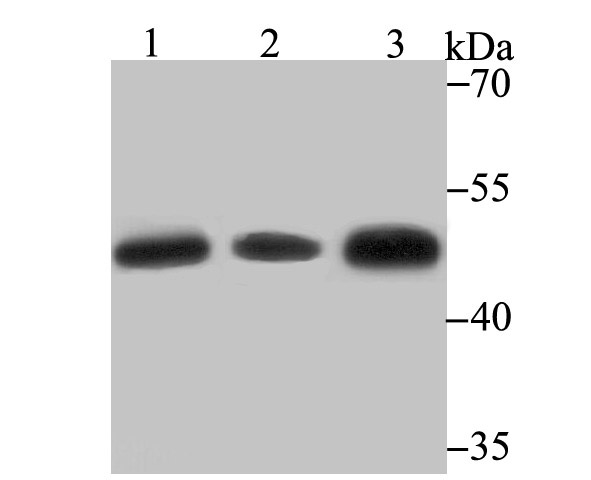
Fig1: Western blot analysis of Glutathione Synthetase on different lysates using anti-Glutathione Synthetase antibody at 1/500 dilution. Positive control: Lane 1: SiHa Lane 2: Mouse colon Lane 3: Rat liverApplication
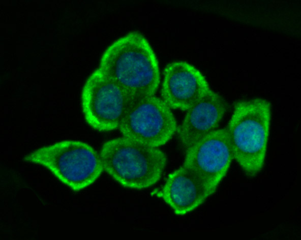
Fig2: ICC staining Glutathione Synthetase in LOVO cells (green). The nuclear counter stain is DAPI (blue). Cells were fixed in paraformaldehyde, permeabilised with 0.25% Triton X100/PBS.Application
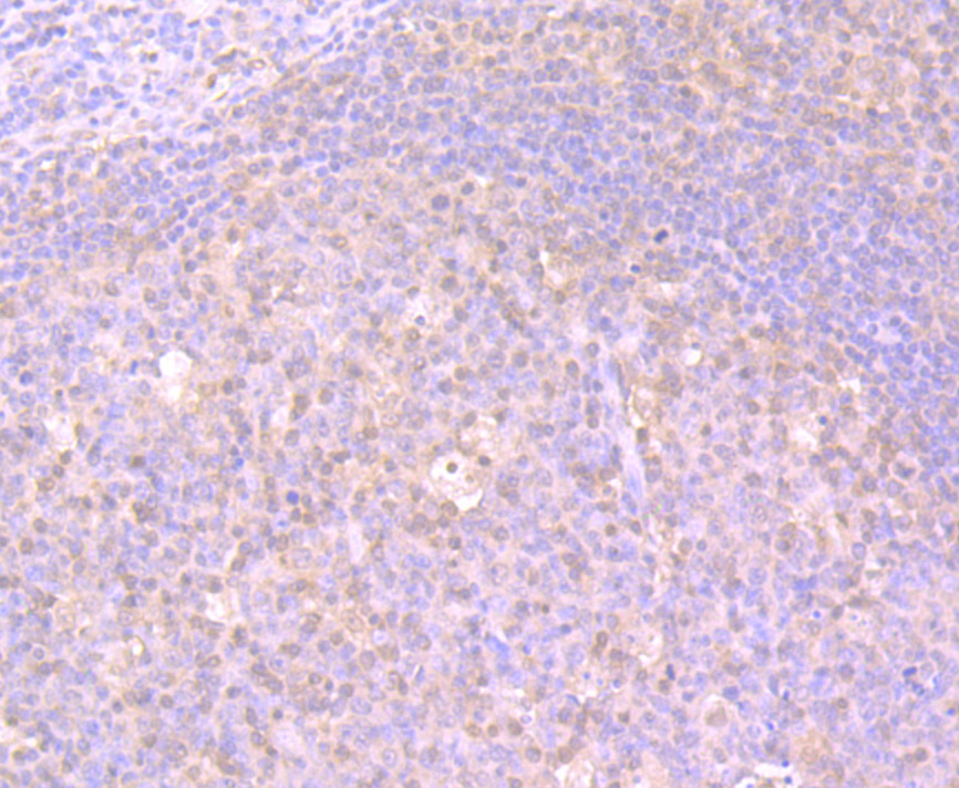
Fig3: Immunohistochemical analysis of paraffin-embedded human tonsil tissue using anti-Glutathione Synthetase antibody. Counter stained with hematoxylin.Application
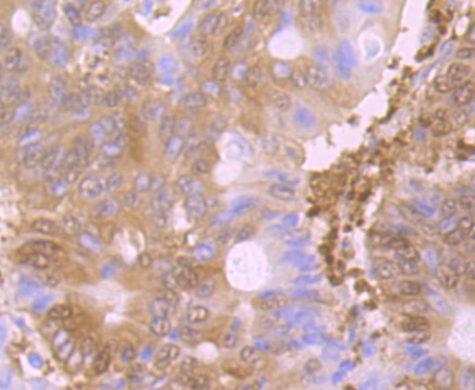
Fig4: Immunohistochemical analysis of paraffin-embedded human colon cancer tissue using anti-Glutathione Synthetase antibody. Counter stained with hematoxylin.Application
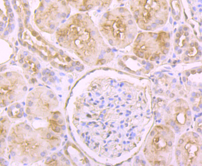
Fig5: Immunohistochemical analysis of paraffin-embedded human kidney tissue using anti-Glutathione Synthetase antibody. Counter stained with hematoxylin.Application
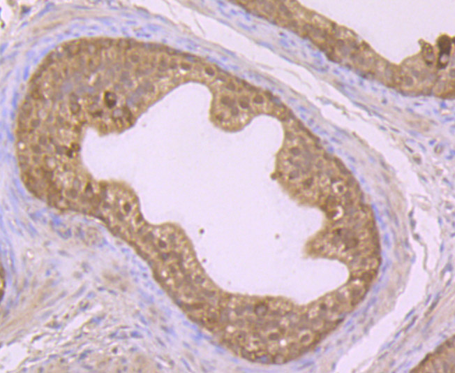
Fig6: Immunohistochemical analysis of paraffin-embedded rat epididymis tissue using anti-Glutathione Synthetase antibody. Counter stained with hematoxylin.Application
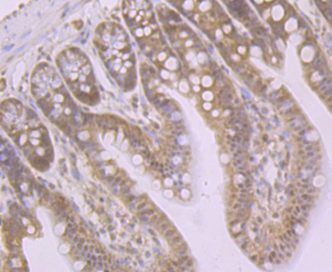
Fig7: Immunohistochemical analysis of paraffin-embedded mouse colon tissue using anti-Glutathione Synthetase antibody. Counter stained with hematoxylin.Application
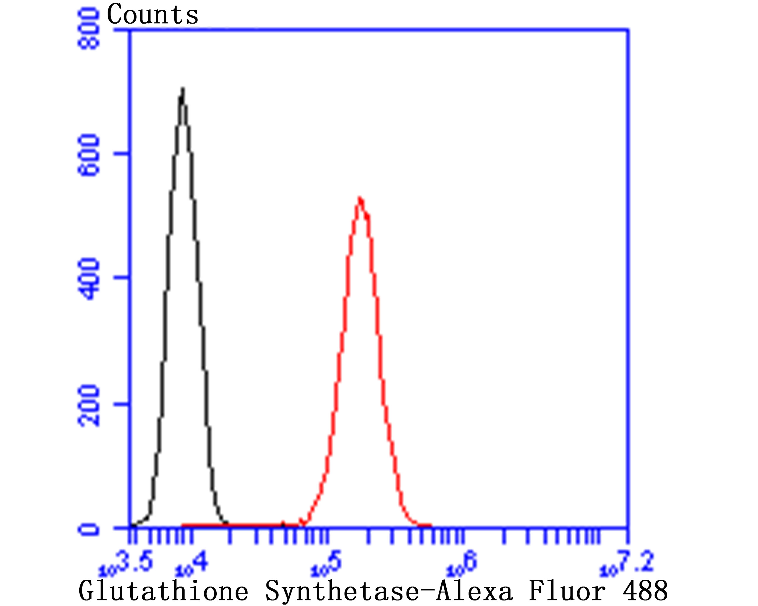
Fig8: Flow cytometric analysis of HepG2 cells with Glutathione Synthetase antibody at 1/100 dilution (red) compared with an unlabelled control (cells without incubation with primary antibody; black). Alexa Fluor 488-conjugated goat anti rabbit IgG was used as the secondary antibody.| Product Name | Glutathione Synthetase [JB95-33] |
|---|---|
| Antibody Type | Primary Antibodies |
| Antigen Alias | epididymis secretory sperm binding protein Li 64p antibody epididymis secretory sperm binding protein Li 88n antibody Glutathione synthase antibody Glutathione synthetase antibody GSH S antibody GSH synthetase antibody GSH-S antibody GSHB_HUMAN antibody GSHS antibody GSS antibody HEL-S-64p antibody HEL-S-88n antibody MGC14098 antibody OTTHUMP00000030711 antibody |
| Product description | GSS (Glutathione synthetase) is a 474 amino acid protein encoded by the gene located at human chromosome 20q11.2. GSS consists of three loops projecting from an antiparallel β-sheet, a parallel β-sheet and a lid of anti-parallel sheets, which provide access to the ATP-binding site. Although Southern blot and gene analysis suggest that GSS may be the only member of a unique family, the crystal structure indicates that GSS belongs to the ATP-GRASP superfamily. GSS is expressed in hemocytes and nucleated cells, including the brain. GSS occurs as a homodimer. There are two steps in the production of Glutathione, begining with γ-GCS and ending with GSS. In an ATP-dependent reaction, GSS produces Glutathione from γ-glutamylcysteine and glycine precursors. Partial hepatectomy, diethyl maleate, buthionine sulfoximine, tert-butylhaydroquinone and thioacetamide increase the ex-pression of GSS, which causes an increase in Glutathione levels. An inherited autosomal recessive disorder, 5-oxoprolinuria (pyroglutamic aciduria), is caused by GSS deficiencies, which leads to central nervous system damage, hemolytic anemia, metabolic acidosis and urinary excretion of 5-oxoproline. |
| Immunogen | Recombinant protein |
| Clonality | Monoclonal |
|---|---|
| Isotype | IgG |
| Host Species | Recombinant rabbit |
| Tested Applications | WBICCIFIHCFC |
| WB:1:500-1:1,000 ICC:1:50-1:200 IHC:1:50-1:200 FC:1:50-1:100 |
|
| Species Reactivity | HumanMouseRat |
| Concentration | 1mg/ml |
| Alternative Names | epididymis secretory sperm binding protein Li 64p antibody epididymis secretory sperm binding protein Li 88n antibody Glutathione synthase antibody Glutathione synthetase antibody GSH S antibody GSH synthetase antibody GSH-S antibody GSHB_HUMAN antibody GSHS antibody GSS antibody HEL-S-64p antibody HEL-S-88n antibody MGC14098 antibody OTTHUMP00000030711 antibody |
|---|---|
| Molecular Weight(MW) | 52 kDa |
| Cellular Localization | Cytosol. Extracellular region or secreted. |
Application
Fig1: Western blot analysis of Glutathione Synthetase on different lysates using anti-Glutathione Synthetase antibody at 1/500 dilution. Positive control: Lane 1: SiHa Lane 2: Mouse colon Lane 3: Rat liver
Application
Fig2: ICC staining Glutathione Synthetase in LOVO cells (green). The nuclear counter stain is DAPI (blue). Cells were fixed in paraformaldehyde, permeabilised with 0.25% Triton X100/PBS.
Application
Fig3: Immunohistochemical analysis of paraffin-embedded human tonsil tissue using anti-Glutathione Synthetase antibody. Counter stained with hematoxylin.
Application
Fig4: Immunohistochemical analysis of paraffin-embedded human colon cancer tissue using anti-Glutathione Synthetase antibody. Counter stained with hematoxylin.
Application
Fig5: Immunohistochemical analysis of paraffin-embedded human kidney tissue using anti-Glutathione Synthetase antibody. Counter stained with hematoxylin.
Application
Fig6: Immunohistochemical analysis of paraffin-embedded rat epididymis tissue using anti-Glutathione Synthetase antibody. Counter stained with hematoxylin.
Application
Fig7: Immunohistochemical analysis of paraffin-embedded mouse colon tissue using anti-Glutathione Synthetase antibody. Counter stained with hematoxylin.
Application
Fig8: Flow cytometric analysis of HepG2 cells with Glutathione Synthetase antibody at 1/100 dilution (red) compared with an unlabelled control (cells without incubation with primary antibody; black). Alexa Fluor 488-conjugated goat anti rabbit IgG was used as the secondary antibody.| Positive Control | SiHa, mouse colon tissue, rat liver tissue lysate, rat epididymis tissue, human tonsil tissue, human colon cancer tissue, human kidney tissue, HepG2, LOVO. |
|---|---|
| Application Notes | WB:1:500-1:1,000 ICC:1:50-1:200 IHC:1:50-1:200 FC:1:50-1:100 |
| Form | Liquid |
|---|---|
| Storage Instructions | Store at +4℃ after thawing. Aliquot store at -20℃ or -80℃. Avoid repeated freeze / thaw cycles. |
| Storage Buffer | 1*TBS (pH7.4), 1%BSA, 40%Glycerol. Preservative: 0.05% Sodium Azide. |


