Application
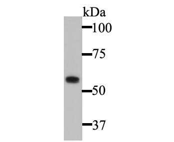
Fig1: Western blot analysis of Cytokeratin 5 on A431 cell lysate. Proteins were transferred to a PVDF membrane and blocked with 5% BSA in PBS for 1 hour at room temperature. The primary antibody was used at a 1:2,000 dilution in 5% BSA at room temperature for 2 hours. Goat Anti-Rabbit IgG - HRP Secondary Antibody (HA1001) at 1:5,000 dilution was used for 1 hour at room temperature.Application
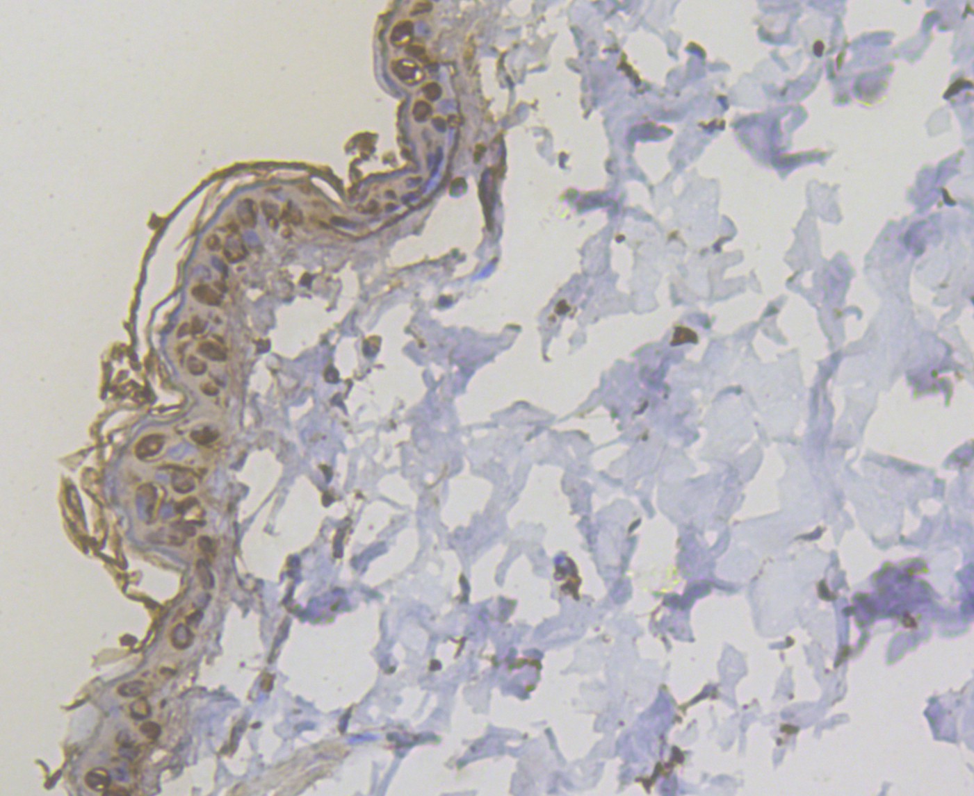
Fig2: Immunohistochemical analysis of paraffin-embedded rat skin tissue using anti-Cytokeratin 5 antibody. The section was pre-treated using heat mediated antigen retrieval with sodium citrate buffer (pH 6.0) for 20 minutes. The tissues were blocked in 5% BSA for 30 minutes at room temperature, washed with ddH2O and PBS, and then probed with the antibody (ER1901-03) at 1/200 dilution, for 30 minutes at room temperature and detected using an HRP conjugated compact polymer system. DAB was used as the chromogen. Counter stained with hematoxylin and mounted with DPX.Application
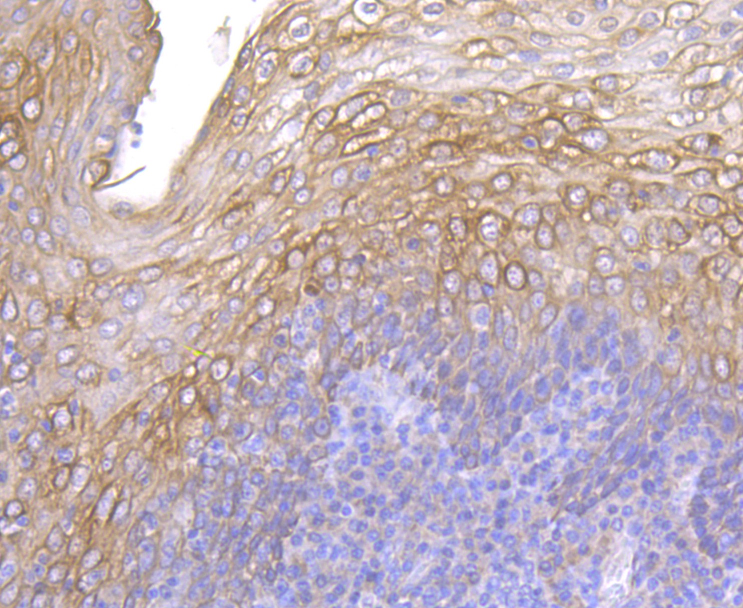
Fig3: Immunohistochemical analysis of paraffin-embedded human tonsil tissue using anti-Cytokeratin 5 antibody. The section was pre-treated using heat mediated antigen retrieval with sodium citrate buffer (pH 6.0) for 20 minutes. The tissues were blocked in 5% BSA for 30 minutes at room temperature, washed with ddH2O and PBS, and then probed with the antibody (ER1901-03) at 1/200 dilution, for 30 minutes at room temperature and detected using an HRP conjugated compact polymer system. DAB was used as the chromogen. Counter stained with hematoxylin and mounted with DPX.Application
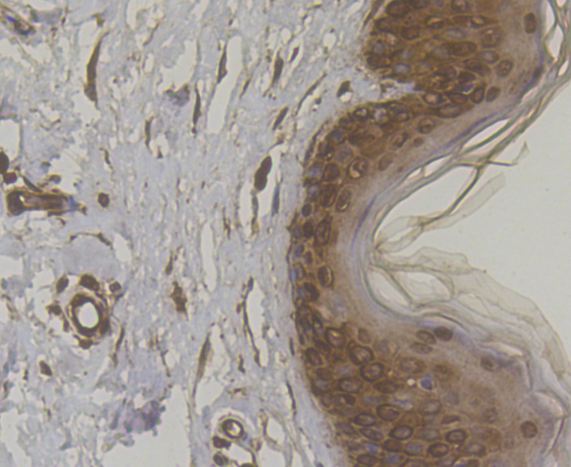
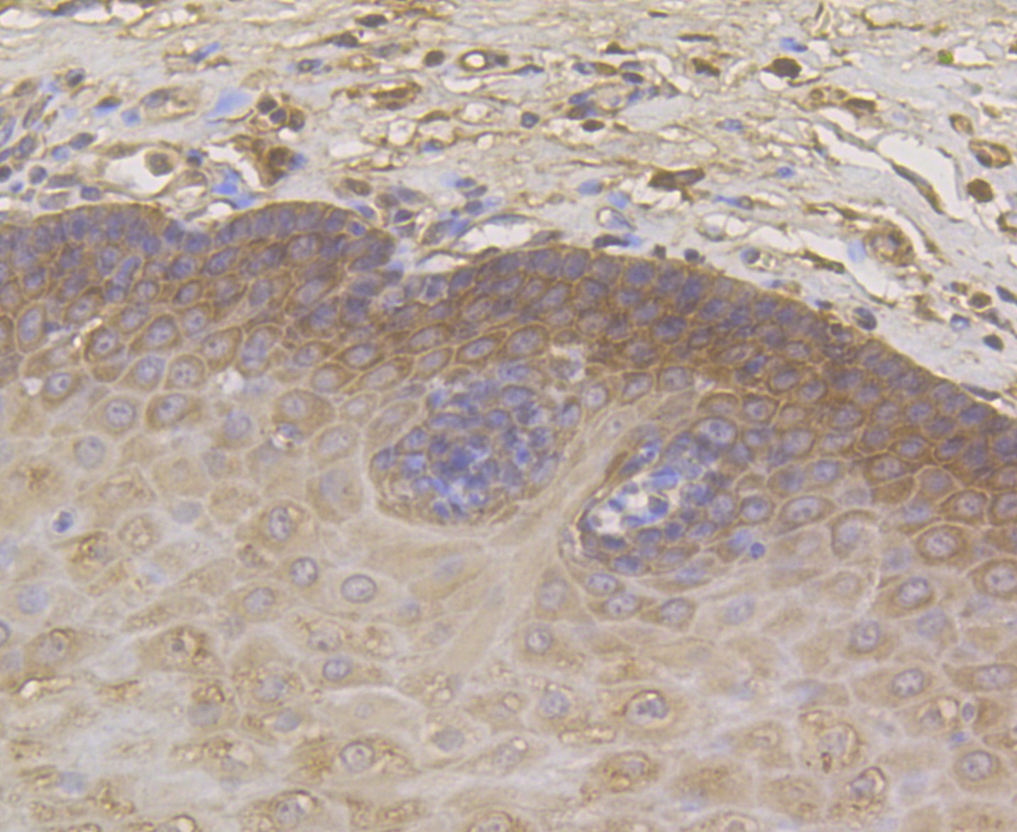
Fig4: Immunohistochemical analysis of paraffin-embedded human skin tissue using anti-Cytokeratin 5 antibody. The section was pre-treated using heat mediated antigen retrieval with sodium citrate buffer (pH 6.0) for 20 minutes. The tissues were blocked in 5% BSA for 30 minutes at room temperature, washed with ddH2O and PBS, and then probed with the antibody (ER1901-03) at 1/200 dilution, for 30 minutes at room temperature and detected using an HRP conjugated compact polymer system. DAB was used as the chromogen. Counter stained with hematoxylin and mounted with DPX.Application
Fig5: Immunohistochemical analysis of paraffin-embedded human esophagus tissue using anti-Cytokeratin 5 antibody. The section was pre-treated using heat mediated antigen retrieval with sodium citrate buffer (pH 6.0) for 20 minutes. The tissues were blocked in 5% BSA for 30 minutes at room temperature, washed with ddH2O and PBS, and then probed with the antibody (ER1901-03) at 1/50 dilution, for 30 minutes at room temperature and detected using an HRP conjugated compact polymer system. DAB was used as the chromogen. Counter stained with hematoxylin and mounted with DPX.Application
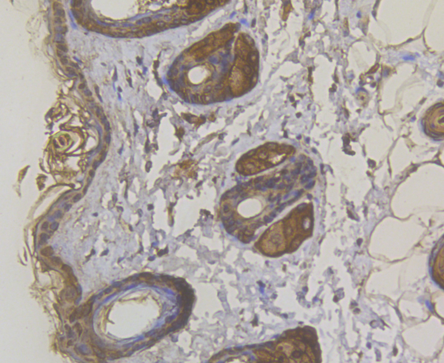
Fig6: Immunohistochemical analysis of paraffin-embedded mouse skin tissue using anti-Cytokeratin 5 antibody. The section was pre-treated using heat mediated antigen retrieval with sodium citrate buffer (pH 6.0) for 20 minutes. The tissues were blocked in 5% BSA for 30 minutes at room temperature, washed with ddH2O and PBS, and then probed with the antibody (ER1901-03) at 1/200 dilution, for 30 minutes at room temperature and detected using an HRP conjugated compact polymer system. DAB was used as the chromogen. Counter stained with hematoxylin and mounted with DPX.| Product Name | Cytokeratin 5 |
|---|---|
| Antibody Type | Primary Antibodies |
| Antigen Alias | 58 kDa cytokeratin antibody CK-5 antibody CK5 antibody Cytokeratin-5 antibody Cytokeratin5 antibody DDD antibody DDD1 antibody EBS2 antibody epidermolysis bullosa simplex 2 Dowling-Meara/Kobner/Weber-Cockayne types antibody K2C5_HUMAN antibody K5 antibody keratin 5 (epidermolysis bullosa simplex, Dowling-Meara/Kobner/Weber-Cockayne types) antibody Keratin 5 antibody Keratin antibody keratin complex 2, basic, gene 5 antibody keratin, type II cytoskeletal 5 antibody Keratin-5 antibody Keratin5 antibody KRT 5 antibody Krt5 antibody KRT5A antibody type II cytoskeletal 5 antibody Type-II keratin Kb5 antibody |
| Product description | The protein encoded by this gene is a member of the keratin gene family. Cytokeratins comprise a diverse group of intermediate filament proteins (IFPs) that are expressed as pairs in both keratinized and non-keratinized epithelial tissue. Cytokeratins play a critical role in differentiation and tissue specialization and function to maintain the overall structural integrity of epithelial cells. Keratin 5 (and K14) are expressed primarily in basal keratinocytes in the epidermis, specifically in the stratified epithelium lining the skin and digestive tract. Keratin intermediate filaments make up the cytoskeletal scaffold within epithelial cells, which contributes to the cell architecture and provides the cells with the ability to withstand mechanical, and non-mechanical, stresses. K5/K14 keratin pairs are able to undergo extensive bundling due to the non-helical tail of K15 acting as a weak cross-linker at the intermediate filament surface. This bundling increases the elasticity, and therefore the mechanical resilience, of the intermediate filaments. K5/K14 intermediate filaments are anchored to the desmosomes of basal cells via desmoplakin and plakophilin-1, connecting the cells to their neighbours. At the hemidesmosome, plectin and BPAG1 associate with transmembrane proteins α6β4 integrin, a type of cell adhesion molecule, and BP180/collagen XVII, linking K5/K14 filaments in the basal cells to the basal lamina. Mutations of the cytokeratin 5 gene (KRT5) have been shown to result in the autosomal dominant disorder epidermolysis bullosa (EB). |
| Immunogen | Synthetic peptide within Human Cytokeratin 5 aa 346-379. |
| Clonality | Polyclonal |
|---|---|
| Isotype | IgG |
| Host Species | Rabbit |
| Tested Applications | WBIHC |
| WB:1:500-1:2,000: IHC:1:50-1:200: |
|
| Species Reactivity | HumanMouseRat |
| Concentration | 1mg/ml |
| Alternative Names | 58 kDa cytokeratin antibody
CK-5 antibody
CK5 antibody
Cytokeratin-5 antibody
Cytokeratin5 antibody
DDD antibody
DDD1 antibody
EBS2 antibody
epidermolysis bullosa simplex 2 Dowling-Meara/Kobner/Weber-Cockayne types antibody
K2C5_HUMAN antibody
K5 antibody
keratin 5 (epidermolysis bullosa simplex Dowling-Meara/Kobner/Weber-Cockayne types) antibody Keratin 5 antibody Keratin antibody keratin complex 2 basic gene 5 antibody keratin type II cytoskeletal 5 antibody Keratin-5 antibody Keratin5 antibody KRT 5 antibody Krt5 antibody KRT5A antibody type II cytoskeletal 5 antibody Type-II keratin Kb5 antibody |
|---|---|
| Molecular Weight(MW) | 62 kDa |
| Cellular Localization | Cytoskeleton. Cytosol. Nucleus. Extracellular region or secreted. |
Application
Fig1: Western blot analysis of Cytokeratin 5 on A431 cell lysate. Proteins were transferred to a PVDF membrane and blocked with 5% BSA in PBS for 1 hour at room temperature. The primary antibody was used at a 1:2,000 dilution in 5% BSA at room temperature for 2 hours. Goat Anti-Rabbit IgG - HRP Secondary Antibody (HA1001) at 1:5,000 dilution was used for 1 hour at room temperature.
Application
Fig2: Immunohistochemical analysis of paraffin-embedded rat skin tissue using anti-Cytokeratin 5 antibody. The section was pre-treated using heat mediated antigen retrieval with sodium citrate buffer (pH 6.0) for 20 minutes. The tissues were blocked in 5% BSA for 30 minutes at room temperature, washed with ddH2O and PBS, and then probed with the antibody (ER1901-03) at 1/200 dilution, for 30 minutes at room temperature and detected using an HRP conjugated compact polymer system. DAB was used as the chromogen. Counter stained with hematoxylin and mounted with DPX.
Application
Fig3: Immunohistochemical analysis of paraffin-embedded human tonsil tissue using anti-Cytokeratin 5 antibody. The section was pre-treated using heat mediated antigen retrieval with sodium citrate buffer (pH 6.0) for 20 minutes. The tissues were blocked in 5% BSA for 30 minutes at room temperature, washed with ddH2O and PBS, and then probed with the antibody (ER1901-03) at 1/200 dilution, for 30 minutes at room temperature and detected using an HRP conjugated compact polymer system. DAB was used as the chromogen. Counter stained with hematoxylin and mounted with DPX.
Application
Fig4: Immunohistochemical analysis of paraffin-embedded human skin tissue using anti-Cytokeratin 5 antibody. The section was pre-treated using heat mediated antigen retrieval with sodium citrate buffer (pH 6.0) for 20 minutes. The tissues were blocked in 5% BSA for 30 minutes at room temperature, washed with ddH2O and PBS, and then probed with the antibody (ER1901-03) at 1/200 dilution, for 30 minutes at room temperature and detected using an HRP conjugated compact polymer system. DAB was used as the chromogen. Counter stained with hematoxylin and mounted with DPX.
Application
Fig5: Immunohistochemical analysis of paraffin-embedded human esophagus tissue using anti-Cytokeratin 5 antibody. The section was pre-treated using heat mediated antigen retrieval with sodium citrate buffer (pH 6.0) for 20 minutes. The tissues were blocked in 5% BSA for 30 minutes at room temperature, washed with ddH2O and PBS, and then probed with the antibody (ER1901-03) at 1/50 dilution, for 30 minutes at room temperature and detected using an HRP conjugated compact polymer system. DAB was used as the chromogen. Counter stained with hematoxylin and mounted with DPX.
Application
Fig6: Immunohistochemical analysis of paraffin-embedded mouse skin tissue using anti-Cytokeratin 5 antibody. The section was pre-treated using heat mediated antigen retrieval with sodium citrate buffer (pH 6.0) for 20 minutes. The tissues were blocked in 5% BSA for 30 minutes at room temperature, washed with ddH2O and PBS, and then probed with the antibody (ER1901-03) at 1/200 dilution, for 30 minutes at room temperature and detected using an HRP conjugated compact polymer system. DAB was used as the chromogen. Counter stained with hematoxylin and mounted with DPX.| Positive Control | A431, rat skin tissue, human tonsil tissue, human skin tissue, human esophagus tissue, mouse skin tissue. |
|---|---|
| Application Notes | WB:1:500-1:2,000: IHC:1:50-1:200: |
| Form | Liquid |
|---|---|
| Storage Instructions | Store at +4℃ after thawing. Aliquot store at -20℃. Avoid repeated freeze / thaw cycles. |
| Storage Buffer | 1*TBS (pH7.4), 1%BSA, 50%Glycerol. Preservative: 0.05% Sodium Azide. |


