Application
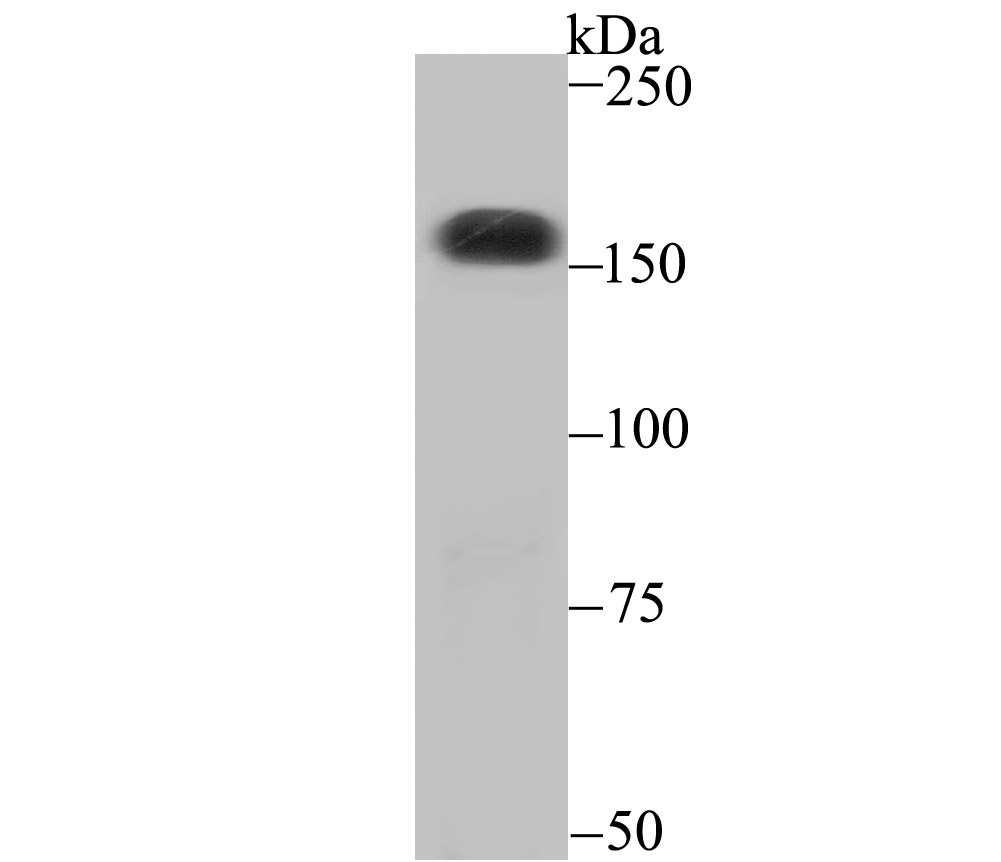
Fig1: Western blot analysis of CPS1 on SiHa cell using anti-CPS1 antibody at 1/500 dilution.Application
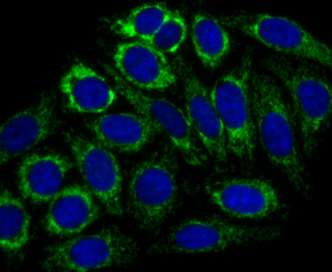
Fig2: ICC staining CPS1 in HepG2 cells (green). The nuclear counter stain is DAPI (blue). Cells were fixed in paraformaldehyde, permeabilised with 0.25% Triton X100/PBS.Application
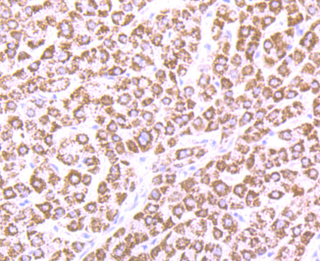
Fig3: Immunohistochemical analysis of paraffin-embedded human liver tissue using anti-CPS1 antibody. Counter stained with hematoxylin.Application
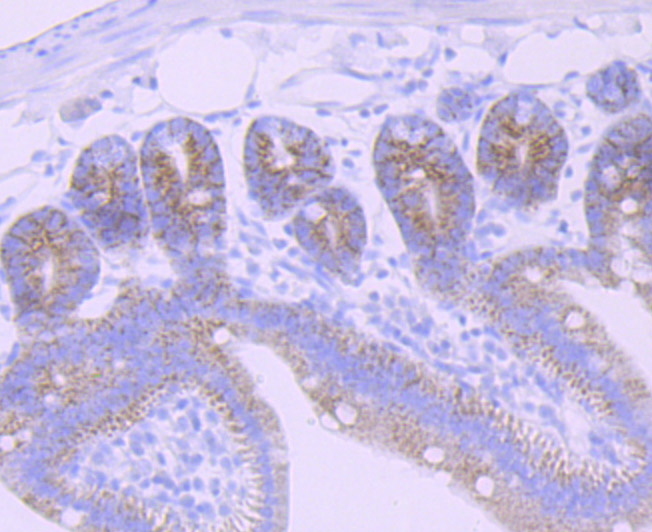
Fig4: Immunohistochemical analysis of paraffin-embedded mouse small intestine tissue using anti-CPS1 antibody. Counter stained with hematoxylin.| Product Name | CPS1 [JB40-33] |
|---|---|
| Antibody Type | Primary Antibodies |
| Antigen Alias | Carbamoyl phosphate synthase [ammonia] antibody Carbamoyl phosphate synthase [ammonia] mitochondrial antibody Carbamoyl phosphate synthase antibody Carbamoyl phosphate synthetase 1 antibody Carbamoyl phosphate synthetase 1 mitochondrial antibody Carbamoyl phosphate synthetase I antibody Carbamoyl-phosphate synthase [ammonia] antibody Carbamoyl-phosphate synthetase I antibody Carbamoylphosphate synthase antibody Carbamoylphosphate synthetase 1 antibody Carbamoylphosphate synthetase I antibody CPS 1 antibody Cps1 antibody CPSase 1 antibody CPSase I antibody CPSASE1 antibody CPSM_HUMAN antibody mitochondrial antibody MS738 antibody |
| Product description | The multicomplex protein, carbamoyl-phosphate synthetase-aspartate carbamoyl transferase-dihydro-orotase (CAD), consists of three distinct proteins, carbamoyl phosphate synthetase 2 (CPS2), aspartate transcarbamylase, and dihydro-orotase, which catalyze the second and third steps of pyrimidine biosynthesis. CAD is allosterically regulated by the phosphorylation of CPS2 by cyclic AMP-dependent protein kinase, and this activation enables CPS2 to catalyze the rate-limiting step of pyrimidine synthesis. CAD is expressed in brain and skeletal muscle. A related protein, carbamoyl phosphate synthetase 1 (CPS1) is expressed in liver. CPS1 catalyzes the rate-limiting step in the urea cycle, and deficiency of CPS1 is an autosomal recessive disorder that causes hyperammonemia. |
| Immunogen | Recombinant protein |
| Clonality | Monoclonal |
|---|---|
| Isotype | IgG |
| Host Species | Recombinant rabbit |
| Tested Applications | WBICCIFIHCIP |
| WB:1:500-1:1,000 ICC:1:50-1:200 IHC:1:50-1:200 |
|
| Species Reactivity | HumanMouseRat |
| Concentration | 1mg/ml |
| Alternative Names | Carbamoyl phosphate synthase [ammonia] antibody Carbamoyl phosphate synthase [ammonia] mitochondrial antibody Carbamoyl phosphate synthase antibody Carbamoyl phosphate synthetase 1 antibody Carbamoyl phosphate synthetase 1 mitochondrial antibody Carbamoyl phosphate synthetase I antibody Carbamoyl-phosphate synthase [ammonia] antibody Carbamoyl-phosphate synthetase I antibody Carbamoylphosphate synthase antibody Carbamoylphosphate synthetase 1 antibody Carbamoylphosphate synthetase I antibody CPS 1 antibody Cps1 antibody CPSase 1 antibody CPSase I antibody CPSASE1 antibody CPSM_HUMAN antibody mitochondrial antibody MS738 antibody |
|---|---|
| Molecular Weight(MW) | 165 kDa |
| Cellular Localization | Mitochondrion. |
| SwissProt ID | P31327 |
|---|
Application
Fig1: Western blot analysis of CPS1 on SiHa cell using anti-CPS1 antibody at 1/500 dilution.
Application
Fig2: ICC staining CPS1 in HepG2 cells (green). The nuclear counter stain is DAPI (blue). Cells were fixed in paraformaldehyde, permeabilised with 0.25% Triton X100/PBS.
Application
Fig3: Immunohistochemical analysis of paraffin-embedded human liver tissue using anti-CPS1 antibody. Counter stained with hematoxylin.
Application
Fig4: Immunohistochemical analysis of paraffin-embedded mouse small intestine tissue using anti-CPS1 antibody. Counter stained with hematoxylin.| Positive Control | HepG2, SiHa, human liver tissue, mouse small intestine tissue. |
|---|---|
| Application Notes | WB:1:500-1:1,000 ICC:1:50-1:200 IHC:1:50-1:200 |
| Form | Liquid |
|---|---|
| Storage Instructions | Store at +4℃ after thawing. Aliquot store at -20℃ or -80℃. Avoid repeated freeze / thaw cycles. |
| Storage Buffer | 1*TBS (pH7.4), 1%BSA, 40%Glycerol. Preservative: 0.05% Sodium Azide. |


