Application
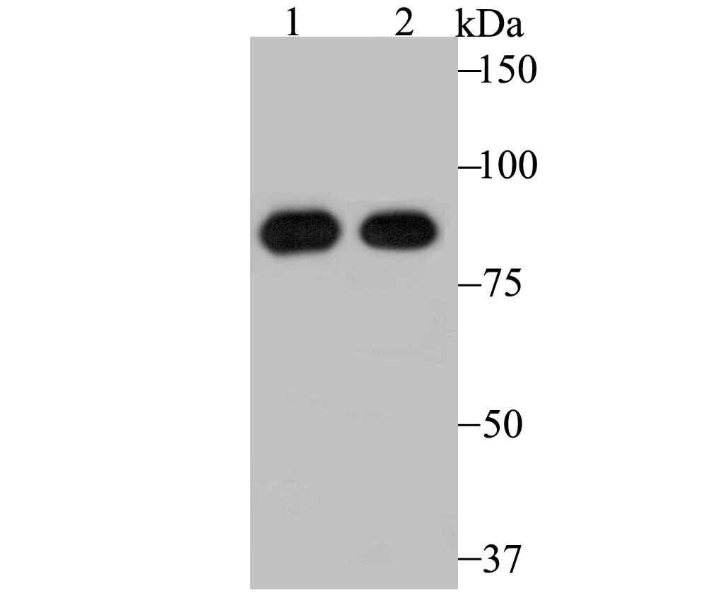
Fig1: Western blot analysis of RRM1 on mouse thymus tissue (1) and A431 cell (2) lysate using anti-RRM1 antibody at 1/500 dilution.Application
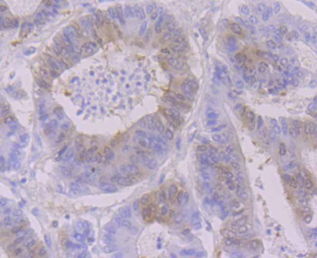
Fig2: Immunohistochemical analysis of paraffin-embedded human colon tissue tissue using anti-RRM1 antibody. Counter stained with hematoxylin.Application
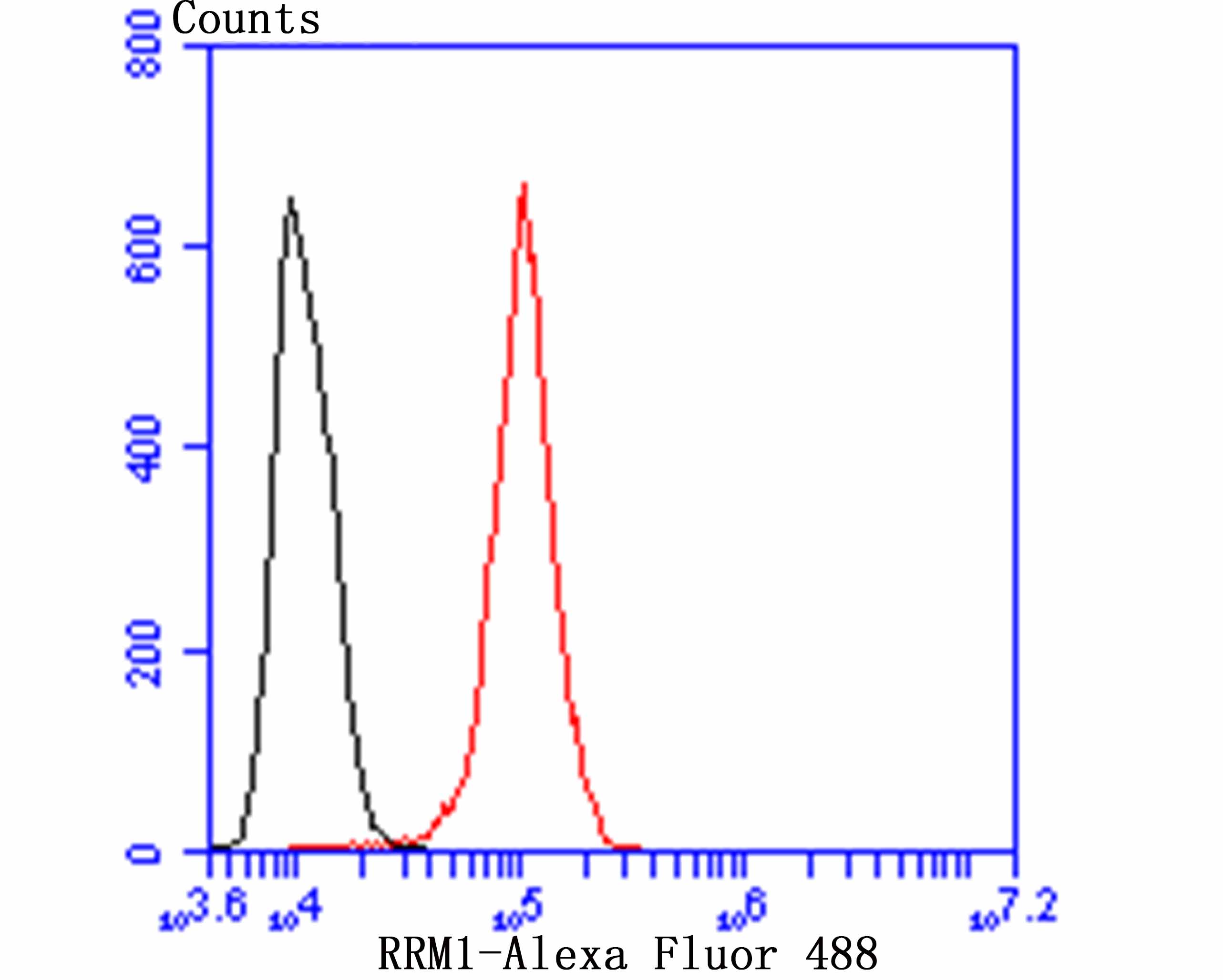
Fig3: Flow cytometric analysis of A431 cells with RRM1 antibody at 1/50 dilution (blue) compared with an unlabelled control (cells without incubation with primary antibody; red). Alexa Fluor 488-conjugated goat anti-rabbit IgG was used as the secondary antibody.| Product Name | RRM1 [JB09-43] |
|---|---|
| Antibody Type | Primary Antibodies |
| Antigen Alias | R1 antibody Ribonucleoside diphosphate reductase large subunit antibody Ribonucleoside diphosphate reductase M1 chain antibody Ribonucleoside diphosphate reductase subunit M1 antibody Ribonucleoside reductase, large subunit antibody Ribonucleoside-diphosphate reductase large subunit antibody Ribonucleoside-diphosphate reductase subunit M1 antibody Ribonucleotide reductase large chain antibody Ribonucleotide reductase large subunit antibody Ribonucleotide reductase M1 antibody Ribonucleotide reductase M1 polypeptide antibody Ribonucleotide reductase R1 subunit antibody Ribonucleotide reductase, M1 subunit antibody RIR 1 antibody RIR1 antibody RIR1_HUMAN antibody RR 1 antibody RR1 antibody RRM 1 antibody RRM1 antibody |
| Product description | Provides the precursors necessary for DNA synthesis. Catalyzes the biosynthesis of deoxyribonucleotides from the corresponding ribonucleotides. Belongs to the ribonucleoside diphosphate reductase large chain family. Contains 1 ATP-cone domain. |
| Immunogen | Recombinant protein |
| Clonality | Monoclonal |
|---|---|
| Isotype | IgG |
| Host Species | Recombinant rabbit |
| Tested Applications | WBIHCFCIP |
| WB:1:500-1:2,000 IHC:1:50-1:100 FC:1:50-1:100 IP:1:10-1:50: |
|
| Species Reactivity | HumanMouse |
| Concentration | 1mg/ml |
| Alternative Names | R1 antibody
Ribonucleoside diphosphate reductase large subunit antibody
Ribonucleoside diphosphate reductase M1 chain antibody
Ribonucleoside diphosphate reductase subunit M1 antibody
Ribonucleoside reductase large subunit antibody Ribonucleoside-diphosphate reductase large subunit antibody Ribonucleoside-diphosphate reductase subunit M1 antibody Ribonucleotide reductase large chain antibody Ribonucleotide reductase large subunit antibody Ribonucleotide reductase M1 antibody Ribonucleotide reductase M1 polypeptide antibody Ribonucleotide reductase R1 subunit antibody Ribonucleotide reductase M1 subunit antibody RIR 1 antibody RIR1 antibody RIR1_HUMAN antibody RR 1 antibody RR1 antibody RRM 1 antibody RRM1 antibody |
|---|---|
| Molecular Weight(MW) | 90 kDa |
| Cellular Localization | Cytoplasm. |
Application
Fig1: Western blot analysis of RRM1 on mouse thymus tissue (1) and A431 cell (2) lysate using anti-RRM1 antibody at 1/500 dilution.
Application
Fig2: Immunohistochemical analysis of paraffin-embedded human colon tissue tissue using anti-RRM1 antibody. Counter stained with hematoxylin.
Application
Fig3: Flow cytometric analysis of A431 cells with RRM1 antibody at 1/50 dilution (blue) compared with an unlabelled control (cells without incubation with primary antibody; red). Alexa Fluor 488-conjugated goat anti-rabbit IgG was used as the secondary antibody.| Positive Control | Mouse thymus tissue lysate, A431, human colon cancer. |
|---|---|
| Application Notes | WB:1:500-1:2,000 IHC:1:50-1:100 FC:1:50-1:100 IP:1:10-1:50: |
| Form | Liquid |
|---|---|
| Storage Instructions | Store at +4℃ after thawing. Aliquot store at -20℃ or -80℃. Avoid repeated freeze / thaw cycles. |
| Storage Buffer | 1*TBS (pH7.4), 1%BSA, 40%Glycerol. Preservative: 0.05% Sodium Azide. |


