Application
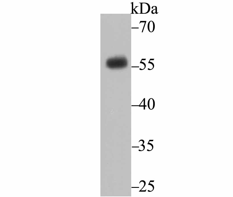
Fig1: Western blot analysis of CD27 on Raji cell using anti-CD27 antibody at 1/500 dilution.Application
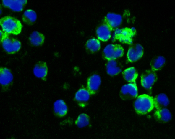
Fig2: ICC staining CD27 in K562 cells (green). The nuclear counter stain is DAPI (blue). Cells were fixed in paraformaldehyde, permeabilised with 0.25% Triton X100/PBS.Application
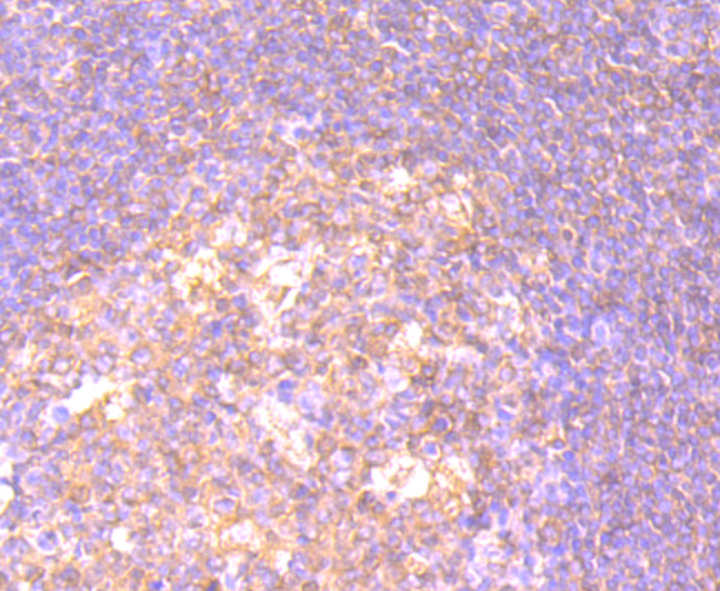
Fig3: Immunohistochemical analysis of paraffin-embedded human tonsil tissue using anti-CD27 antibody. Counter stained with hematoxylin.Application
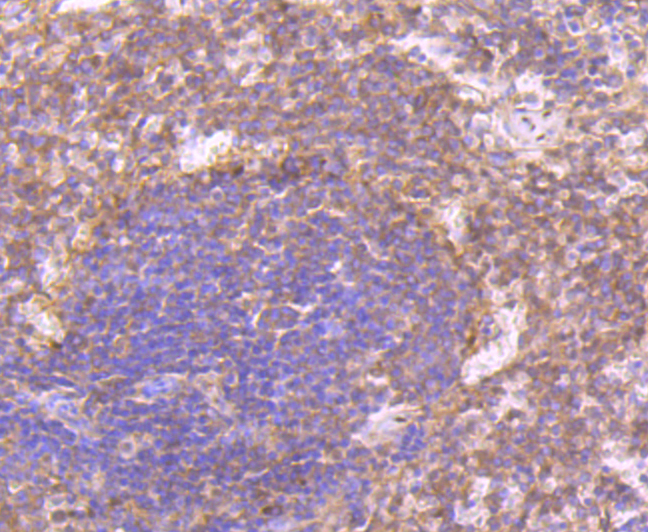
Fig4: Immunohistochemical analysis of paraffin-embedded rat spleen tissue using anti-CD27 antibody. Counter stained with hematoxylin.Application
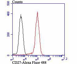
Fig5: Flow cytometric analysis of Daudi cells with CD27 antibody at 1/100 dilution (red) compared with an unlabelled control (cells without incubation with primary antibody; black). Alexa Fluor 488-conjugated goat anti rabbit IgG was used as the secondary antibody.| Product Name | CD27 [JB40-98] |
|---|---|
| Antibody Type | Primary Antibodies |
| Antigen Alias | CD 27 antibody CD27 antibody CD27 antigen antibody CD27 molecule antibody CD27_HUMAN antibody CD27L receptor antibody LPFS2 antibody MGC20393 antibody OTTHUMP00000238557 antibody S152 antibody T cell activation antigen CD27 antibody T cell antivation antigen S152 antibody T-cell activation antigen CD27 antibody T14 antibody TNFRSF 7 antibody TNFRSF7 antibody TNFSF7 antibody Tp 55 antibody Tp55 antibody Tumor necrosis factor receptor superfamily member 7 antibody |
| Product description | The tumor necrosis factor (TNF) receptor family is composed of several type I integral membrane glycoproteins that exhibit homology in their cysteine-rich extracellular domains. Members of this family include FAS, OX40, CD27 and CD30. Ligands for these receptors are often type II transmembrane glycoproteins, as is the case for CD27 and CD30. CD27 is a homodimeric lymphocyte-specific surface antigen present on T and B lymphocytes. Activation of the CD3 complex via the T cell receptor for antigen leads to an increase in CD27 expression. Together, CD27 and its ligand, CD27L, generate co-stimulatory signals required for complete T cell activation. CD30 is a surface marker for neoplastic cells of the Hodgkin's lymphoma and related hematologic malignancies. CD30L has been shown to enhance the proliferation of the Hodgkin's cell line HDLM-2, but exerts antiproliferative effects on large cell anaplastic lymphoma cell lines. |
| Immunogen | Intracellular domain corresponding to Human CD27. |
| Clonality | Monoclonal |
|---|---|
| Isotype | IgG |
| Host Species | Recombinant rabbit |
| Tested Applications | WBICCIHCFC |
| WB:1:500-1:1,000 IHC:1:50-1:200 ICC:1:50-1:200 FC:1:50-1:100 |
|
| Species Reactivity | HumanRat |
| Concentration | 1mg/ml |
| Alternative Names | CD 27 antibody CD27 antibody CD27 antigen antibody CD27 molecule antibody CD27_HUMAN antibody CD27L receptor antibody LPFS2 antibody MGC20393 antibody OTTHUMP00000238557 antibody S152 antibody T cell activation antigen CD27 antibody T cell antivation antigen S152 antibody T-cell activation antigen CD27 antibody T14 antibody TNFRSF 7 antibody TNFRSF7 antibody TNFSF7 antibody Tp 55 antibody Tp55 antibody Tumor necrosis factor receptor superfamily member 7 antibody |
|---|---|
| Molecular Weight(MW) | Predicted band size: 29 kDa |
| Cellular Localization | Membrane. |
| SwissProt ID | P26842 |
|---|
Application
Fig1: Western blot analysis of CD27 on Raji cell using anti-CD27 antibody at 1/500 dilution.
Application
Fig2: ICC staining CD27 in K562 cells (green). The nuclear counter stain is DAPI (blue). Cells were fixed in paraformaldehyde, permeabilised with 0.25% Triton X100/PBS.
Application
Fig3: Immunohistochemical analysis of paraffin-embedded human tonsil tissue using anti-CD27 antibody. Counter stained with hematoxylin.
Application
Fig4: Immunohistochemical analysis of paraffin-embedded rat spleen tissue using anti-CD27 antibody. Counter stained with hematoxylin.
Application
Fig5: Flow cytometric analysis of Daudi cells with CD27 antibody at 1/100 dilution (red) compared with an unlabelled control (cells without incubation with primary antibody; black). Alexa Fluor 488-conjugated goat anti rabbit IgG was used as the secondary antibody.| Positive Control | Raji, Daudi, K562, human tonsil tissue, rat spleen tissue. |
|---|---|
| Application Notes | WB:1:500-1:1,000 IHC:1:50-1:200 ICC:1:50-1:200 FC:1:50-1:100 |
| Form | Liquid |
|---|---|
| Storage Instructions | Store at +4℃ after thawing. Aliquot store at -20℃ or -80℃. Avoid repeated freeze / thaw cycles. |
| Storage Buffer | 1*TBS (pH7.4), 1%BSA, 40%Glycerol. Preservative: 0.05% Sodium Azide. |


