Application
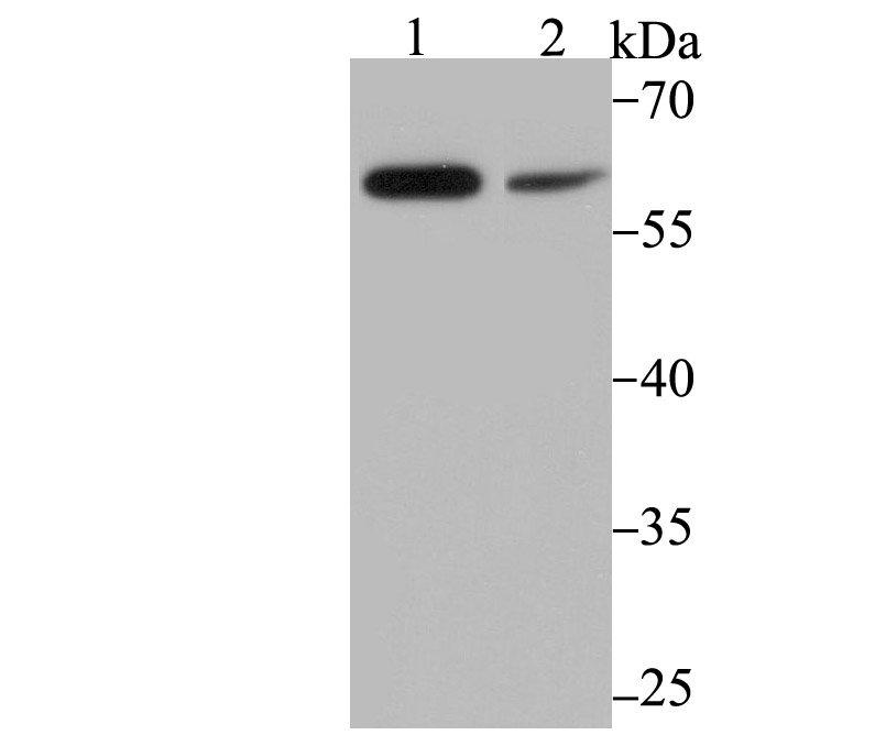
Fig1: Western blot analysis of PAK2 on mouse thymus tissue (1) and MCF-7 cell (2) lysate using anti-PAK2 antibody at 1/500 dilution.Application
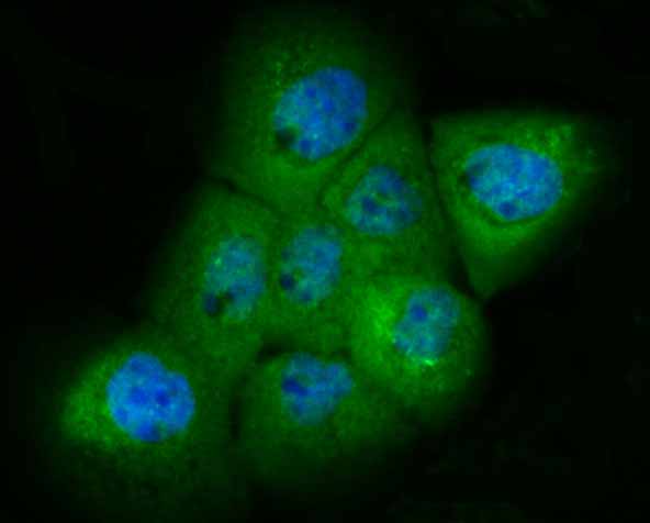
Fig2: ICC staining PAK2 in A431 cells (green). The nuclear counter stain is DAPI (blue). Cells were fixed in paraformaldehyde, permeabilised with 0.25% Triton X100/PBS.Application
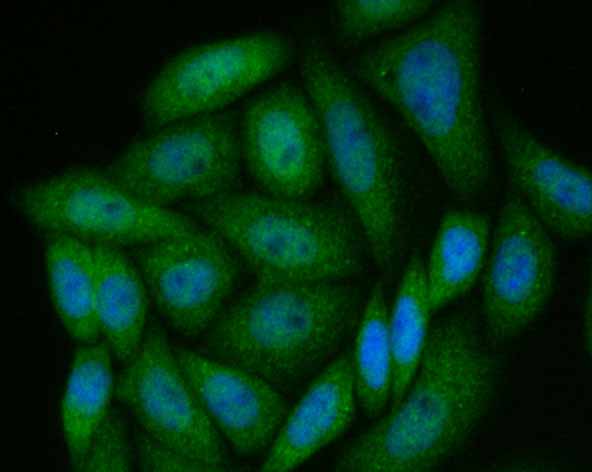
Fig3: ICC staining PAK2 in SiHa cells (green). The nuclear counter stain is DAPI (blue). Cells were fixed in paraformaldehyde, permeabilised with 0.25% Triton X100/PBS.Application
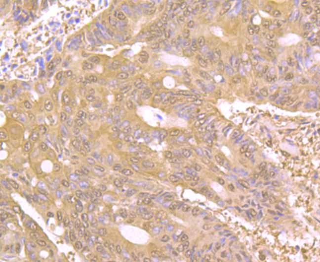
Fig4: Immunohistochemical analysis of paraffin-embedded human colon cancer tissue using anti-PAK2 antibody. Counter stained with hematoxylin.Application
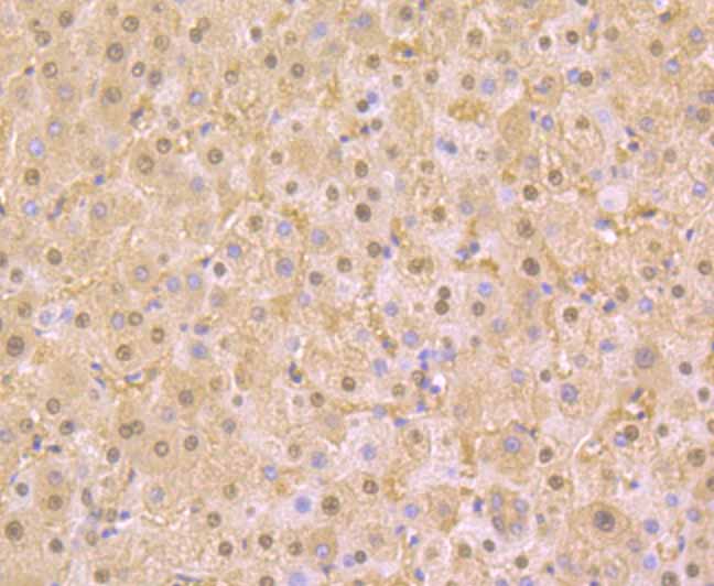
Fig5: Immunohistochemical analysis of paraffin-embedded human liver tissue using anti-PAK2 antibody. Counter stained with hematoxylin.Application
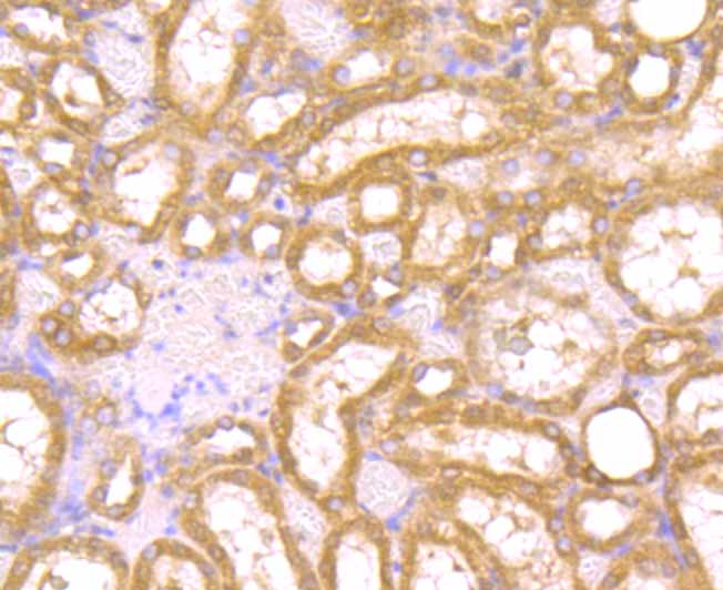
Fig6: Immunohistochemical analysis of paraffin-embedded rat kidney tissue using anti-PAK2 antibody. Counter stained with hematoxylin.Application
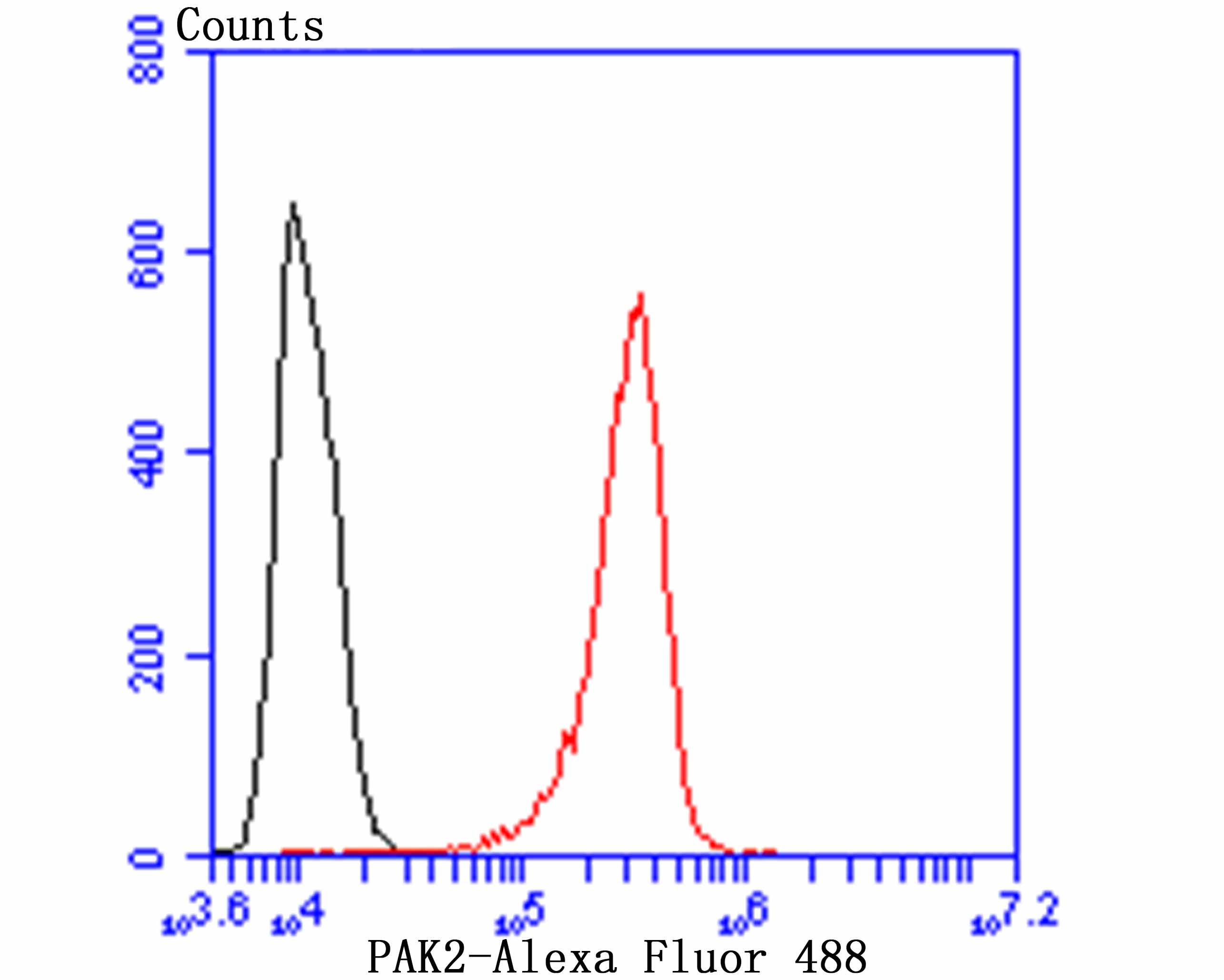
Fig7: Flow cytometric analysis of A431 cells with PAK2 antibody at 1/100 dilution (red) compared with an unlabelled control (cells without incubation with primary antibody; black). Alexa Fluor 488-conjugated goat anti rabbit IgG was used as the secondary antibody.| Product Name | PAK2 [JB41-84] |
|---|---|
| Antibody Type | Primary Antibodies |
| Antigen Alias | C-t-PAK2 antibody CB422 antibody EC 2.7.11.1 antibody Gamma PAK antibody Gamma-PAK antibody hPAK65 antibody Kinase antibody p21 (CDKN1A) activated kinase 2 antibody p21 (CDKN1A)-activated kinase 2a antibody p21 activated kinase 2 antibody p21 protein (Cdc42/Rac)-activated kinase 2 antibody p21 protein Cdc42 Rac activated kinase 2 antibody p21-activated kinase 2 antibody p21-activated kinase, 65-KD antibody p21-activated protein kinase I antibody p21CDKN1A activated kinase 2 antibody p27 antibody p34 antibody p58 antibody p65PAK antibody PAK 2 antibody PAK-2 antibody PAK-2p34 antibody Pak2 antibody PAK2_HUMAN antibody PAK65 antibody PAKgamma antibody S6 H4 kinase antibody S6/H4 kinase antibody Serine threonine protein kinase PAK 2 antibody Serine/threonine protein kinase PAK 2 antibody |
| Product description | Three recently identified isoforms of serine/threonine kinases, designated αPAK p68, βPAK p65 and γPAK p62, have been shown to exhibit a high degree of sequence homology with the S. cerevisiae kinase STE20, involved in pheromone signaling. The α, β, and γPAK isoforms complex specifically with Rac1 and Cdc42 in their active GTP bound state, inhibiting their intrinsic GTPase activity leading to their autophosphorylation. Once phosphorylated and their affinity for Rac/Cdc42 reduced, the PAK isoforms disassociate from the complex to seek downstream substrates. One such putative substrate is MEK kinase, an upstream effector of MEK4 which is involved in the JNK signaling pathway. While the PAK isoforms interact in a GTP-dependent manner with Rac1 and Cdc42, they do not interact with Rho. |
| Immunogen | Recombinant protein |
| Clonality | Monoclonal |
|---|---|
| Isotype | IgG |
| Host Species | Recombinant rabbit |
| Tested Applications | WBICCIFIHCFC |
| WB:1:500-1:1,000 IHC:1:100-1:500 ICC:1:100-1:500 FC:1:50-1:100 |
|
| Species Reactivity | HumanMouseRat |
| Concentration | 1mg/ml |
| Alternative Names | C-t-PAK2 antibody
CB422 antibody
EC 2.7.11.1 antibody
Gamma PAK antibody
Gamma-PAK antibody
hPAK65 antibody
Kinase antibody
p21 (CDKN1A) activated kinase 2 antibody
p21 (CDKN1A)-activated kinase 2a antibody
p21 activated kinase 2 antibody
p21 protein (Cdc42/Rac)-activated kinase 2 antibody
p21 protein Cdc42 Rac activated kinase 2 antibody
p21-activated kinase 2 antibody
p21-activated kinase 65-KD antibody p21-activated protein kinase I antibody p21CDKN1A activated kinase 2 antibody p27 antibody p34 antibody p58 antibody p65PAK antibody PAK 2 antibody PAK-2 antibody PAK-2p34 antibody Pak2 antibody PAK2_HUMAN antibody PAK65 antibody PAKgamma antibody S6 H4 kinase antibody S6/H4 kinase antibody Serine threonine protein kinase PAK 2 antibody Serine/threonine protein kinase PAK 2 antibody |
|---|---|
| Molecular Weight(MW) | 58 kDa |
| Cellular Localization | Cytoplasm. Nucleus. |
Application
Fig1: Western blot analysis of PAK2 on mouse thymus tissue (1) and MCF-7 cell (2) lysate using anti-PAK2 antibody at 1/500 dilution.
Application
Fig2: ICC staining PAK2 in A431 cells (green). The nuclear counter stain is DAPI (blue). Cells were fixed in paraformaldehyde, permeabilised with 0.25% Triton X100/PBS.
Application
Fig3: ICC staining PAK2 in SiHa cells (green). The nuclear counter stain is DAPI (blue). Cells were fixed in paraformaldehyde, permeabilised with 0.25% Triton X100/PBS.
Application
Fig4: Immunohistochemical analysis of paraffin-embedded human colon cancer tissue using anti-PAK2 antibody. Counter stained with hematoxylin.
Application
Fig5: Immunohistochemical analysis of paraffin-embedded human liver tissue using anti-PAK2 antibody. Counter stained with hematoxylin.
Application
Fig6: Immunohistochemical analysis of paraffin-embedded rat kidney tissue using anti-PAK2 antibody. Counter stained with hematoxylin.
Application
Fig7: Flow cytometric analysis of A431 cells with PAK2 antibody at 1/100 dilution (red) compared with an unlabelled control (cells without incubation with primary antibody; black). Alexa Fluor 488-conjugated goat anti rabbit IgG was used as the secondary antibody.| Positive Control | Mouse thymus tissue lysate, MCF-7, A431, SiHa, human colon cancer tissue, human liver tissue, rat kidney tissue. |
|---|---|
| Application Notes | WB:1:500-1:1,000 IHC:1:100-1:500 ICC:1:100-1:500 FC:1:50-1:100 |
| Form | Liquid |
|---|---|
| Storage Instructions | Store at +4℃ after thawing. Aliquot store at -20℃ or -80℃. Avoid repeated freeze / thaw cycles. |
| Storage Buffer | 1*TBS (pH7.4), 1%BSA, 40%Glycerol. Preservative: 0.05% Sodium Azide. |


