Application
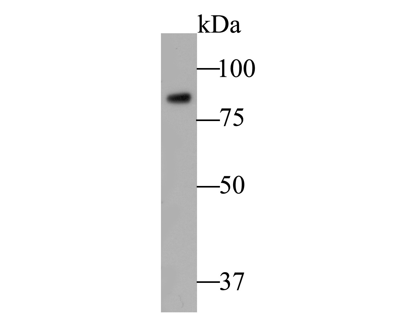
Fig1: Western blot analysis of SSRP1 on Daudi cell lysate. Proteins were transferred to a PVDF membrane and blocked with 5% BSA in PBS for 1 hour at room temperature. The primary antibody (ER1901-13, 1/500) was used in 5% BSA at room temperature for 2 hours. Goat Anti-Rabbit IgG - HRP Secondary Antibody (HA1001) at 1:5,000 dilution was used for 1 hour at room temperature.Application
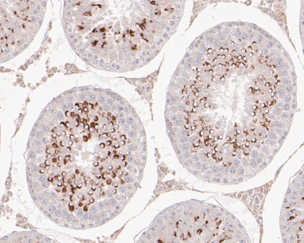
Fig2: Immunohistochemical analysis of paraffin-embedded rat testis tissue using anti-SSRP1 antibody. The section was pre-treated using heat mediated antigen retrieval with sodium citrate buffer (pH 6.0) for 20 minutes. The tissues were blocked in 5% BSA for 30 minutes at room temperature, washed with ddH2O and PBS, and then probed with the primary antibody (ER1901-13, 1/50) for 30 minutes at room temperature. The detection was performed using an HRP conjugated compact polymer system. DAB was used as the chromogen. Tissues were counterstained with hematoxylin and mounted with DPX.Application
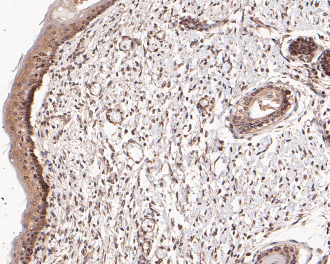
Fig3: Immunohistochemical analysis of paraffin-embedded human skin tissue using anti-SSRP1 antibody. The section was pre-treated using heat mediated antigen retrieval with sodium citrate buffer (pH 6.0) for 20 minutes. The tissues were blocked in 5% BSA for 30 minutes at room temperature, washed with ddH2O and PBS, and then probed with the primary antibody (ER1901-13, 1/50) for 30 minutes at room temperature. The detection was performed using an HRP conjugated compact polymer system. DAB was used as the chromogen. Tissues were counterstained with hematoxylin and mounted with DPX.Application
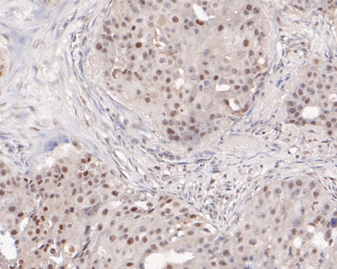
Fig4: Immunohistochemical analysis of paraffin-embedded human breast cancer tissue using anti-SSRP1 antibody. The section was pre-treated using heat mediated antigen retrieval with sodium citrate buffer (pH 6.0) for 20 minutes. The tissues were blocked in 5% BSA for 30 minutes at room temperature, washed with ddH2O and PBS, and then probed with the primary antibody (ER1901-13, 1/50) for 30 minutes at room temperature. The detection was performed using an HRP conjugated compact polymer system. DAB was used as the chromogen. Tissues were counterstained with hematoxylin and mounted with DPX.Application
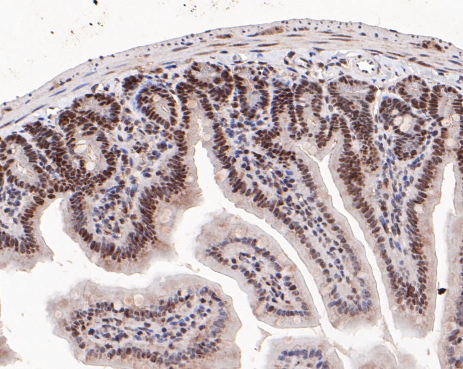
Fig5: Immunohistochemical analysis of paraffin-embedded mouse colon tissue using anti-SSRP1 antibody. The section was pre-treated using heat mediated antigen retrieval with sodium citrate buffer (pH 6.0) for 20 minutes. The tissues were blocked in 5% BSA for 30 minutes at room temperature, washed with ddH2O and PBS, and then probed with the primary antibody (ER1901-13, 1/50) for 30 minutes at room temperature. The detection was performed using an HRP conjugated compact polymer system. DAB was used as the chromogen. Tissues were counterstained with hematoxylin and mounted with DPX.Application
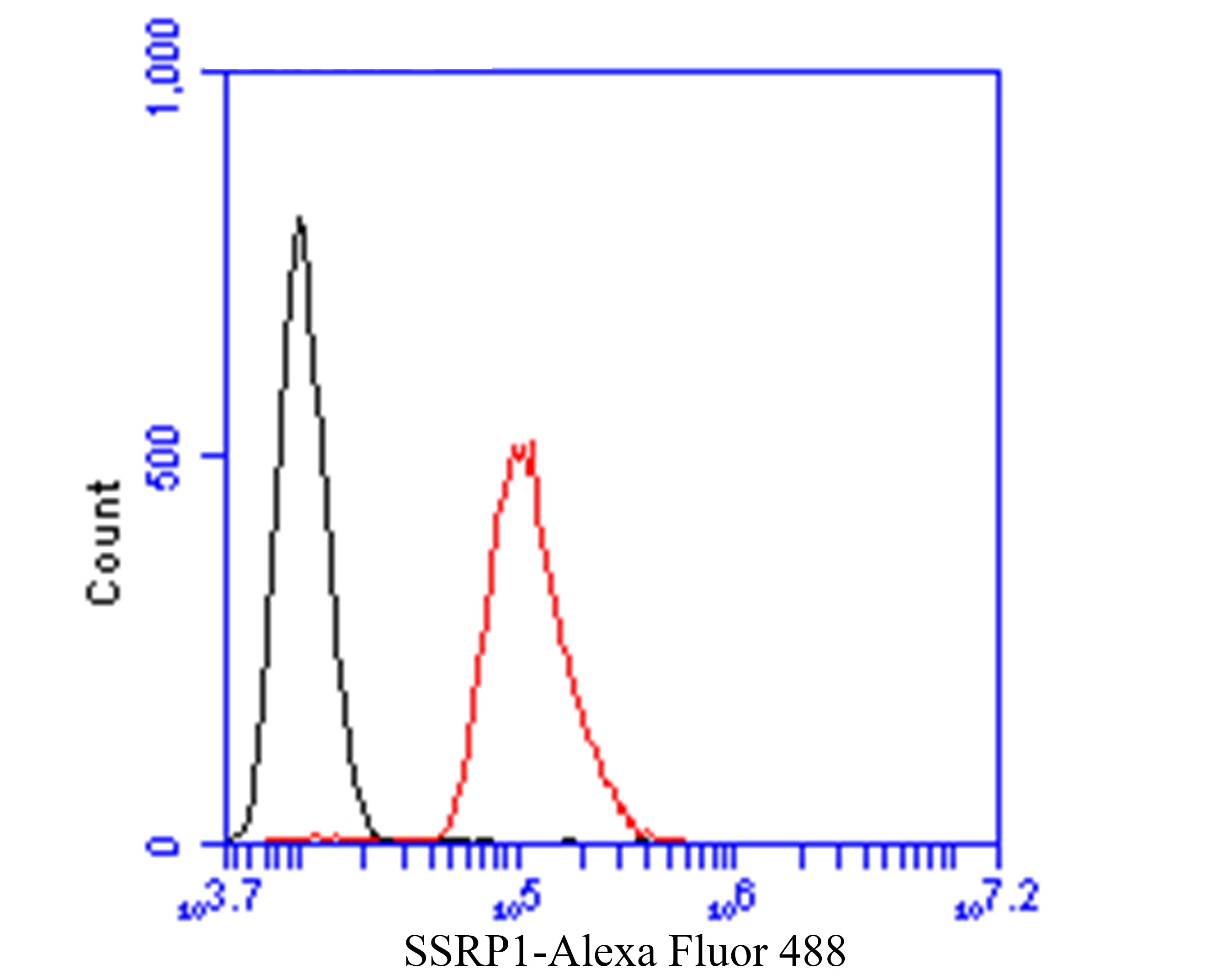
Fig6: Flow cytometric analysis of SSRP1 was done on SH-SY5Y cells. The cells were fixed, permeabilized and stained with the primary antibody (ER1901-13, 1/50) (red). After incubation of the primary antibody at room temperature for an hour, the cells were stained with a Alexa Fluor 488-conjugated Goat anti-Rabbit IgG Secondary antibody at 1/1000 dilution for 30 minutes.Unlabelled sample was used as a control (cells without incubation with primary antibody; black).| Product Name | SSRP1 |
|---|---|
| Antibody Type | Primary Antibodies |
| Antigen Alias | Chromatin-specific transcription elongation factor 80 kDa subunit antibody Cisplatin DNA SSRP antibody Facilitates chromatin remodeling 80 kDa subunit antibody Facilitates chromatin transcription complex 80 kDa subunit antibody Facilitates chromatin transcription complex subunit SSRP1 antibody FACT 80 kDa subunit antibody FACT complex subunit SSRP1 antibody FACT80 antibody FACTp80 antibody hSSRP1 antibody Recombination signal sequence recognition protein 1 antibody Recombination signal sequence recognition protein antibody SSRP 1 antibody SSRP1 antibody SSRP1_HUMAN antibody Structure specific recognition protein 1 antibody Structure-specific recognition protein 1 antibody T160 antibody |
| Product description | Component of the FACT complex, a general chromatin factor that acts to reorganize nucleosomes. The FACT complex is involved in multiple processes that require DNA as a template such as mRNA elongation, DNA replication and DNA repair. During transcription elongation the FACT complex acts as a histone chaperone that both destabilizes and restores nucleosomal structure. It facilitates the passage of RNA polymerase II and transcription by promoting the dissociation of one histone H2A-H2B dimer from the nucleosome, then subsequently promotes the reestablishment of the nucleosome following the passage of RNA polymerase II. The FACT complex is probably also involved in phosphorylation of 'Ser-392' of p53/TP53 via its association with CK2 (casein kinase II). Binds specifically to double-stranded DNA and at low levels to DNA modified by the antitumor agent cisplatin. May potentiate cisplatin-induced cell death by blocking replication and repair of modified DNA. Also acts as a transcriptional coactivator for p63/TP63. |
| Immunogen | Recombinant protein within human SSRP1 aa 180-400. |
| Clonality | Polyclonal |
|---|---|
| Isotype | IgG |
| Host Species | Rabbit |
| Tested Applications | WBIHC-PFC |
| WB:1:500-1:1000: IHC-P:1:50-1:200: FC:1:50-1:100: |
|
| Species Reactivity | HumanMouseRat |
| Concentration | 1mg/ml |
| Alternative Names | Chromatin-specific transcription elongation factor 80 kDa subunit antibody
Cisplatin DNA SSRP antibody
Facilitates chromatin remodeling 80 kDa subunit antibody Facilitates chromatin transcription complex 80 kDa subunit antibody Facilitates chromatin transcription complex subunit SSRP1 antibody FACT 80 kDa subunit antibody FACT complex subunit SSRP1 antibody FACT80 antibody FACTp80 antibody hSSRP1 antibody Recombination signal sequence recognition protein 1 antibody Recombination signal sequence recognition protein antibody SSRP 1 antibody SSRP1 antibody SSRP1_HUMAN antibody Structure specific recognition protein 1 antibody Structure-specific recognition protein 1 antibody T160 antibody |
|---|---|
| Molecular Weight(MW) | 81 kDa |
| Cellular Localization | Nucleus. |
Application
Fig1: Western blot analysis of SSRP1 on Daudi cell lysate. Proteins were transferred to a PVDF membrane and blocked with 5% BSA in PBS for 1 hour at room temperature. The primary antibody (ER1901-13, 1/500) was used in 5% BSA at room temperature for 2 hours. Goat Anti-Rabbit IgG - HRP Secondary Antibody (HA1001) at 1:5,000 dilution was used for 1 hour at room temperature.
Application
Fig2: Immunohistochemical analysis of paraffin-embedded rat testis tissue using anti-SSRP1 antibody. The section was pre-treated using heat mediated antigen retrieval with sodium citrate buffer (pH 6.0) for 20 minutes. The tissues were blocked in 5% BSA for 30 minutes at room temperature, washed with ddH2O and PBS, and then probed with the primary antibody (ER1901-13, 1/50) for 30 minutes at room temperature. The detection was performed using an HRP conjugated compact polymer system. DAB was used as the chromogen. Tissues were counterstained with hematoxylin and mounted with DPX.
Application
Fig3: Immunohistochemical analysis of paraffin-embedded human skin tissue using anti-SSRP1 antibody. The section was pre-treated using heat mediated antigen retrieval with sodium citrate buffer (pH 6.0) for 20 minutes. The tissues were blocked in 5% BSA for 30 minutes at room temperature, washed with ddH2O and PBS, and then probed with the primary antibody (ER1901-13, 1/50) for 30 minutes at room temperature. The detection was performed using an HRP conjugated compact polymer system. DAB was used as the chromogen. Tissues were counterstained with hematoxylin and mounted with DPX.
Application
Fig4: Immunohistochemical analysis of paraffin-embedded human breast cancer tissue using anti-SSRP1 antibody. The section was pre-treated using heat mediated antigen retrieval with sodium citrate buffer (pH 6.0) for 20 minutes. The tissues were blocked in 5% BSA for 30 minutes at room temperature, washed with ddH2O and PBS, and then probed with the primary antibody (ER1901-13, 1/50) for 30 minutes at room temperature. The detection was performed using an HRP conjugated compact polymer system. DAB was used as the chromogen. Tissues were counterstained with hematoxylin and mounted with DPX.
Application
Fig5: Immunohistochemical analysis of paraffin-embedded mouse colon tissue using anti-SSRP1 antibody. The section was pre-treated using heat mediated antigen retrieval with sodium citrate buffer (pH 6.0) for 20 minutes. The tissues were blocked in 5% BSA for 30 minutes at room temperature, washed with ddH2O and PBS, and then probed with the primary antibody (ER1901-13, 1/50) for 30 minutes at room temperature. The detection was performed using an HRP conjugated compact polymer system. DAB was used as the chromogen. Tissues were counterstained with hematoxylin and mounted with DPX.
Application
Fig6: Flow cytometric analysis of SSRP1 was done on SH-SY5Y cells. The cells were fixed, permeabilized and stained with the primary antibody (ER1901-13, 1/50) (red). After incubation of the primary antibody at room temperature for an hour, the cells were stained with a Alexa Fluor 488-conjugated Goat anti-Rabbit IgG Secondary antibody at 1/1000 dilution for 30 minutes.Unlabelled sample was used as a control (cells without incubation with primary antibody; black).| Positive Control | Daudi cell lysates, rat testis tissue, human skin tissue, human breast cancer tissue, mouse colon tissue, SH-SY5Y. |
|---|---|
| Application Notes | WB:1:500-1:1000: IHC-P:1:50-1:200: FC:1:50-1:100: |
| Form | Liquid |
|---|---|
| Storage Instructions | Store at +4℃ after thawing. Aliquot store at -20℃. Avoid repeated freeze / thaw cycles. |
| Storage Buffer | 1*TBS (pH7.4), 1%BSA, 50%Glycerol. Preservative: 0.05% Sodium Azide. |


