Application
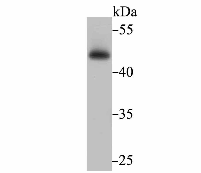
Fig1: Western blot analysis of Flotillin 1 on PC-3M cell lysate using anti-Flotillin 1 antibody at 1/500 dilution.Application
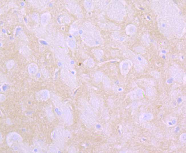
Fig2: Immunohistochemical analysis of paraffin-embedded rat brain tissue using anti-Flotillin 1 antibody. Counter stained with hematoxylin.Application
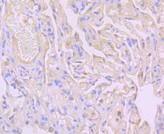
Fig3: Immunohistochemical analysis of paraffin-embedded human lung cancer tissue using anti-Flotillin 1 antibody. Counter stained with hematoxylin.Application
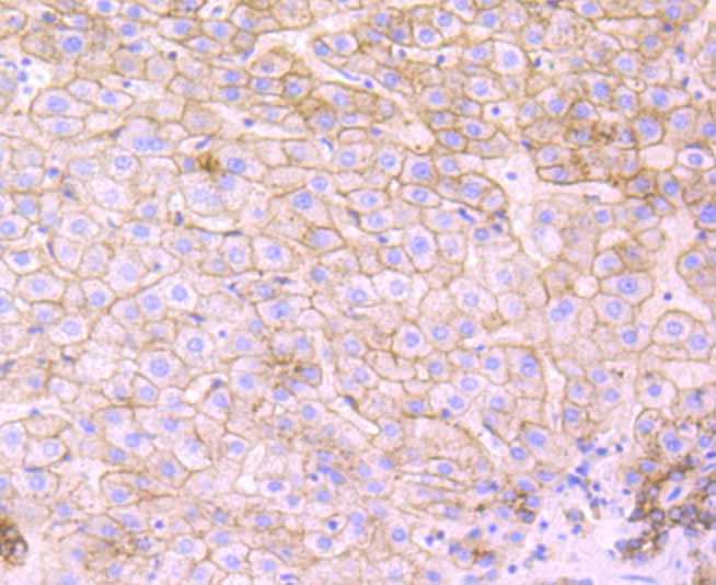
Fig4: Immunohistochemical analysis of paraffin-embedded human liver tissue using anti-Flotillin 1 antibody. Counter stained with hematoxylin.Application
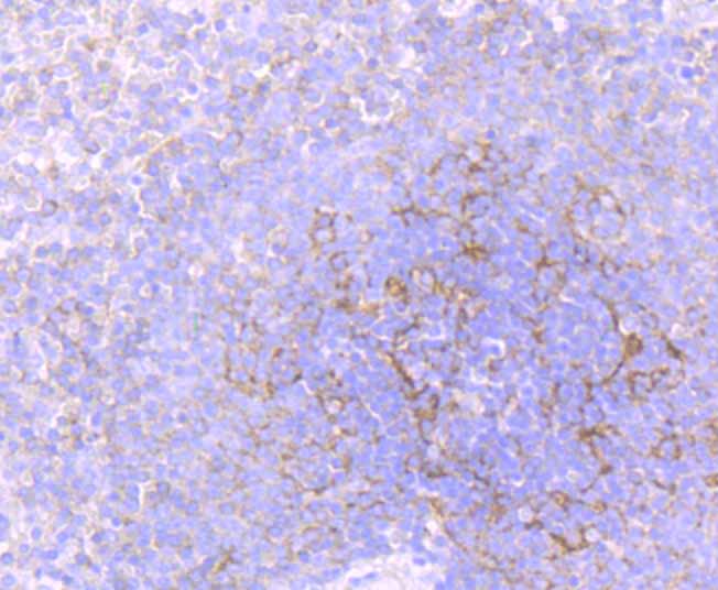
Fig5: Immunohistochemical analysis of paraffin-embedded human spleen tissue using anti-Flotillin 1 antibody. Counter stained with hematoxylin.Application
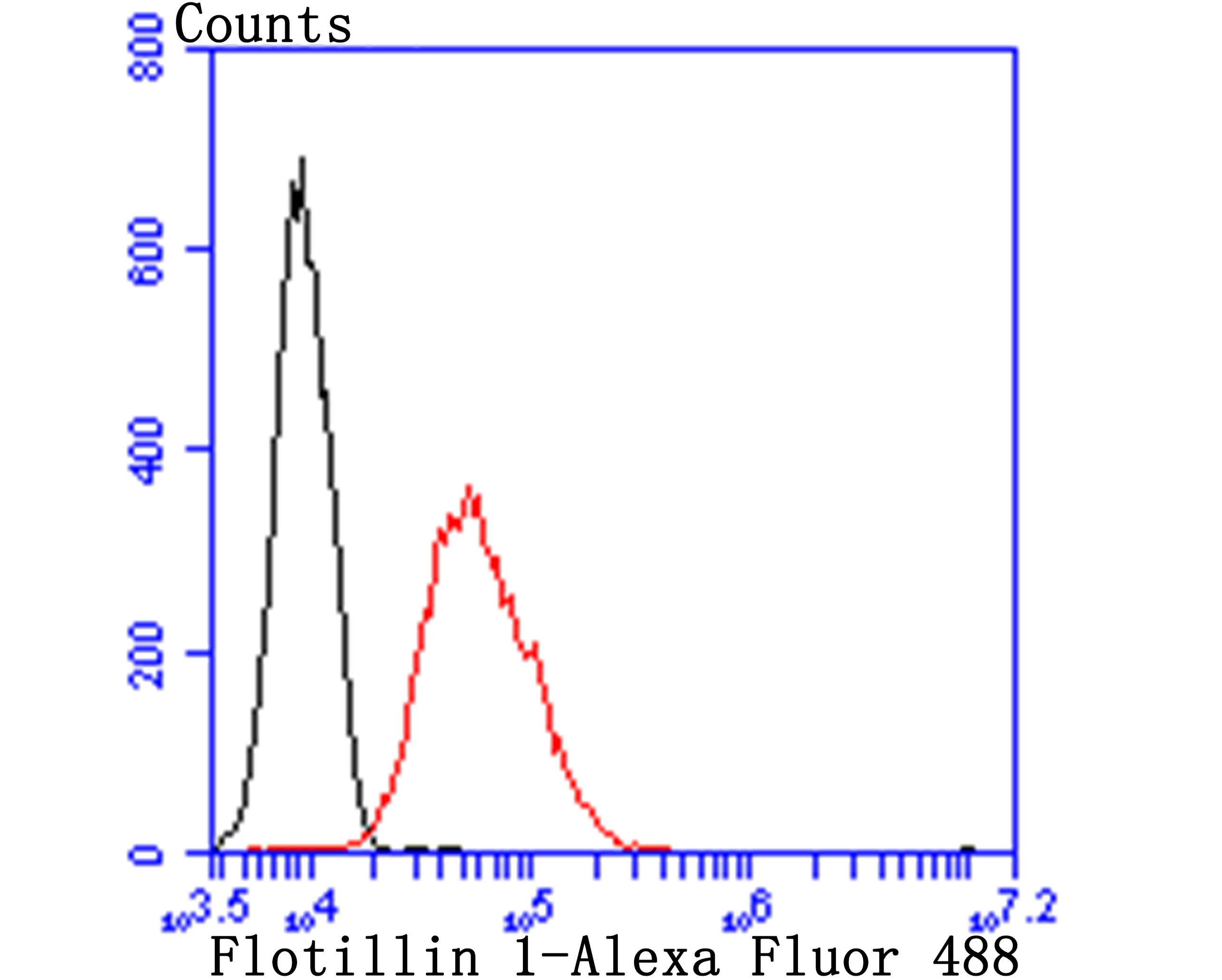
Fig6: Flow cytometric analysis of HUVEC cells with Flotillin 1 antibody at 1/100 dilution (red) compared with an unlabelled control (cells without incubation with primary antibody; black). Alexa Fluor 488-conjugated goat anti-rabbit IgG was used as the secondary antibody.| Product Name | Flotillin 1 [JB19-45] |
|---|---|
| Antibody Type | Primary Antibodies |
| Antigen Alias | FLOT 1 antibody FLOT1 antibody FLOT1_HUMAN antibody Flotillin-1 antibody Flotillin1 antibody Integral membrane component of caveolae antibody Reggie 2 antibody |
| Product description | Lipid rafts are sphingolipid- and cholesterol-rich membrane microdomains that are insoluble in nonionic detergents. Lipid rafts are important for numerous cellular processes, including signal transduction, membrane trafficking and molecular sorting. Flotillins are lipid raft components in neurons and caveloae-associated proteins in A498 kidney cells. Flotillin-1 belongs to the band 7.2/stomatin protein family, whose members are characterized by the presence of a hydrophobic N-terminal region that is predicted to form a single, outside to inside, transmembrane domain. Flotillin-1 and -2 have complementary tissue distributions and their expression levels are independently regulated. At the cellular level, Flotillin-2 is ubiquitously expressed, whereas Flotillin-1 is expressed in A498 kidney cells, muscle cell lines and fibroblasts. Flotillins form a ternary complex with CAP and Cbl, directing the localization of the CAP-Cbl complex to a lipid raft subdomain of the plasma membrane. Association of ER-X with Flotillin localizes ER-X within plasma membrane caveloae and mediates rapid oestrogen activation of the MAP kinase cascade. The expression of the flotillins is also correlated to the progression of Alzhemier pathology. |
| Immunogen | Recombinant protein |
| Clonality | Monoclonal |
|---|---|
| Isotype | IgG |
| Host Species | Recombinant rabbit |
| Tested Applications | WBIHCFC |
| WB:1:500-1:1,000 IHC:1:50-1:100 FC:1:50-1:100 |
|
| Species Reactivity | HumanMouseRat |
| Concentration | 1mg/ml |
| Alternative Names | FLOT 1 antibody FLOT1 antibody FLOT1_HUMAN antibody Flotillin-1 antibody Flotillin1 antibody Integral membrane component of caveolae antibody Reggie 2 antibody |
|---|---|
| Molecular Weight(MW) | 47 kDa |
| Cellular Localization | Cell membrane. Endosome. |
Application
Fig1: Western blot analysis of Flotillin 1 on PC-3M cell lysate using anti-Flotillin 1 antibody at 1/500 dilution.
Application
Fig2: Immunohistochemical analysis of paraffin-embedded rat brain tissue using anti-Flotillin 1 antibody. Counter stained with hematoxylin.
Application
Fig3: Immunohistochemical analysis of paraffin-embedded human lung cancer tissue using anti-Flotillin 1 antibody. Counter stained with hematoxylin.
Application
Fig4: Immunohistochemical analysis of paraffin-embedded human liver tissue using anti-Flotillin 1 antibody. Counter stained with hematoxylin.
Application
Fig5: Immunohistochemical analysis of paraffin-embedded human spleen tissue using anti-Flotillin 1 antibody. Counter stained with hematoxylin.
Application
Fig6: Flow cytometric analysis of HUVEC cells with Flotillin 1 antibody at 1/100 dilution (red) compared with an unlabelled control (cells without incubation with primary antibody; black). Alexa Fluor 488-conjugated goat anti-rabbit IgG was used as the secondary antibody.| Positive Control | PC-3M, rat brain tissue, human lung cancer tissue, human liver tissue, human spleen tissue, HUVEC. |
|---|---|
| Application Notes | WB:1:500-1:1,000 IHC:1:50-1:100 FC:1:50-1:100 |
| Form | Liquid |
|---|---|
| Storage Instructions | Store at +4℃ after thawing. Aliquot store at -20℃ or -80℃. Avoid repeated freeze / thaw cycles. |
| Storage Buffer | 1*TBS (pH7.4), 1%BSA, 40%Glycerol. Preservative: 0.05% Sodium Azide. |


