Application
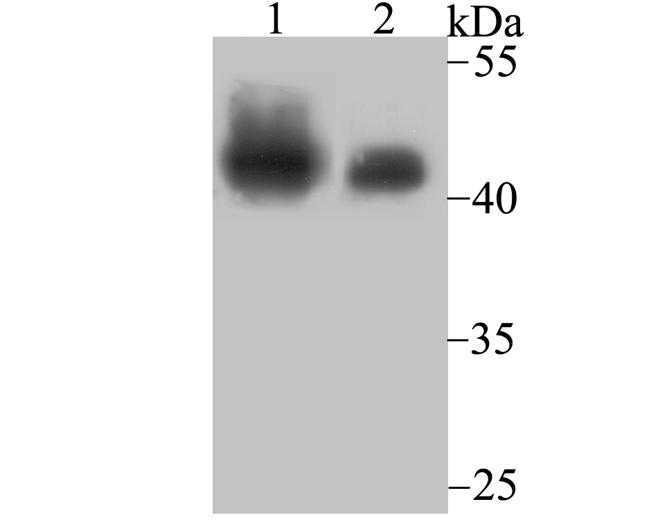
Fig1: Western blot analysis of SNF5 on Daudi cell and mouse lymph node tissue lysates using anti-SNF5 antibody at 1/500 dilution.Application
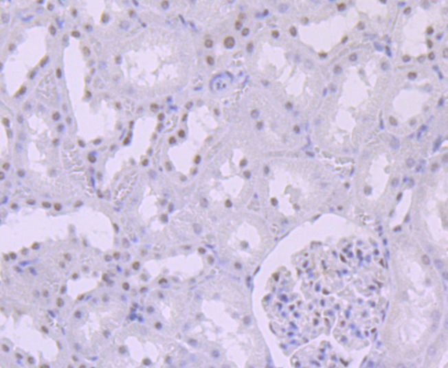
Fig2: Immunohistochemical analysis of paraffin-embedded rat kidney tissue using anti-SNF5 antibody. Counter stained with hematoxylin.Application
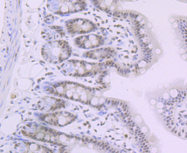
Fig3: Immunohistochemical analysis of paraffin-embedded mouse colon tissue using anti-SNF5 antibody. Counter stained with hematoxylin.Application
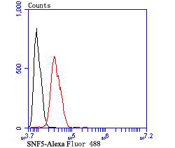
Fig4: Flow cytometric analysis of SH-SY-5Y cells with SNF5 antibody at 1/100 dilution (red) compared with an unlabelled control (cells without incubation with primary antibody; black). Alexa Fluor 488-conjugated goat anti-rabbit IgG was used as the secondary antibody.| Product Name | SNF5 [JB49-18] |
|---|---|
| Antibody Type | Primary Antibodies |
| Antigen Alias | BAF47 antibody BRG1-associated factor 47 antibody hSNF5 antibody INI1 antibody Integrase interactor 1 protein antibody Malignant rhabdoid tumor suppressor antibody RDT antibody RTPS1 antibody Sfh1p antibody SMARCB1 antibody SNF5 homolog antibody SNF5_HUMAN antibody SNF5L1 antibody Snr1 antibody Sucrose nonfermenting yeast homolog like 1 antibody SWI/SNF complex component SNF5 antibody SWI/SNF-related matrix-associated actin-dependent regulator of chromatin subfamily B member 1 antibody SWI10 antibody Transcription factor TYE4 antibody Transcription regulatory protein SNF5 antibody TYE4 antibody |
| Product description | The SWI-SNF complex is involved in the activation of transcription via the remodeling of nucleosome structure in an ATP-dependent manner. Brm (also designated SNF2α) and Brg-1 (also designated SNF2β) are the ATPase subunits of the mammalian SWI-SNF complex. Brm, Brg-1, Ini1 (integrase interactor 1, also designated SNF5), BAF155 (also designated SRG3) and BAF170 are thought to comprise the functional core of the SWI-SNF complex. Addition of Ini1, BAF155 and BAF170 to Brg-1 appears to increase remodeling activity. Other complex subunits are thought to play regulatory roles. hSNF2L and hSNF2H both appear to be homologs of Drosophila ISWI, a Brm related ATPase that is present in chromatin remodeling complexes other than SWI/SNF, including the NURF (nucleosome remodeling factor). |
| Immunogen | Synthetic peptide of C terminal Human SNF5. |
| Clonality | Monoclonal |
|---|---|
| Isotype | IgG |
| Host Species | Recombinant rabbit |
| Tested Applications | WBIHCFC |
| WB:1:500-1:1,000 IHC:1:50-1:100 FC:1:50-1:100 |
|
| Species Reactivity | HumanMouseRat |
| Concentration | 1mg/ml |
| Alternative Names | BAF47 antibody BRG1-associated factor 47 antibody hSNF5 antibody INI1 antibody Integrase interactor 1 protein antibody Malignant rhabdoid tumor suppressor antibody RDT antibody RTPS1 antibody Sfh1p antibody SMARCB1 antibody SNF5 homolog antibody SNF5_HUMAN antibody SNF5L1 antibody Snr1 antibody Sucrose nonfermenting yeast homolog like 1 antibody SWI/SNF complex component SNF5 antibody SWI/SNF-related matrix-associated actin-dependent regulator of chromatin subfamily B member 1 antibody SWI10 antibody Transcription factor TYE4 antibody Transcription regulatory protein SNF5 antibody TYE4 antibody |
|---|---|
| Molecular Weight(MW) | 44 kDa |
| Cellular Localization | Nucleus. |
Application
Fig1: Western blot analysis of SNF5 on Daudi cell and mouse lymph node tissue lysates using anti-SNF5 antibody at 1/500 dilution.
Application
Fig2: Immunohistochemical analysis of paraffin-embedded rat kidney tissue using anti-SNF5 antibody. Counter stained with hematoxylin.
Application
Fig3: Immunohistochemical analysis of paraffin-embedded mouse colon tissue using anti-SNF5 antibody. Counter stained with hematoxylin.
Application
Fig4: Flow cytometric analysis of SH-SY-5Y cells with SNF5 antibody at 1/100 dilution (red) compared with an unlabelled control (cells without incubation with primary antibody; black). Alexa Fluor 488-conjugated goat anti-rabbit IgG was used as the secondary antibody.| Positive Control | Daudi, mouse lymph node tissue lysate, rat kidney tissue, mouse colon tissue, SH-SY-5Y. |
|---|---|
| Application Notes | WB:1:500-1:1,000 IHC:1:50-1:100 FC:1:50-1:100 |
| Form | Liquid |
|---|---|
| Storage Instructions | Store at +4℃ after thawing. Aliquot store at -20℃ or -80℃. Avoid repeated freeze / thaw cycles. |
| Storage Buffer | 1*TBS (pH7.4), 1%BSA, 40%Glycerol. Preservative: 0.05% Sodium Azide. |


