Application
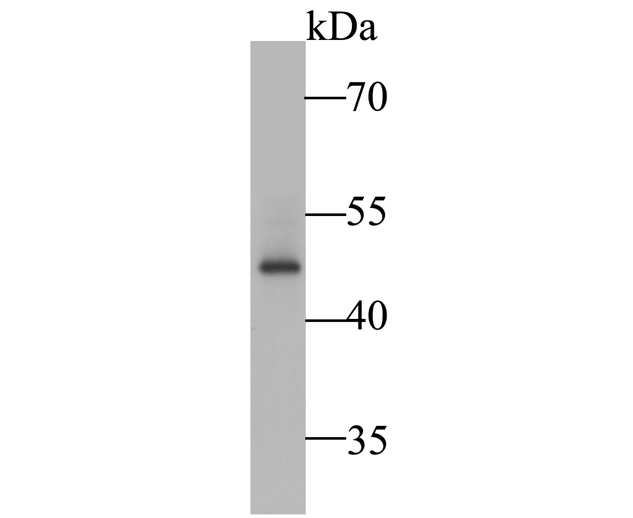
Fig1: Western blot analysis of SSB on K562 cell lysate. Proteins were transferred to a PVDF membrane and blocked with 5% BSA in PBS for 1 hour at room temperature. The primary antibody (ER1901-16, 1/5,000) was used in 5% BSA at room temperature for 2 hours. Goat Anti-Rabbit IgG - HRP Secondary Antibody (HA1001) at 1:5,000 dilution was used for 1 hour at room temperature.Application
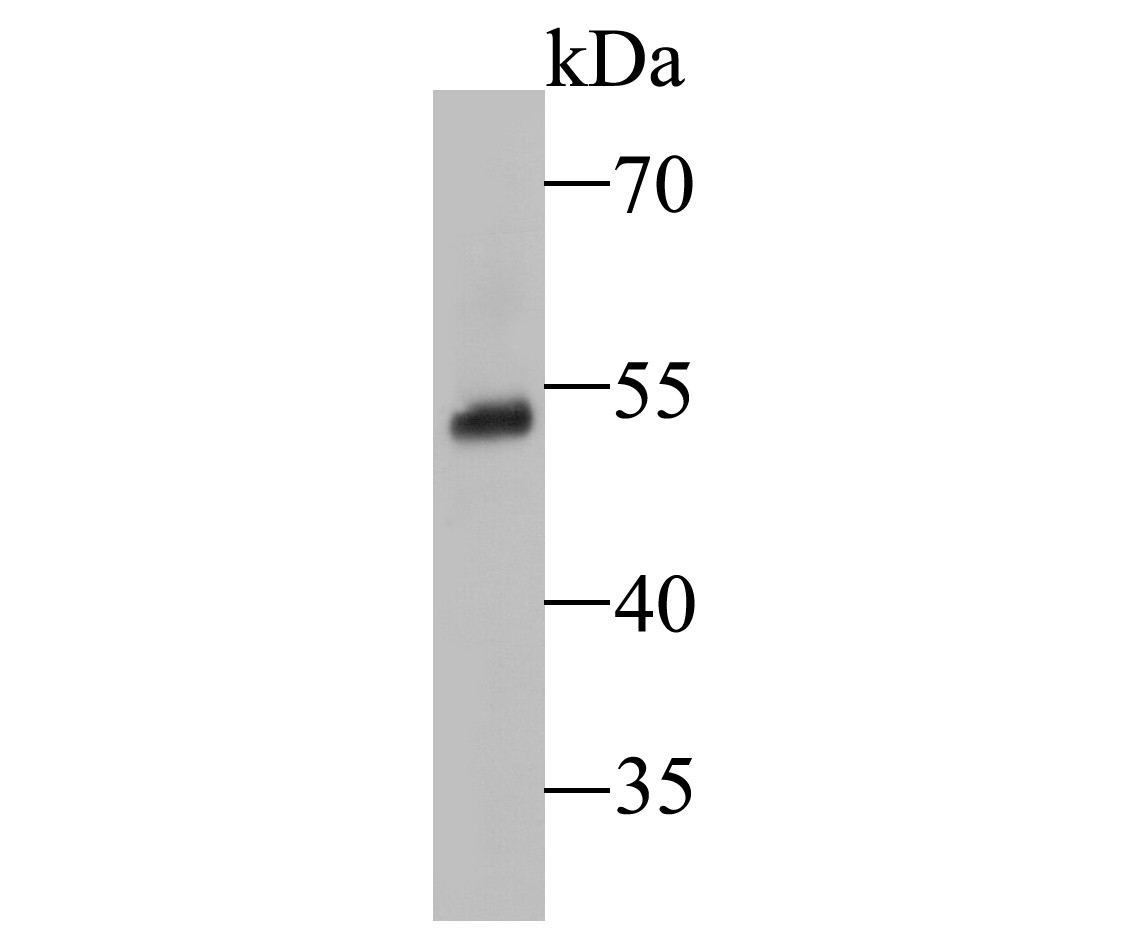
Fig2: Western blot analysis of SSB on human lung tissue lysate. Proteins were transferred to a PVDF membrane and blocked with 5% BSA in PBS for 1 hour at room temperature. The primary antibody (ER1901-16, 1/1,000) was used in 5% BSA at room temperature for 2 hours. Goat Anti-Rabbit IgG - HRP Secondary Antibody (HA1001) at 1:5,000 dilution was used for 1 hour at room temperature.Application
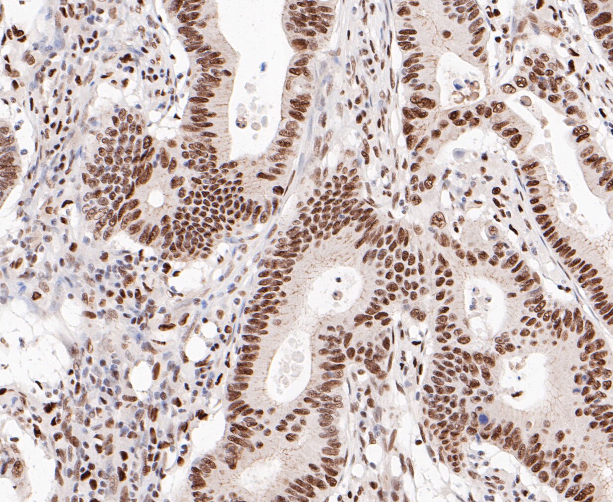
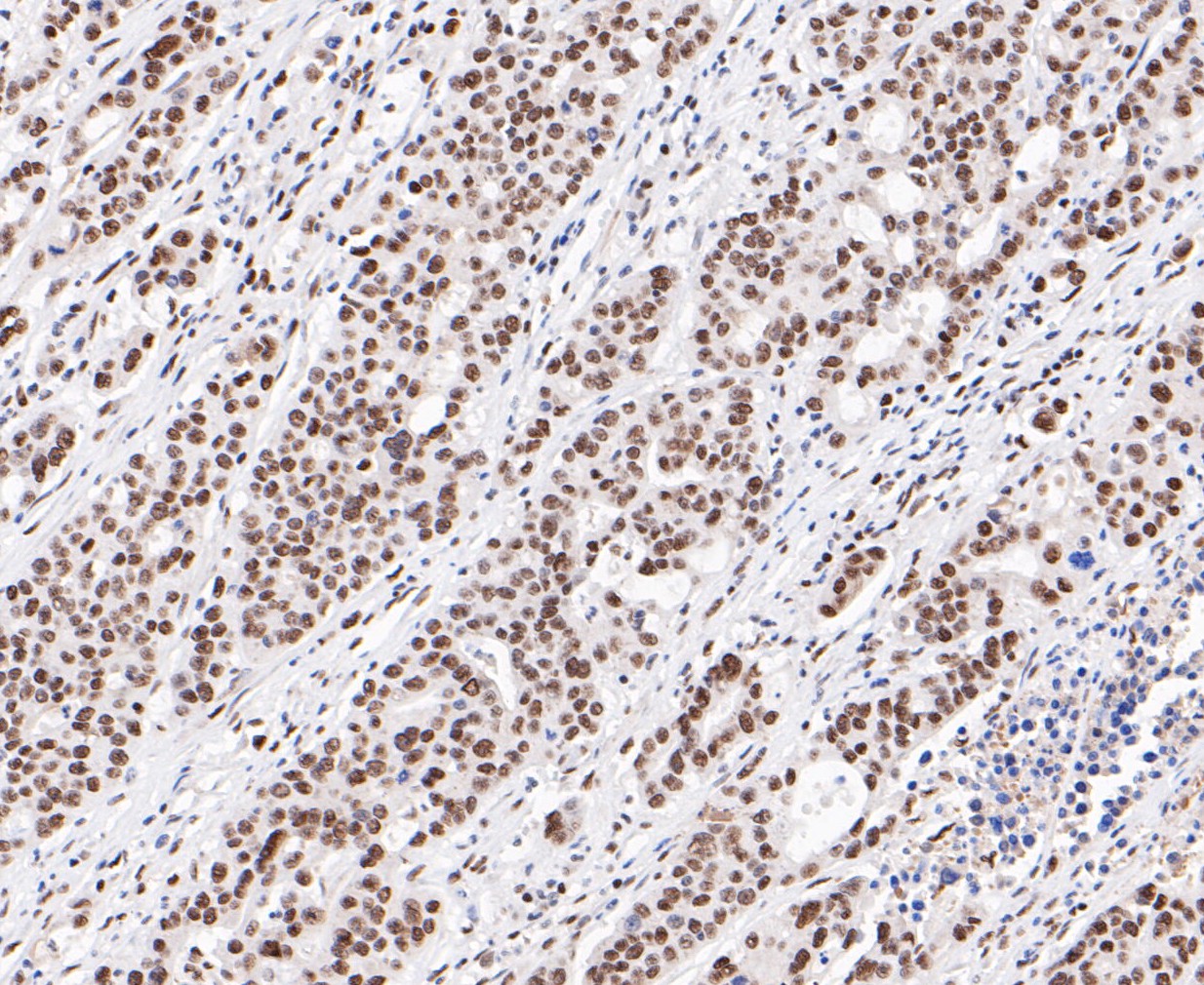
Fig3: Immunohistochemical analysis of paraffin-embedded human colon cancer tissue using anti-SSB antibody. The section was pre-treated using heat mediated antigen retrieval with sodium citrate buffer (pH 6.0) for 20 minutes. The tissues were blocked in 5% BSA for 30 minutes at room temperature, washed with ddH2O and PBS, and then probed with the primary antibody (ER1901-16, 1/800) for 30 minutes at room temperature. The detection was performed using an HRP conjugated compact polymer system. DAB was used as the chromogen. Tissues were counterstained with hematoxylin and mounted with DPX.Application
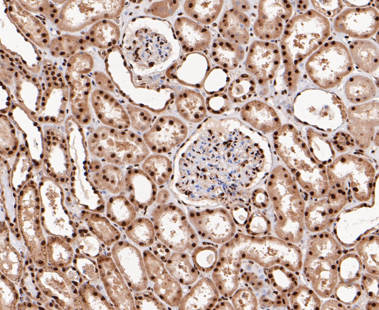
Fig4: Immunohistochemical analysis of paraffin-embedded human kidney tissue using anti-SSB antibody. The section was pre-treated using heat mediated antigen retrieval with sodium citrate buffer (pH 6.0) for 20 minutes. The tissues were blocked in 5% BSA for 30 minutes at room temperature, washed with ddH2O and PBS, and then probed with the primary antibody (ER1901-16, 1/50) for 30 minutes at room temperature. The detection was performed using an HRP conjugated compact polymer system. DAB was used as the chromogen. Tissues were counterstained with hematoxylin and mounted with DPX.Application
Fig5: Immunohistochemical analysis of paraffin-embedded human stomach cancer tissue using anti-SSB antibody. The section was pre-treated using heat mediated antigen retrieval with sodium citrate buffer (pH 6.0) for 20 minutes. The tissues were blocked in 5% BSA for 30 minutes at room temperature, washed with ddH2O and PBS, and then probed with the primary antibody (ER1901-16, 1/800) for 30 minutes at room temperature. The detection was performed using an HRP conjugated compact polymer system. DAB was used as the chromogen. Tissues were counterstained with hematoxylin and mounted with DPX.Application
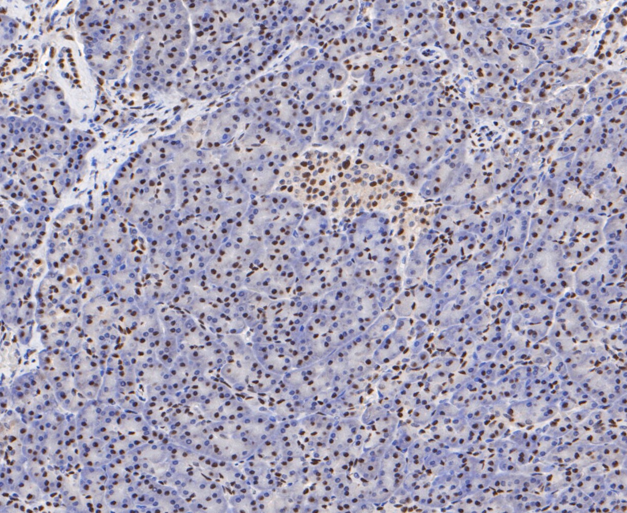
Fig6: Immunohistochemical analysis of paraffin-embedded human pancreas tissue using anti-SSB antibody. The section was pre-treated using heat mediated antigen retrieval with sodium citrate buffer (pH 6.0) for 20 minutes. The tissues were blocked in 5% BSA for 30 minutes at room temperature, washed with ddH2O and PBS, and then probed with the primary antibody (ER1901-16, 1/50) for 30 minutes at room temperature. The detection was performed using an HRP conjugated compact polymer system. DAB was used as the chromogen. Tissues were counterstained with hematoxylin and mounted with DPX.Application
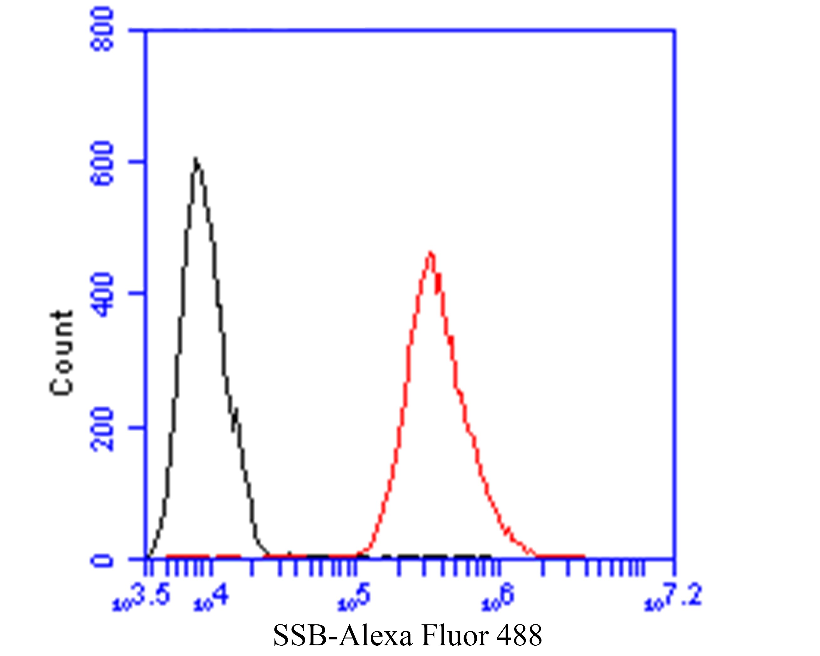
Fig7: Flow cytometric analysis of SSB was done on HCT116 cells. The cells were fixed, permeabilized and stained with the primary antibody (ER1901-16, 1/50) (red). After incubation of the primary antibody at room temperature for an hour, the cells were stained with a Alexa Fluor 488-conjugated Goat anti-Rabbit IgG Secondary antibody at 1/1000 dilution for 30 minutes.Unlabelled sample was used as a control (cells without incubation with primary antibody; black).| Product Name | SSB |
|---|---|
| Antibody Type | Primary Antibodies |
| Antigen Alias | Autoantigen La antibody La antibody La autoantigen antibody La autoantigen homolog antibody La protein antibody La ribonucleoprotein antibody La ribonucleoprotein domain family member 3 antibody LA_HUMAN antibody LARP3 antibody Lupus La antigen antibody Lupus La protein antibody Lupus La protein homolog antibody MGC118101 antibody MGC93380 antibody mRNA for autoantigen antibody OTTMUSP00000014043 antibody RP23 273G23.1 antibody Sjoegren syndrome type B antigen antibody Sjogren syndrome antigen B (autoantigen La) antibody Sjogren syndrome antigen B antibody SS B antibody SS-B antibody SS-B/La protein antibody SSB antibody |
| Product description | The protein encoded by this gene is involved in diverse aspects of RNA metabolism, including binding and protecting poly(U) termini of nascent RNA polymerase III transcripts from exonuclease digestion, processing 5' and 3' ends of pre-tRNA precursors, acting as an RNA chaperone, and binding viral RNAs associated with hepatitis C virus. Autoantibodies reacting with this protein are found in the sera of patients with Sjogren syndrome and systemic lupus erythematosus. Alternative promoter usage results in two different transcript variants which encode the same protein. In case of Coxsackievirus B3 infection, binds to the viral internal ribosome entry site (IRES) and stimulates the IRES-mediated translation. |
| Immunogen | Recombinant protein within Human SSB aa 165-346. |
| Clonality | Polyclonal |
|---|---|
| Isotype | IgG |
| Host Species | Rabbit |
| Tested Applications | WBIHCFC |
| WB:1:500-1:2,000: IHC:1:50-1:800: FC:1:50-1:100: |
|
| Species Reactivity | Human |
| Concentration | 1mg/ml |
| Alternative Names | Autoantigen La antibody
La antibody
La autoantigen antibody La autoantigen homolog antibody La protein antibody La ribonucleoprotein antibody La ribonucleoprotein domain family member 3 antibody LA_HUMAN antibody LARP3 antibody Lupus La antigen antibody Lupus La protein antibody Lupus La protein homolog antibody MGC118101 antibody MGC93380 antibody mRNA for autoantigen antibody OTTMUSP00000014043 antibody RP23 273G23.1 antibody Sjoegren syndrome type B antigen antibody Sjogren syndrome antigen B (autoantigen La) antibody Sjogren syndrome antigen B antibody SS B antibody SS-B antibody SS-B/La protein antibody SSB antibody |
|---|---|
| Molecular Weight(MW) | 47 kDa |
| Cellular Localization | Nucleus. |
| SwissProt ID | P05455 |
|---|
Application
Fig1: Western blot analysis of SSB on K562 cell lysate. Proteins were transferred to a PVDF membrane and blocked with 5% BSA in PBS for 1 hour at room temperature. The primary antibody (ER1901-16, 1/5,000) was used in 5% BSA at room temperature for 2 hours. Goat Anti-Rabbit IgG - HRP Secondary Antibody (HA1001) at 1:5,000 dilution was used for 1 hour at room temperature.
Application
Fig2: Western blot analysis of SSB on human lung tissue lysate. Proteins were transferred to a PVDF membrane and blocked with 5% BSA in PBS for 1 hour at room temperature. The primary antibody (ER1901-16, 1/1,000) was used in 5% BSA at room temperature for 2 hours. Goat Anti-Rabbit IgG - HRP Secondary Antibody (HA1001) at 1:5,000 dilution was used for 1 hour at room temperature.
Application
Fig3: Immunohistochemical analysis of paraffin-embedded human colon cancer tissue using anti-SSB antibody. The section was pre-treated using heat mediated antigen retrieval with sodium citrate buffer (pH 6.0) for 20 minutes. The tissues were blocked in 5% BSA for 30 minutes at room temperature, washed with ddH2O and PBS, and then probed with the primary antibody (ER1901-16, 1/800) for 30 minutes at room temperature. The detection was performed using an HRP conjugated compact polymer system. DAB was used as the chromogen. Tissues were counterstained with hematoxylin and mounted with DPX.
Application
Fig4: Immunohistochemical analysis of paraffin-embedded human kidney tissue using anti-SSB antibody. The section was pre-treated using heat mediated antigen retrieval with sodium citrate buffer (pH 6.0) for 20 minutes. The tissues were blocked in 5% BSA for 30 minutes at room temperature, washed with ddH2O and PBS, and then probed with the primary antibody (ER1901-16, 1/50) for 30 minutes at room temperature. The detection was performed using an HRP conjugated compact polymer system. DAB was used as the chromogen. Tissues were counterstained with hematoxylin and mounted with DPX.
Application
Fig5: Immunohistochemical analysis of paraffin-embedded human stomach cancer tissue using anti-SSB antibody. The section was pre-treated using heat mediated antigen retrieval with sodium citrate buffer (pH 6.0) for 20 minutes. The tissues were blocked in 5% BSA for 30 minutes at room temperature, washed with ddH2O and PBS, and then probed with the primary antibody (ER1901-16, 1/800) for 30 minutes at room temperature. The detection was performed using an HRP conjugated compact polymer system. DAB was used as the chromogen. Tissues were counterstained with hematoxylin and mounted with DPX.
Application
Fig6: Immunohistochemical analysis of paraffin-embedded human pancreas tissue using anti-SSB antibody. The section was pre-treated using heat mediated antigen retrieval with sodium citrate buffer (pH 6.0) for 20 minutes. The tissues were blocked in 5% BSA for 30 minutes at room temperature, washed with ddH2O and PBS, and then probed with the primary antibody (ER1901-16, 1/50) for 30 minutes at room temperature. The detection was performed using an HRP conjugated compact polymer system. DAB was used as the chromogen. Tissues were counterstained with hematoxylin and mounted with DPX.
Application
Fig7: Flow cytometric analysis of SSB was done on HCT116 cells. The cells were fixed, permeabilized and stained with the primary antibody (ER1901-16, 1/50) (red). After incubation of the primary antibody at room temperature for an hour, the cells were stained with a Alexa Fluor 488-conjugated Goat anti-Rabbit IgG Secondary antibody at 1/1000 dilution for 30 minutes.Unlabelled sample was used as a control (cells without incubation with primary antibody; black).| Positive Control | K562, human lung tissue, human colon cancer tissue, human kidney tissue, human stomach cancer tissue, human pancreas tissue. |
|---|---|
| Application Notes | WB:1:500-1:2,000: IHC:1:50-1:800: FC:1:50-1:100: |
| Form | Liquid |
|---|---|
| Storage Instructions | Store at +4℃ after thawing. Aliquot store at -20℃. Avoid repeated freeze / thaw cycles. |
| Storage Buffer | 1*TBS (pH7.4), 1%BSA, 50%Glycerol. Preservative: 0.05% Sodium Azide. |


