Application
Fig1: Western blot analysis of CD46 on Siha and PC-3M cell lysates using anti-CD46 antibody at 1/1,000 dilution.Application
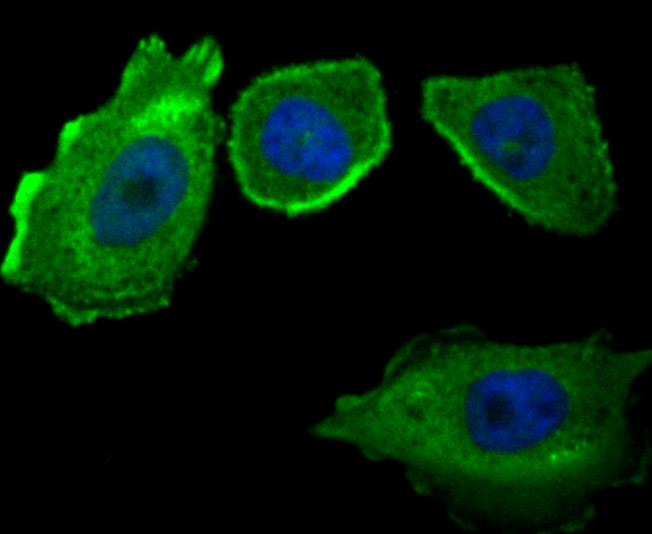
Fig2: ICC staining CD46 in PC-3M cells (green). The nuclear counter stain is DAPI (blue). Cells were fixed in paraformaldehyde, permeabilised with 0.25% Triton X100/PBS.Application
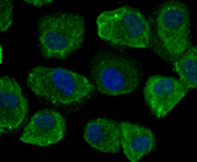
Fig3: ICC staining CD46 in HUVEC cells (green). The nuclear counter stain is DAPI (blue). Cells were fixed in paraformaldehyde, permeabilised with 0.25% Triton X100/PBS.Application
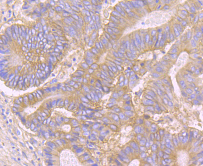
Fig4: Immunohistochemical analysis of paraffin-embedded human colon cancer tissue using anti-CD46 antibody. Counter stained with hematoxylin.Application
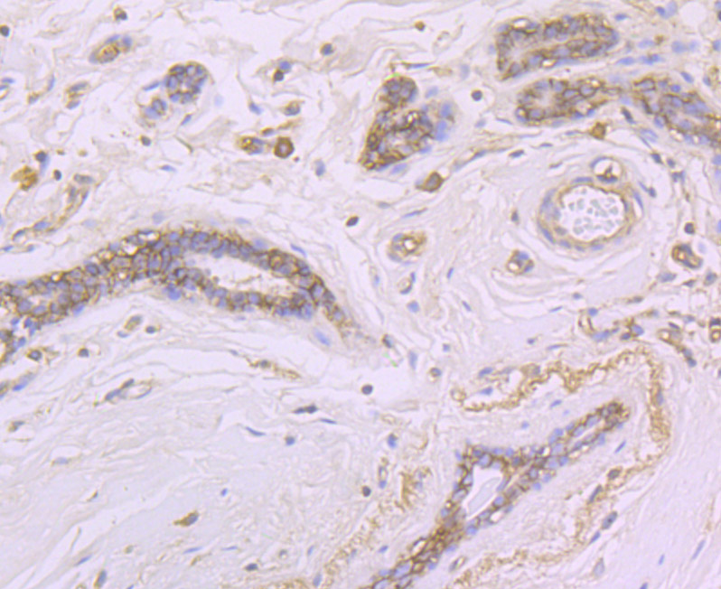
Fig5: Immunohistochemical analysis of paraffin-embedded human breast tissue using anti-CD46 antibody. Counter stained with hematoxylin.Application
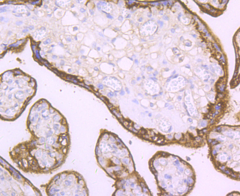
Fig6: Immunohistochemical analysis of paraffin-embedded human placenta tissue using anti-CD46 antibody. Counter stained with hematoxylin.Application
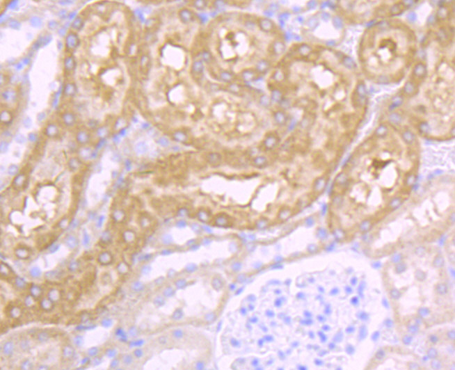
Fig7: Immunohistochemical analysis of paraffin-embedded rat kidney tissue using anti-CD46 antibody. Counter stained with hematoxylin.| Product Name | CD46 [JB25-49] |
|---|---|
| Antibody Type | Primary Antibodies |
| Antigen Alias | AHUS2 antibody Antigen defined by monoclonal antibody TRA 2 10 antibody Antigen identified by monoclonal antibody TRA 2 10 antibody CD46 antibody CD46 antigen antibody CD46 antigen complement regulatory protein antibody CD46 molecule antibody CD46 molecule complement regulatory protein antibody Complement membrane cofactor protein antibody MCP antibody MCP_HUMAN antibody Measles virus receptor antibody membrane cofactor protein (CD46, trophoblast-lymphocyte cross-reactive antigen) antibody Membrane cofactor protein antibody MGC26544 antibody MIC10 antibody TLX antibody TRA2.10 antibody Trophoblast leucocyte common antigen antibody Trophoblast leukocyte common antigen antibody Trophoblast lymphocyte cross reactive antigen antibody |
| Product description | CD46, also called membrane cofactor protein (MCP), is a transmembrane glycoprotein that exists as a non-disulfide-linked dimer. CD46 regulates the complement cascade by inhibiting C3b and C4b deposited on self tissue. CD46 is a cofactor that binds to C3b and C4b, allowing their degradation by a plasma serine protease called factor I. This function resides in the complement control protein repeats (CCPs), with CCP1-4 essential for regulation. CD46 is widely distributed on thymocytes, T cells, B cells, monocytes, granulocytes, NK cells, platelets, endothelial cells, epithelial cells, fibroblasts, placenta and sperm, but not on erythrocytes. It is the major high affinity receptor for measles virus and human herpes virus. Mouse cells ubiquitously express CRRY, which is a functional ortholog of human decay-accelerating factor (DAF; CD55) and membrane cofactor protein (MCP; CD46). |
| Immunogen | Recombinant protein |
| Clonality | Monoclonal |
|---|---|
| Isotype | IgG |
| Host Species | Recombinant rabbit |
| Tested Applications | WBICCIHC |
| WB:1:1,000-1:5,000 ICC:1:50-1:200 IHC:1:50-1:200 |
|
| Species Reactivity | HumanRat |
| Concentration | 1mg/ml |
| Alternative Names | AHUS2 antibody
Antigen defined by monoclonal antibody TRA 2 10 antibody
Antigen identified by monoclonal antibody TRA 2 10 antibody
CD46 antibody
CD46 antigen antibody
CD46 antigen complement regulatory protein antibody
CD46 molecule antibody
CD46 molecule complement regulatory protein antibody
Complement membrane cofactor protein antibody
MCP antibody
MCP_HUMAN antibody
Measles virus receptor antibody
membrane cofactor protein (CD46 trophoblast-lymphocyte cross-reactive antigen) antibody Membrane cofactor protein antibody MGC26544 antibody MIC10 antibody TLX antibody TRA2.10 antibody Trophoblast leucocyte common antigen antibody Trophoblast leukocyte common antigen antibody Trophoblast lymphocyte cross reactive antigen antibody |
|---|---|
| Molecular Weight(MW) | 55 kDa |
| Cellular Localization | Cytoplasmic vesicle. Membrane. |
Application
Fig1: Western blot analysis of CD46 on Siha and PC-3M cell lysates using anti-CD46 antibody at 1/1,000 dilution.
Application
Fig2: ICC staining CD46 in PC-3M cells (green). The nuclear counter stain is DAPI (blue). Cells were fixed in paraformaldehyde, permeabilised with 0.25% Triton X100/PBS.
Application
Fig3: ICC staining CD46 in HUVEC cells (green). The nuclear counter stain is DAPI (blue). Cells were fixed in paraformaldehyde, permeabilised with 0.25% Triton X100/PBS.
Application
Fig4: Immunohistochemical analysis of paraffin-embedded human colon cancer tissue using anti-CD46 antibody. Counter stained with hematoxylin.
Application
Fig5: Immunohistochemical analysis of paraffin-embedded human breast tissue using anti-CD46 antibody. Counter stained with hematoxylin.
Application
Fig6: Immunohistochemical analysis of paraffin-embedded human placenta tissue using anti-CD46 antibody. Counter stained with hematoxylin.
Application
Fig7: Immunohistochemical analysis of paraffin-embedded rat kidney tissue using anti-CD46 antibody. Counter stained with hematoxylin.| Positive Control | Siha, HUVEC, PC-3M, rat kidney tissue, human colon cancer tissue, human breast tissue, human placenta tissue. |
|---|---|
| Application Notes | WB:1:1,000-1:5,000 ICC:1:50-1:200 IHC:1:50-1:200 |
| Form | Liquid |
|---|---|
| Storage Instructions | Store at +4℃ after thawing. Aliquot store at -20℃ or -80℃. Avoid repeated freeze / thaw cycles. |
| Storage Buffer | 1*TBS (pH7.4), 1%BSA, 40%Glycerol. Preservative: 0.05% Sodium Azide. |


