Application
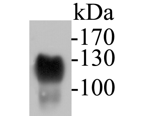
Fig1: Western blot analysis of CD43 on human thymus tissue lysates. Proteins were transferred to a PVDF membrane and blocked with 5% BSA in PBS for 1 hour at room temperature. The primary antibody (ER1901-28, 1/500) was used in 5% BSA at room temperature for 2 hours. Goat Anti-Rabbit IgG - HRP Secondary Antibody (HA1001) at 1:5,000 dilution was used for 1 hour at room temperature.Application
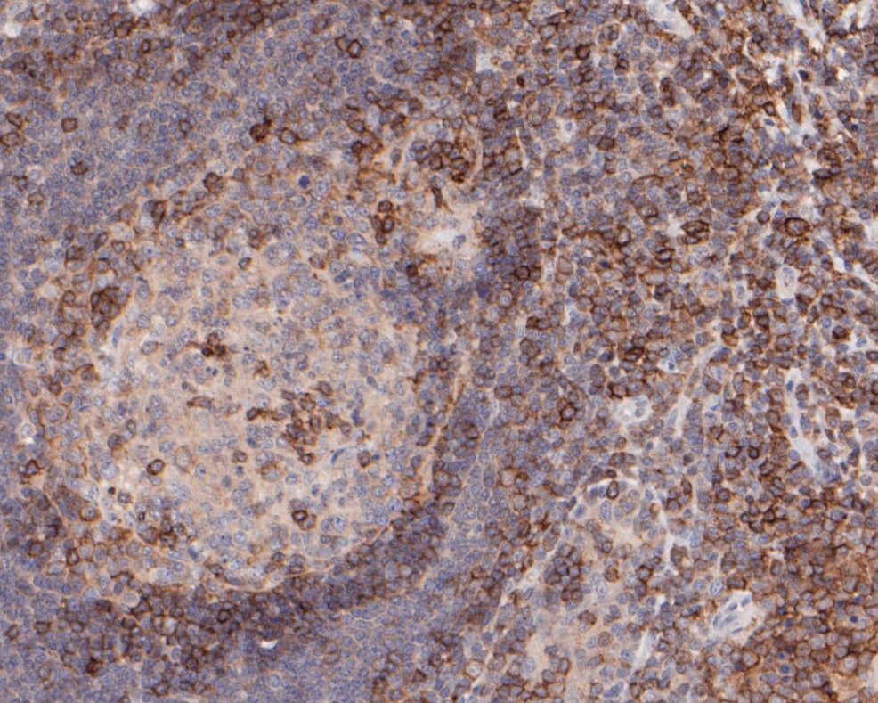
Fig2: Immunohistochemical analysis of paraffin-embedded human tonsil tissue using anti-CD43 antibody. The section was pre-treated using heat mediated antigen retrieval with sodium citrate buffer (pH 6.0) for 20 minutes. The tissues were blocked in 5% BSA for 30 minutes at room temperature, washed with ddH2O and PBS, and then probed with the primary antibody (ER1901-28, 1/100) for 30 minutes at room temperature. The detection was performed using an HRP conjugated compact polymer system. DAB was used as the chromogen. Tissues were counterstained with hematoxylin and mounted with DPX.Application
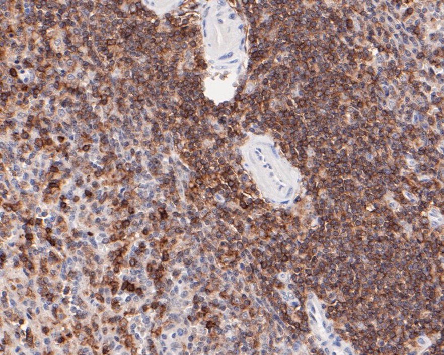
Fig3: Immunohistochemical analysis of paraffin-embedded human spleen tissue using anti-CD43 antibody. The section was pre-treated using heat mediated antigen retrieval with sodium citrate buffer (pH 6.0) for 20 minutes. The tissues were blocked in 5% BSA for 30 minutes at room temperature, washed with ddH2O and PBS, and then probed with the primary antibody (ER1901-28, 1/100) for 30 minutes at room temperature. The detection was performed using an HRP conjugated compact polymer system. DAB was used as the chromogen. Tissues were counterstained with hematoxylin and mounted with DPX.Application
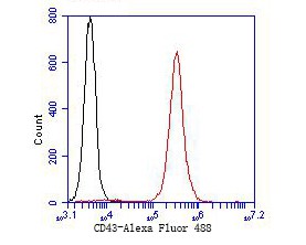
Fig4: Flow cytometric analysis of CD43 was done on Jurkat cells. The cells were fixed, permeabilized and stained with the primary antibody (ER1901-28, 1/50) (red). After incubation of the primary antibody at room temperature for an hour, the cells were stained with a Alexa Fluor 488-conjugated goat anti-rabbit IgG Secondary antibody at 1/1000 dilution for 30 minutes.Unlabelled sample was used as a control (cells without incubation with primary antibody; black).| Product Name | CD43 |
|---|---|
| Antibody Type | Primary Antibodies |
| Antigen Alias | CD 43 antibody CD43 antibody CD43 antigen antibody Galactoglycoprotein antibody GALGP antibody GPL 115 antibody GPL115 antibody Human gene for sialophorin antibody Leucocyte sialoglycoprotein antibody LEUK_HUMAN antibody Leukocyte large sialoglycoprotein antibody Leukocyte sialoglycoprotein antibody Leukosialin antibody LSN antibody Ly-48 antibody sialophorin (gpL115, leukosialin, CD43) antibody Sialophorin antibody Spn antibody |
| Product description | Predominant cell surface sialoprotein of leukocytes which regulates multiple T-cell functions, including T-cell activation, proliferation, differentiation, trafficking and migration. Positively regulates T-cell trafficking to lymph-nodes via its association with ERM proteins (EZR, RDX and MSN) (By similarity). Negatively regulates Th2 cell differentiation and predisposes the differentiation of T-cells towards a Th1 lineage commitment. Promotes the expression of IFN-gamma by T-cells during T-cell receptor (TCR) activation of naive cells and induces the expression of IFN-gamma by CD4+ T-cells and to a lesser extent by CD8+ T-cells (PubMed:18036228). Plays a role in preparing T-cells for cytokine sensing and differentiation into effector cells by inducing the expression of cytokine receptors IFNGR and IL4R, promoting IFNGR and IL4R signaling and by mediating the clustering of IFNGR with TCR (PubMed:24328034). Acts as a major E-selectin ligand responsible for Th17 cell rolling on activated vasculature and recruitment during inflammation. Mediates Th17 cells, but not Th1 cells, adhesion to E-selectin. Acts as a T-cell counter-receptor for SIGLEC1 (By similarity).CD43 cytoplasmic tail: Protects cells from apoptotic signals, promoting cell survival. |
| Immunogen | Synthetic peptide within C-terminal human CD43. |
| Clonality | Polyclonal |
|---|---|
| Isotype | IgG |
| Host Species | Rabbit |
| Tested Applications | WBIHCFC |
| WB:1:500-1:2,000: IHC-P:1:50-1:200: FC:1:50-1:100: |
|
| Species Reactivity | Human |
| Concentration | 1mg/mL |
| Alternative Names | CD 43 antibody
CD43 antibody
CD43 antigen antibody Galactoglycoprotein antibody GALGP antibody GPL 115 antibody GPL115 antibody Human gene for sialophorin antibody Leucocyte sialoglycoprotein antibody LEUK_HUMAN antibody Leukocyte large sialoglycoprotein antibody Leukocyte sialoglycoprotein antibody Leukosialin antibody LSN antibody Ly-48 antibody sialophorin (gpL115 leukosialin CD43) antibody Sialophorin antibody Spn antibody |
|---|---|
| Molecular Weight(MW) | 40 kDa (Predicted band size) |
| Cellular Localization | Membrane, microvillus, uropodium, nucleus. |
| SwissProt ID | P16150 |
|---|
Application
Fig1: Western blot analysis of CD43 on human thymus tissue lysates. Proteins were transferred to a PVDF membrane and blocked with 5% BSA in PBS for 1 hour at room temperature. The primary antibody (ER1901-28, 1/500) was used in 5% BSA at room temperature for 2 hours. Goat Anti-Rabbit IgG - HRP Secondary Antibody (HA1001) at 1:5,000 dilution was used for 1 hour at room temperature.
Application
Fig2: Immunohistochemical analysis of paraffin-embedded human tonsil tissue using anti-CD43 antibody. The section was pre-treated using heat mediated antigen retrieval with sodium citrate buffer (pH 6.0) for 20 minutes. The tissues were blocked in 5% BSA for 30 minutes at room temperature, washed with ddH2O and PBS, and then probed with the primary antibody (ER1901-28, 1/100) for 30 minutes at room temperature. The detection was performed using an HRP conjugated compact polymer system. DAB was used as the chromogen. Tissues were counterstained with hematoxylin and mounted with DPX.
Application
Fig3: Immunohistochemical analysis of paraffin-embedded human spleen tissue using anti-CD43 antibody. The section was pre-treated using heat mediated antigen retrieval with sodium citrate buffer (pH 6.0) for 20 minutes. The tissues were blocked in 5% BSA for 30 minutes at room temperature, washed with ddH2O and PBS, and then probed with the primary antibody (ER1901-28, 1/100) for 30 minutes at room temperature. The detection was performed using an HRP conjugated compact polymer system. DAB was used as the chromogen. Tissues were counterstained with hematoxylin and mounted with DPX.
Application
Fig4: Flow cytometric analysis of CD43 was done on Jurkat cells. The cells were fixed, permeabilized and stained with the primary antibody (ER1901-28, 1/50) (red). After incubation of the primary antibody at room temperature for an hour, the cells were stained with a Alexa Fluor 488-conjugated goat anti-rabbit IgG Secondary antibody at 1/1000 dilution for 30 minutes.Unlabelled sample was used as a control (cells without incubation with primary antibody; black).| Positive Control | Human thymus tissue lysates, human tonsil tissue, human spleen tissue, Jurkat. |
|---|---|
| Application Notes | WB:1:500-1:2,000: IHC-P:1:50-1:200: FC:1:50-1:100: |
| Form | Liquid |
|---|---|
| Storage Instructions | Store at +4℃ after thawing. Aliquot store at -20℃. Avoid repeated freeze / thaw cycles. |
| Storage Buffer | 1*TBS (pH7.4), 1%BSA, 50%Glycerol. Preservative: 0.05% Sodium Azide. |


