Application
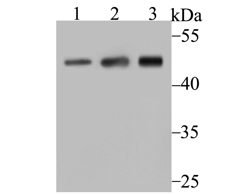
Fig1: Western blot analysis of Arp3 on different lysates using anti-Arp3 antibody at 1/500 dilution. Positive control: Lane 1: A431 Lane 2: Mouse placenta Lane 3: Mouse thymusApplication
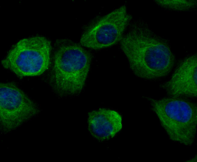
Fig2: ICC staining Arp3 in HUVEC cells (green). The nuclear counter stain is DAPI (blue). Cells were fixed in paraformaldehyde, permeabilised with 0.25% Triton X100/PBS.Application
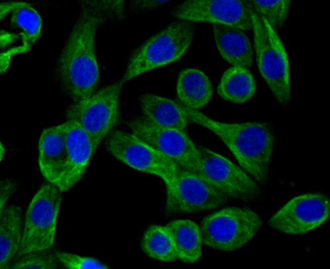
Fig3: ICC staining Arp3 in LOVO cells (green). The nuclear counter stain is DAPI (blue). Cells were fixed in paraformaldehyde, permeabilised with 0.25% Triton X100/PBS.Application
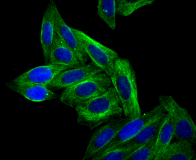
Fig4: ICC staining Arp3 in SiHa cells (green). The nuclear counter stain is DAPI (blue). Cells were fixed in paraformaldehyde, permeabilised with 0.25% Triton X100/PBS.Application
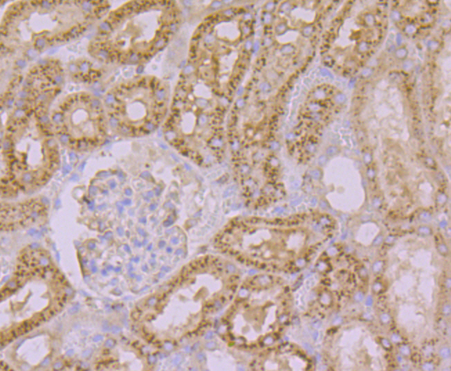
Fig5: Immunohistochemical analysis of paraffin-embedded rat kidney tissue using anti-Arp3 antibody. Counter stained with hematoxylin.Application
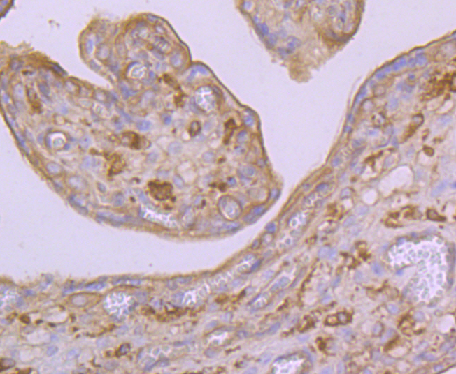
Fig6: Immunohistochemical analysis of paraffin-embedded human placenta tissue using anti-Arp3 antibody. Counter stained with hematoxylin.Application
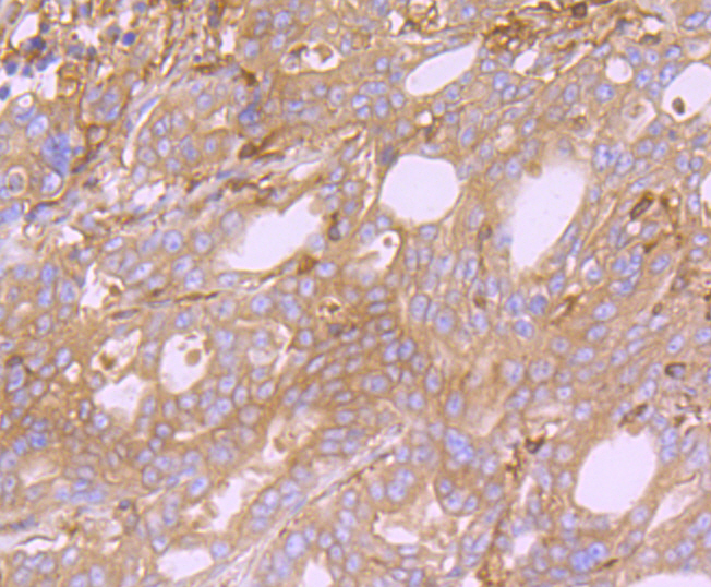
Fig7: Immunohistochemical analysis of paraffin-embedded human stomach cancer tissue using anti-Arp3 antibody. Counter stained with hematoxylin.Application
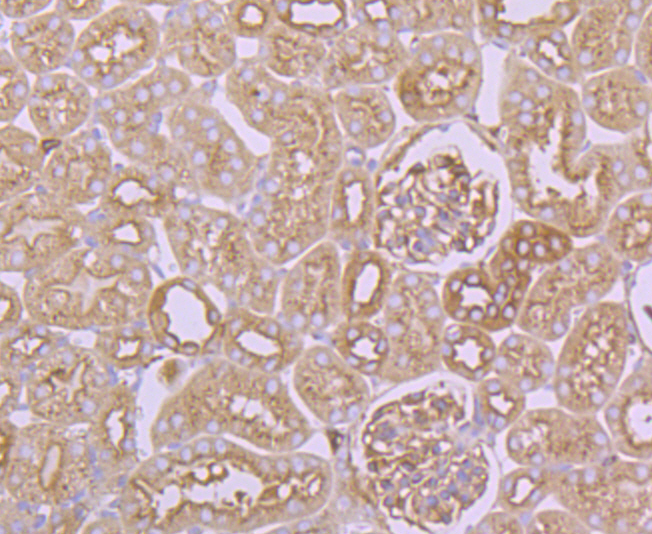
Fig8: Immunohistochemical analysis of paraffin-embedded mouse kidney tissue using anti-Arp3 antibody. Counter stained with hematoxylin.Application
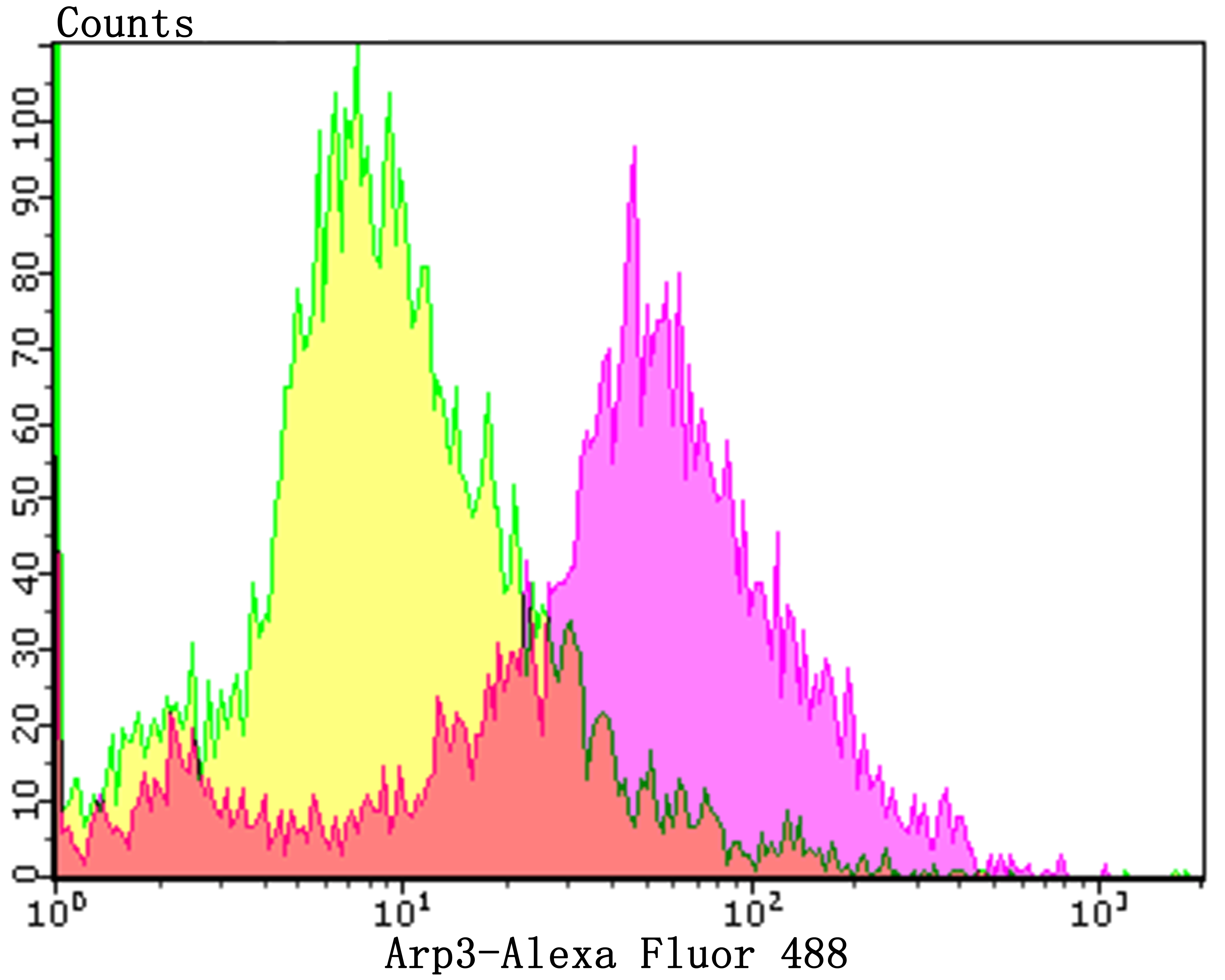
Fig9: Flow cytometric analysis of HL-60 cells with Arp3 antibody at 1/100 dilution (purple) compared with an unlabelled control (cells without incubation with primary antibody; yellow).Alexa Fluor 488-conjugated goat anti-rabbit IgG was used as the secondary antibody.| Product Name | Arp3 [JB33-44] |
|---|---|
| Antibody Type | Primary Antibodies |
| Antigen Alias | Actin like protein 3 antibody Actin related protein 3 antibody ACTR3 antibody ARP3 actin related protein 3 homolog (yeast) antibody |
| Product description | Actin polymerization is required for a variety of cell functions, including chemotaxis, cell migration, cell adhesion, and platelet activation. Cells trigger actin polymerization through either the de novo nucleation of filaments from monomeric actin, the severing of existing filaments to create uncapped barbed ends, or the uncapping existing barbed ends. The nucleation of actin is a rate-limiting and unfavorable reaction in actin polymerization and therefore requires the involvement of the Arp2/3 complex, which helps create new filaments and promotes the end-to-side cross-linking of actin filaments into the branching meshwork. The Arp2/3 complex consists of the actin-related proteins Arp2 and Arp3, and various other accessory proteins. The Arp2/3 complex promotes actin nucleation by binding the pointed end of actin filaments, or by associating with the side of an existing filament, and nucleates growth in the barbed direction. In addition, the Arp2/3 complex also mediates actin cytoskeletal outgrowths that are regulated by the Rho family of small GTPases. In response to GTP-binding Cdc42, the Arp2/3 complex binds the Cdc42 substrates, namely the WASP proteins, and initiates the formation of lamellipodia and filopodia. |
| Immunogen | Recombinant protein of C-terminal human Arp3. |
| Clonality | Monoclonal |
|---|---|
| Isotype | IgG |
| Host Species | Recombinant rabbit |
| Tested Applications | WBICCIFIHCFC |
| WB:1:500-1:1,000 ICC:1:50-1:200 IHC:1:50-1:200 FC:1:50-1:100 |
|
| Species Reactivity | HumanMouseRat |
| Concentration | 1mg/ml |
| Alternative Names | Actin like protein 3 antibody Actin related protein 3 antibody ACTR3 antibody ARP3 actin related protein 3 homolog (yeast) antibody |
|---|---|
| Molecular Weight(MW) | 47 kDa |
| Cellular Localization | Cell projection. Cytoskeleton. |
Application
Fig1: Western blot analysis of Arp3 on different lysates using anti-Arp3 antibody at 1/500 dilution. Positive control: Lane 1: A431 Lane 2: Mouse placenta Lane 3: Mouse thymus
Application
Fig2: ICC staining Arp3 in HUVEC cells (green). The nuclear counter stain is DAPI (blue). Cells were fixed in paraformaldehyde, permeabilised with 0.25% Triton X100/PBS.
Application
Fig3: ICC staining Arp3 in LOVO cells (green). The nuclear counter stain is DAPI (blue). Cells were fixed in paraformaldehyde, permeabilised with 0.25% Triton X100/PBS.
Application
Fig4: ICC staining Arp3 in SiHa cells (green). The nuclear counter stain is DAPI (blue). Cells were fixed in paraformaldehyde, permeabilised with 0.25% Triton X100/PBS.
Application
Fig5: Immunohistochemical analysis of paraffin-embedded rat kidney tissue using anti-Arp3 antibody. Counter stained with hematoxylin.
Application
Fig6: Immunohistochemical analysis of paraffin-embedded human placenta tissue using anti-Arp3 antibody. Counter stained with hematoxylin.
Application
Fig7: Immunohistochemical analysis of paraffin-embedded human stomach cancer tissue using anti-Arp3 antibody. Counter stained with hematoxylin.
Application
Fig8: Immunohistochemical analysis of paraffin-embedded mouse kidney tissue using anti-Arp3 antibody. Counter stained with hematoxylin.
Application
Fig9: Flow cytometric analysis of HL-60 cells with Arp3 antibody at 1/100 dilution (purple) compared with an unlabelled control (cells without incubation with primary antibody; yellow).Alexa Fluor 488-conjugated goat anti-rabbit IgG was used as the secondary antibody.| Positive Control | A431, mouse placenta tissue lysate, mouse thymus tissue lysate, HUVEC, LOVO, SiHa, rat kidney tissue, human placenta tissue, human stomach cancer tissue, mouse kidney tissue, HL-60. |
|---|---|
| Application Notes | WB:1:500-1:1,000 ICC:1:50-1:200 IHC:1:50-1:200 FC:1:50-1:100 |
| Form | Liquid |
|---|---|
| Storage Instructions | Store at +4℃ after thawing. Aliquot store at -20℃. Avoid repeated freeze / thaw cycles. |
| Storage Buffer | 1*TBS (pH7.4), 1%BSA, 40%Glycerol. Preservative: 0.05% Sodium Azide. |


