Application
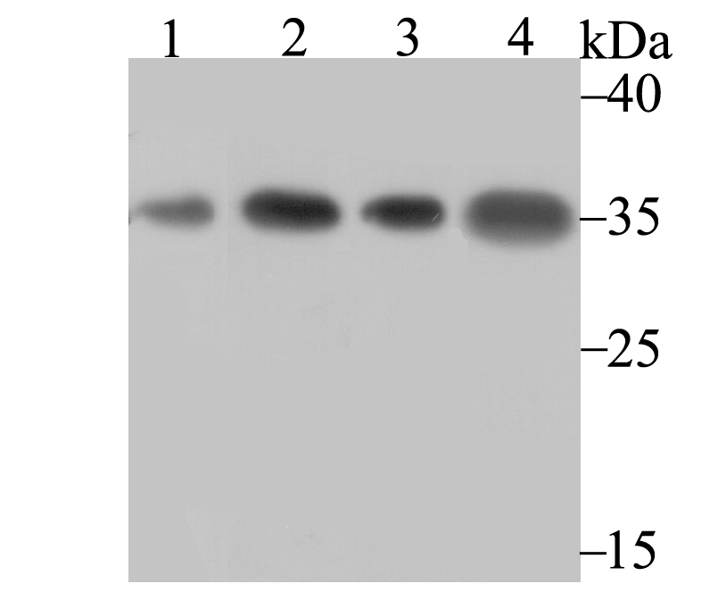
Fig1: Western blot analysis of U1A on different lysates using anti-U1A antibody at 1/500 dilution. Positive control: Lane 1: Rat brain Lane 2: K562 Lane 3: A431 Lane 4: Mouse kidneyApplication
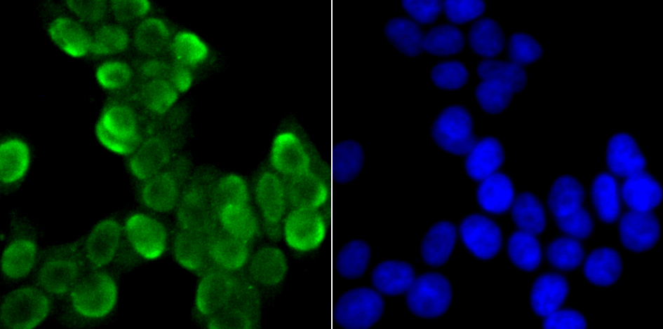
Fig2: ICC staining U1A in 293T cells (green). The nuclear counter stain is DAPI (blue). Cells were fixed in paraformaldehyde, permeabilised with 0.25% Triton X100/PBS.Application
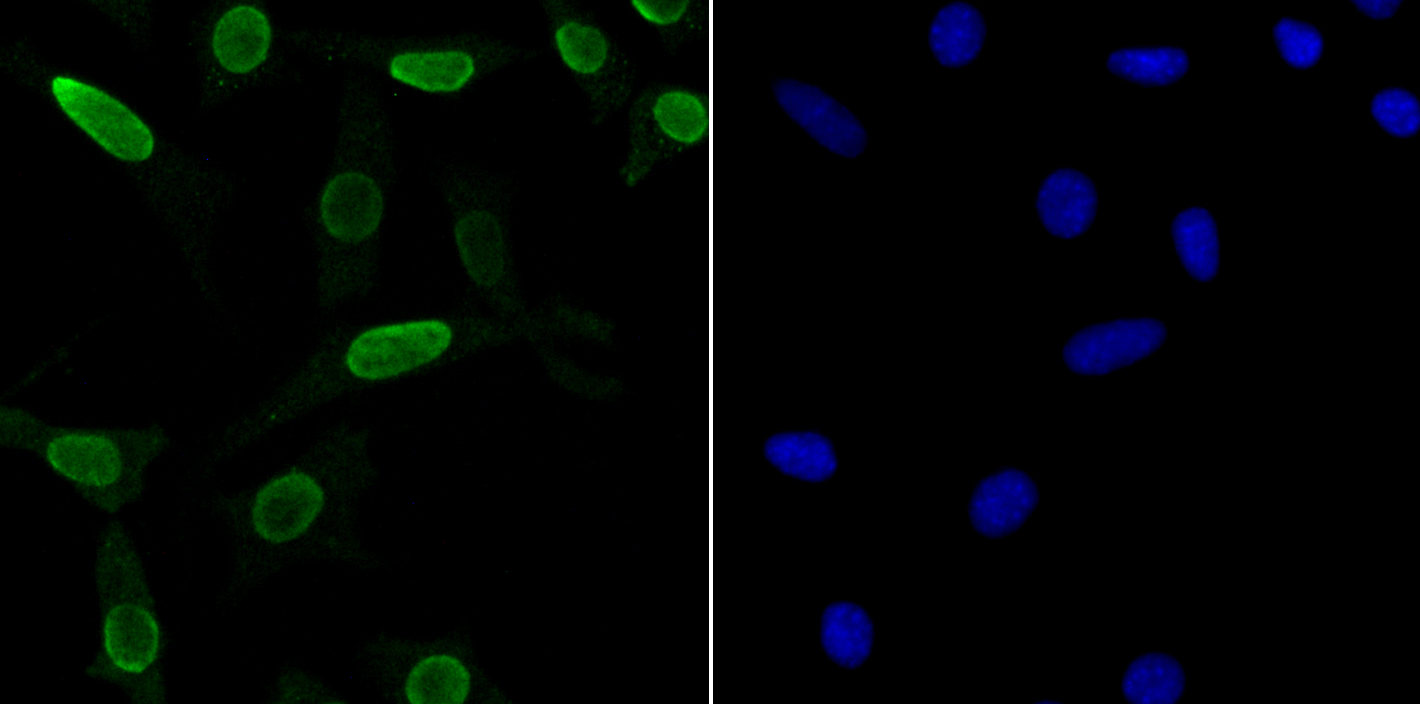
Fig3: ICC staining U1A in SH-SY-5Y cells (green). The nuclear counter stain is DAPI (blue). Cells were fixed in paraformaldehyde, permeabilised with 0.25% Triton X100/PBS.Application
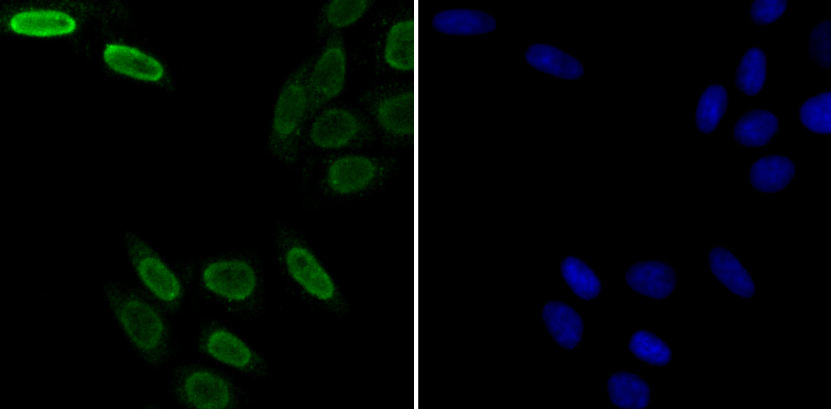
Fig4: ICC staining U1A in SiHa cells (green). The nuclear counter stain is DAPI (blue). Cells were fixed in paraformaldehyde, permeabilised with 0.25% Triton X100/PBS.Application
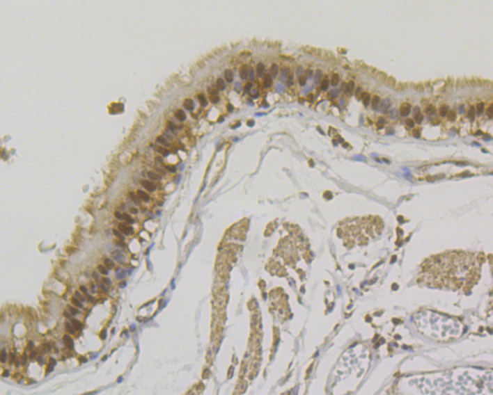
Fig5: Immunohistochemical analysis of paraffin-embedded human lung cancer tissue using anti-U1A antibody. Counter stained with hematoxylin.Application
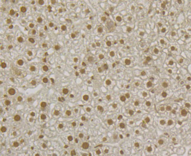
Fig6: Immunohistochemical analysis of paraffin-embedded mouse liver tissue using anti-U1A antibody. Counter stained with hematoxylin.| Product Name | U1A [JB33-48] |
|---|---|
| Antibody Type | Primary Antibodies |
| Antigen Alias | fc19d01 antibody Mud1 antibody small nuclear ribonucleoprotein polypeptide A antibody snRNP A antibody snRNP protein A antibody SNRPA antibody SNRPA_HUMAN antibody U1 small nuclear ribonucleoprotein A antibody U1 small nuclear RNP specific A antibody U1 snRNP A antibody U1 snRNP specific protein A antibody U1-A antibody U1A antibody wu:fc19d01 antibody zgc:77810 antibody |
| Product description | SNRPA (small nuclear ribonucleoprotein polypeptide A), also known as U1A (U1 snRNP protein A), is a component of the RNA spliceosome, a complex of proteins that are required for the precise excision of introns from pre-messenger RNA (pre-mRNA). Localizing to the nucleus, SNRPA contains two RRM (RNA recognition motif) domains, namely RRM1 and RRM2, and RRM1 specifically associates with the stem loop II of U1 snRNA (small nuclear RNA). In addition to functioning as a component of the U1 snRNP, SNRPA negatively regulates polyadenylation of SNRPA pre-mRNA, thereby negatively regulating itself. By inhibiting the addition of a polyA tail that would allow the pre-mRNA to mature, SNRPA causes the nuclear exosome degradation of the SNRPA pre-mRNA. At least 16% of cellular SNRPA also functions in an snRNP-free form (SF-A) that complexes with a group of non-snRNP proteins. |
| Immunogen | Recombinant protein corresponding to N-terminal human U1A. |
| Clonality | Monoclonal |
|---|---|
| Isotype | IgG |
| Host Species | Recombinant rabbit |
| Tested Applications | WBICCIFIHCIP |
| WB:1:500-1:1,000 ICC:1:50-1:200 IHC:1:50-1:200 IP:1:50-1:100 |
|
| Species Reactivity | HumanMouseRat |
| Concentration | 1mg/ml |
| Alternative Names | fc19d01 antibody Mud1 antibody small nuclear ribonucleoprotein polypeptide A antibody snRNP A antibody snRNP protein A antibody SNRPA antibody SNRPA_HUMAN antibody U1 small nuclear ribonucleoprotein A antibody U1 small nuclear RNP specific A antibody U1 snRNP A antibody U1 snRNP specific protein A antibody U1-A antibody U1A antibody wu:fc19d01 antibody zgc:77810 antibody |
|---|---|
| Molecular Weight(MW) | 31 kDa |
| Cellular Localization | Nucleus. |
Application
Fig1: Western blot analysis of U1A on different lysates using anti-U1A antibody at 1/500 dilution. Positive control: Lane 1: Rat brain Lane 2: K562 Lane 3: A431 Lane 4: Mouse kidney
Application
Fig2: ICC staining U1A in 293T cells (green). The nuclear counter stain is DAPI (blue). Cells were fixed in paraformaldehyde, permeabilised with 0.25% Triton X100/PBS.
Application
Fig3: ICC staining U1A in SH-SY-5Y cells (green). The nuclear counter stain is DAPI (blue). Cells were fixed in paraformaldehyde, permeabilised with 0.25% Triton X100/PBS.
Application
Fig4: ICC staining U1A in SiHa cells (green). The nuclear counter stain is DAPI (blue). Cells were fixed in paraformaldehyde, permeabilised with 0.25% Triton X100/PBS.
Application
Fig5: Immunohistochemical analysis of paraffin-embedded human lung cancer tissue using anti-U1A antibody. Counter stained with hematoxylin.
Application
Fig6: Immunohistochemical analysis of paraffin-embedded mouse liver tissue using anti-U1A antibody. Counter stained with hematoxylin.| Positive Control | Rat brain tissue lysate, K562, A431, mouse kidney tissue lysate, 293T, SH-SY-5Y, SiHa, human lung cancer tissue, mouse liver tissue. |
|---|---|
| Application Notes | WB:1:500-1:1,000 ICC:1:50-1:200 IHC:1:50-1:200 IP:1:50-1:100 |
| Form | Liquid |
|---|---|
| Storage Instructions | Store at +4℃ after thawing. Aliquot store at -20℃. Avoid repeated freeze / thaw cycles. |
| Storage Buffer | 1*TBS (pH7.4), 1%BSA, 40%Glycerol. Preservative: 0.05% Sodium Azide. |


