Application
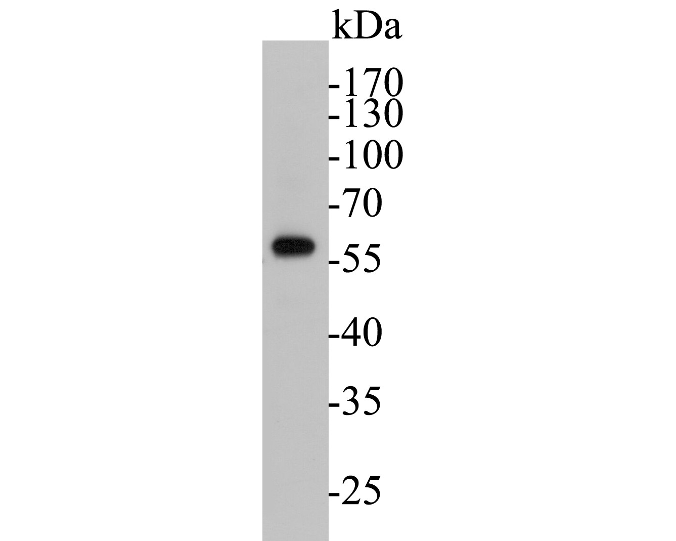
Fig1: Western blot analysis of AKT3 on SH-SY5Y cell lysates. Proteins were transferred to a PVDF membrane and blocked with 5% BSA in PBS for 1 hour at room temperature. The primary antibody (ER1901-33, 1/500) was used in 5% BSA at room temperature for 2 hours. Goat Anti-Rabbit IgG - HRP Secondary Antibody (HA1001) at 1:5,000 dilution was used for 1 hour at room temperature.Application
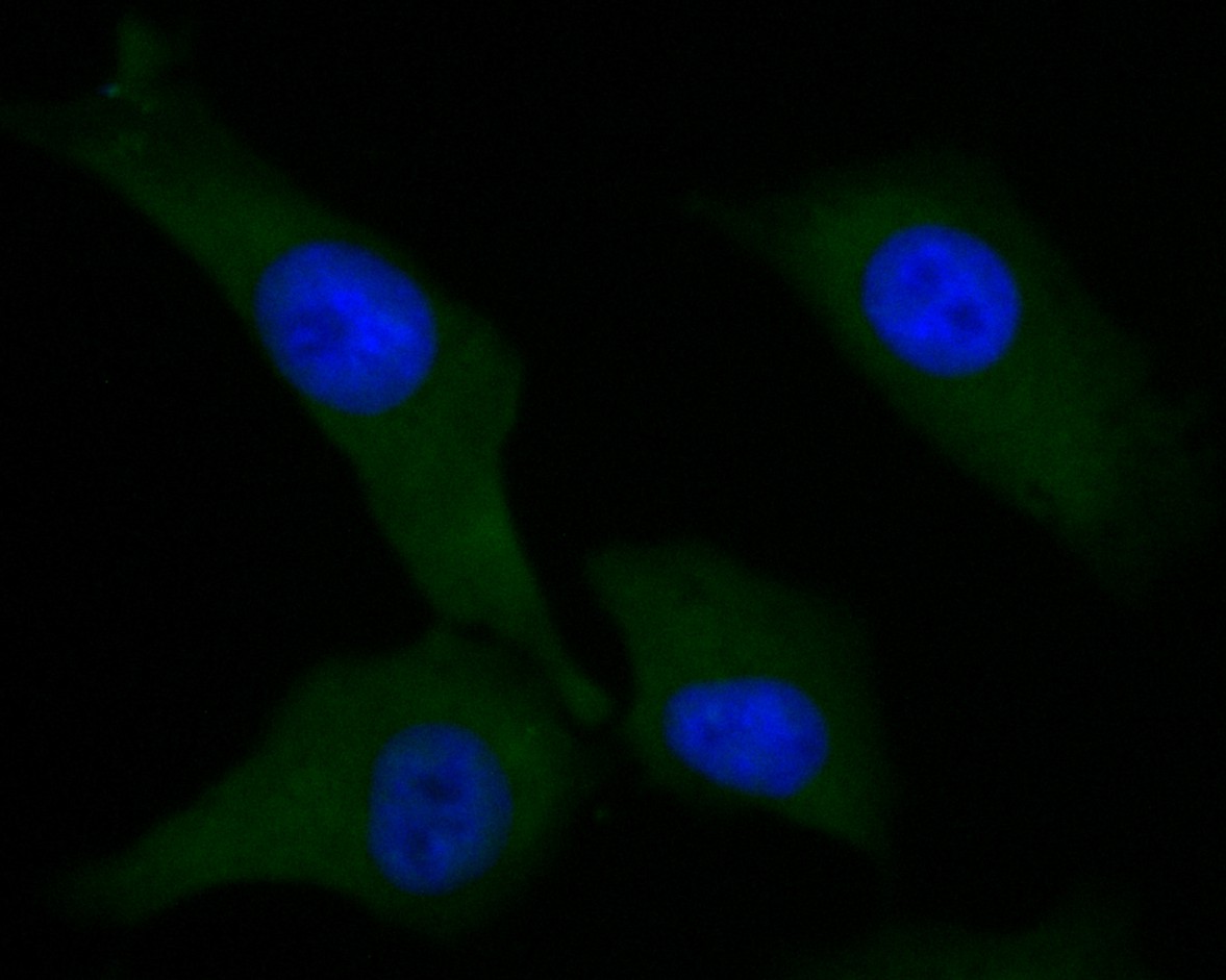
Fig2: ICC staining of AKT3 in A549 cells (green). Formalin fixed cells were permeabilized with 0.1% Triton X-100 in TBS for 10 minutes at room temperature and blocked with 1% Blocker BSA for 15 minutes at room temperature. Cells were probed with the primary antibody (ER1901-33, 1/50) for 1 hour at room temperature, washed with PBS. Alexa Fluor®488 Goat anti-Rabbit IgG was used as the secondary antibody at 1/1,000 dilution. The nuclear counter stain is DAPI (blue).Application
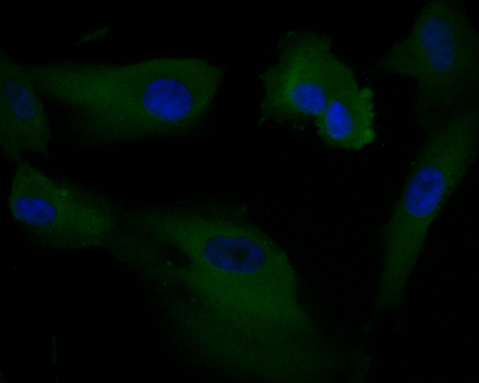
Fig3: ICC staining of AKT3 in EA.hy926 cells (green). Formalin fixed cells were permeabilized with 0.1% Triton X-100 in TBS for 10 minutes at room temperature and blocked with 1% Blocker BSA for 15 minutes at room temperature. Cells were probed with the primary antibody (ER1901-33, 1/50) for 1 hour at room temperature, washed with PBS. Alexa Fluor®488 Goat anti-Rabbit IgG was used as the secondary antibody at 1/1,000 dilution. The nuclear counter stain is DAPI (blue).Application
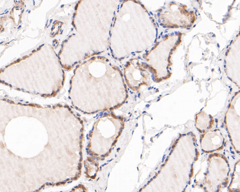
Fig4: Immunohistochemical analysis of paraffin-embedded human thyroid tissue using anti-AKT3 antibody. The section was pre-treated using heat mediated antigen retrieval with sodium citrate buffer (pH 6.0) for 20 minutes. The tissues were blocked in 5% BSA for 30 minutes at room temperature, washed with ddH2O and PBS, and then probed with the primary antibody (ER1901-33, 1/200) for 30 minutes at room temperature. The detection was performed using an HRP conjugated compact polymer system. DAB was used as the chromogen. Tissues were counterstained with hematoxylin and mounted with DPX.Application
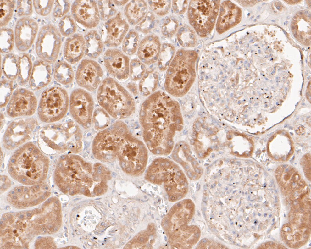
Fig5: Immunohistochemical analysis of paraffin-embedded human kidney tissue using anti-AKT3 antibody. The section was pre-treated using heat mediated antigen retrieval with sodium citrate buffer (pH 6.0) for 20 minutes. The tissues were blocked in 5% BSA for 30 minutes at room temperature, washed with ddH2O and PBS, and then probed with the primary antibody (ER1901-33, 1/200) for 30 minutes at room temperature. The detection was performed using an HRP conjugated compact polymer system. DAB was used as the chromogen. Tissues were counterstained with hematoxylin and mounted with DPX.Application
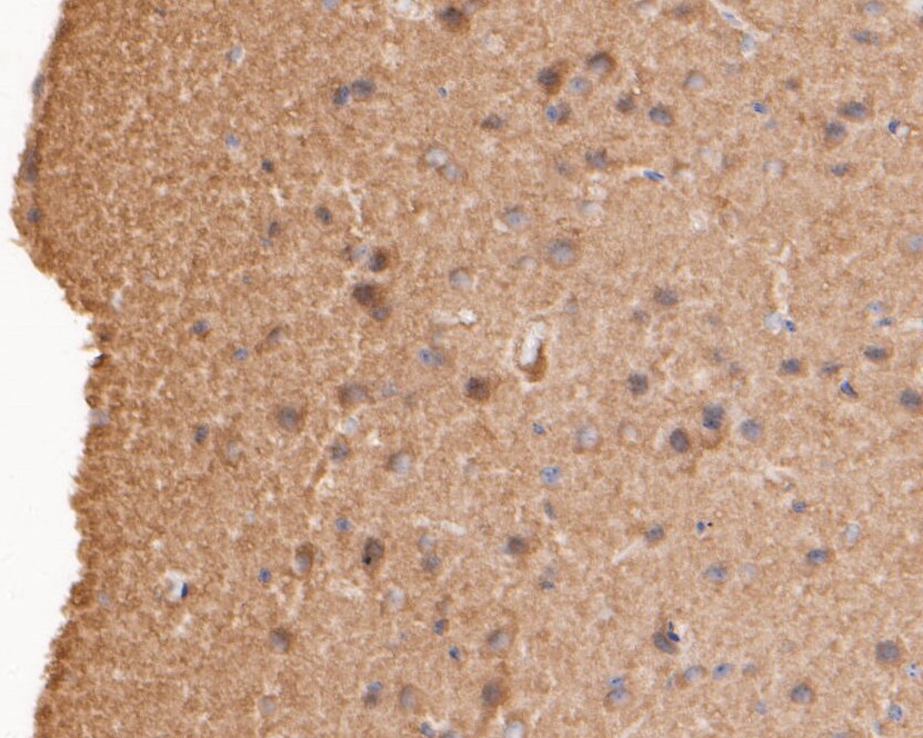
Fig6: Immunohistochemical analysis of paraffin-embedded mouse brain tissue using anti-AKT3 antibody. The section was pre-treated using heat mediated antigen retrieval with sodium citrate buffer (pH 6.0) for 20 minutes. The tissues were blocked in 5% BSA for 30 minutes at room temperature, washed with ddH2O and PBS, and then probed with the primary antibody (ER1901-33, 1/200) for 30 minutes at room temperature. The detection was performed using an HRP conjugated compact polymer system. DAB was used as the chromogen. Tissues were counterstained with hematoxylin and mounted with DPX.Application
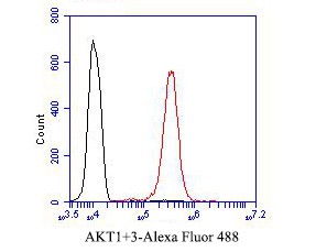
Fig7: Flow cytometric analysis of AKT3 was done on EA.hy926 cells. The cells were fixed, permeabilized and stained with the primary antibody (ER1901-33, 1/50) (red). After incubation of the primary antibody at room temperature for an hour, the cells were stained with a Alexa Fluor 488-conjugated Goat anti-Rabbit IgG Secondary antibody at 1/1000 dilution for 30 minutes.Unlabelled sample was used as a control (cells without incubation with primary antibody; black).| Product Name | AKT3 |
|---|---|
| Antibody Type | Primary Antibodies |
| Antigen Alias | Akt3 antibody AKT3 kinase antibody AKT3_HUMAN antibody DKFZp434N0250 antibody MPPH antibody PKB gamma antibody PKBG antibody PRKBG antibody Protein kinase Akt-3 antibody Protein Kinase AKT3 antibody Protein kinase B gamma antibody RAC gamma antibody RAC gamma serine/threonine protein kinase antibody RAC-gamma serine/threonine-protein kinase antibody RAC-PK-gamma antibody RACPK Gamma antibody Serine threonine protein kinase Akt 3 antibody Serine threonine protein kinase Akt3 antibody STK 2 antibody STK-2 antibody STK2 antibody V akt murine thymoma viral oncogene homolog 3 (protein kinase B, gamma) antibody V akt murine thymoma viral oncogene homolog 3 antibody V akt murine thymoma viral oncogene homolog 3 protein kinase B gamma antibody |
| Product description | AKT3 is one of 3 closely related serine/threonine-protein kinases (AKT1, AKT2 and AKT3) called the AKT kinase, and which regulate many processes including metabolism, proliferation, cell survival, growth and angiogenesis. This is mediated through serine and/or threonine phosphorylation of a range of downstream substrates. Over 100 substrate candidates have been reported so far, but for most of them, no isoform specificity has been reported. AKT3 is the least studied AKT isoform. It plays an important role in brain development and is crucial for the viability of malignant glioma cells. AKT3 isoform may also be the key molecule in up-regulation and down-regulation of MMP13 via IL13. Required for the coordination of mitochondrial biogenesis with growth factor-induced increases in cellular energy demands. Down-regulation by RNA interference reduces the expression of the phosphorylated form of BAD, resulting in the induction of caspase-dependent apoptosis. |
| Immunogen | Recombinant protein within human AKT3 aa 200-400. |
| Clonality | Polyclonal |
|---|---|
| Isotype | IgG |
| Host Species | Rabbit |
| Tested Applications | WBICCIHC-PFC |
| WB:1:500-1:2,000: ICC:1:50-1:100: IHC-P:1:50-1:200: FC:1:50-1:100: |
|
| Species Reactivity | HumanMouse |
| Concentration | 1mg/ml |
| Alternative Names | Akt3 antibody
AKT3 kinase antibody
AKT3_HUMAN antibody DKFZp434N0250 antibody MPPH antibody PKB gamma antibody PKBG antibody PRKBG antibody Protein kinase Akt-3 antibody Protein Kinase AKT3 antibody Protein kinase B gamma antibody RAC gamma antibody RAC gamma serine/threonine protein kinase antibody RAC-gamma serine/threonine-protein kinase antibody RAC-PK-gamma antibody RACPK Gamma antibody Serine threonine protein kinase Akt 3 antibody Serine threonine protein kinase Akt3 antibody STK 2 antibody STK-2 antibody STK2 antibody V akt murine thymoma viral oncogene homolog 3 (protein kinase B gamma) antibody V akt murine thymoma viral oncogene homolog 3 antibody V akt murine thymoma viral oncogene homolog 3 protein kinase B gamma antibody |
|---|---|
| Molecular Weight(MW) | 56 kDa |
| Cellular Localization | Nucleus, Cytoplasm, Membrane. |
Application
Fig1: Western blot analysis of AKT3 on SH-SY5Y cell lysates. Proteins were transferred to a PVDF membrane and blocked with 5% BSA in PBS for 1 hour at room temperature. The primary antibody (ER1901-33, 1/500) was used in 5% BSA at room temperature for 2 hours. Goat Anti-Rabbit IgG - HRP Secondary Antibody (HA1001) at 1:5,000 dilution was used for 1 hour at room temperature.
Application
Fig2: ICC staining of AKT3 in A549 cells (green). Formalin fixed cells were permeabilized with 0.1% Triton X-100 in TBS for 10 minutes at room temperature and blocked with 1% Blocker BSA for 15 minutes at room temperature. Cells were probed with the primary antibody (ER1901-33, 1/50) for 1 hour at room temperature, washed with PBS. Alexa Fluor®488 Goat anti-Rabbit IgG was used as the secondary antibody at 1/1,000 dilution. The nuclear counter stain is DAPI (blue).
Application
Fig3: ICC staining of AKT3 in EA.hy926 cells (green). Formalin fixed cells were permeabilized with 0.1% Triton X-100 in TBS for 10 minutes at room temperature and blocked with 1% Blocker BSA for 15 minutes at room temperature. Cells were probed with the primary antibody (ER1901-33, 1/50) for 1 hour at room temperature, washed with PBS. Alexa Fluor®488 Goat anti-Rabbit IgG was used as the secondary antibody at 1/1,000 dilution. The nuclear counter stain is DAPI (blue).
Application
Fig4: Immunohistochemical analysis of paraffin-embedded human thyroid tissue using anti-AKT3 antibody. The section was pre-treated using heat mediated antigen retrieval with sodium citrate buffer (pH 6.0) for 20 minutes. The tissues were blocked in 5% BSA for 30 minutes at room temperature, washed with ddH2O and PBS, and then probed with the primary antibody (ER1901-33, 1/200) for 30 minutes at room temperature. The detection was performed using an HRP conjugated compact polymer system. DAB was used as the chromogen. Tissues were counterstained with hematoxylin and mounted with DPX.
Application
Fig5: Immunohistochemical analysis of paraffin-embedded human kidney tissue using anti-AKT3 antibody. The section was pre-treated using heat mediated antigen retrieval with sodium citrate buffer (pH 6.0) for 20 minutes. The tissues were blocked in 5% BSA for 30 minutes at room temperature, washed with ddH2O and PBS, and then probed with the primary antibody (ER1901-33, 1/200) for 30 minutes at room temperature. The detection was performed using an HRP conjugated compact polymer system. DAB was used as the chromogen. Tissues were counterstained with hematoxylin and mounted with DPX.
Application
Fig6: Immunohistochemical analysis of paraffin-embedded mouse brain tissue using anti-AKT3 antibody. The section was pre-treated using heat mediated antigen retrieval with sodium citrate buffer (pH 6.0) for 20 minutes. The tissues were blocked in 5% BSA for 30 minutes at room temperature, washed with ddH2O and PBS, and then probed with the primary antibody (ER1901-33, 1/200) for 30 minutes at room temperature. The detection was performed using an HRP conjugated compact polymer system. DAB was used as the chromogen. Tissues were counterstained with hematoxylin and mounted with DPX.
Application
Fig7: Flow cytometric analysis of AKT3 was done on EA.hy926 cells. The cells were fixed, permeabilized and stained with the primary antibody (ER1901-33, 1/50) (red). After incubation of the primary antibody at room temperature for an hour, the cells were stained with a Alexa Fluor 488-conjugated Goat anti-Rabbit IgG Secondary antibody at 1/1000 dilution for 30 minutes.Unlabelled sample was used as a control (cells without incubation with primary antibody; black).| Positive Control | SH-SY5Y cell lysates, A549, EA.hy926, human thyroid tissue, human kidney tissue, mouse brain tissue. |
|---|---|
| Application Notes | WB:1:500-1:2,000: ICC:1:50-1:100: IHC-P:1:50-1:200: FC:1:50-1:100: |
| Form | Liquid |
|---|---|
| Storage Instructions | Store at +4℃ after thawing. Aliquot store at -20℃. Avoid repeated freeze / thaw cycles. |
| Storage Buffer | 1*TBS (pH7.4), 1%BSA, 50%Glycerol. Preservative: 0.05% Sodium Azide. |


