Application
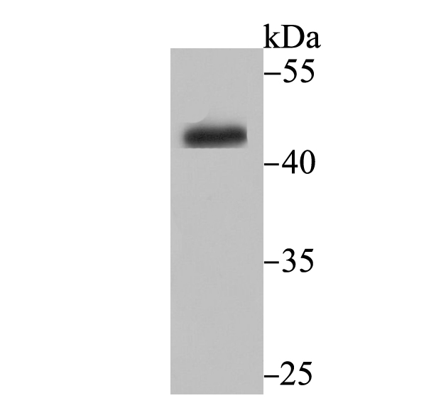
Fig1: Western blot analysis of CYP26A1 on HepG2 cell lysate using anti-CYP26A1 antibody at 1/500 dilution.Application
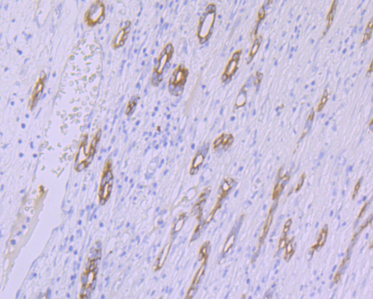
Fig2: Immunohistochemical analysis of paraffin-embedded human liver tissue using anti-CYP26A1 antibody. Counter stained with hematoxylin.Application
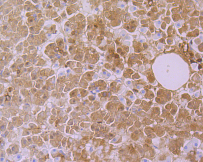
Fig3: Immunohistochemical analysis of paraffin-embedded human pancreas tissue using anti-CYP26A1 antibody. Counter stained with hematoxylin.| Product Name | CYP26A1 [JG33-98] |
|---|---|
| Antibody Type | Primary Antibodies |
| Antigen Alias | CP26 antibody CP26A_HUMAN antibody CYP26 antibody CYP26A 1 antibody cyp26a1 antibody Cytochrome P450 26A1 antibody Cytochrome P450 family 26 subfamily A polypeptide 1 antibody Cytochrome P450 retinoic acid-inactivating 1 antibody cytochrome P450, family 26, subfamily A, polypeptide 1 antibody cytochrome P450, subfamily XXVIA, polypeptide 1 antibody Cytochrome P450RAI antibody hP450RAI antibody OTTHUMP00000020102 antibody OTTHUMP00000020103 antibody P450 antibody P450 retinoic acid inactivating 1 antibody P450RAI antibody P450RAI1 antibody Retinoic acid 4 hydroxylase antibody Retinoic acid 4-hydroxylase antibody Retinoic acid metabolizing cytochrome antibody Retinoic acid-metabolizing cytochrome antibody |
| Product description | The cytochrome P450 proteins (CYPs) are monooxygenases that catalyze many reactions involved in drug metabolism and synthesis of cholesterol, steroids, and other lipids. P450 enzymes are classified into subfamilies based on their sequence similarities. CYP26A1, also referred to as retinoic acid-4-hydroxylase, is a major retinoic acid catabolic enzyme. CYP26A1 plays an important role in protecting tailbud tissues from inappropriate exposure to retinoic acid. CYP26A1 transcription is epigenetically regulated by nuclear retinoic acid receptor β2. Mutations in the gene encoding for CYP26A1 are associated with caudal agenesis and spina bifida, imperforate anus, agenesis of the caudal portions of the digestive and urogenital tracts, and malformed lumbosacral skeletal elements. CYP26A1 is upregulated in adenomatous polyposis coli mouse adenomas, human FAP adenomas, human sporadic colon carcinomas, and in the intestine of adenomatous polyposis coli (apcmcr) mutant zebrafish embryos. |
| Immunogen | Recombinant protein corresponding to human CYP26A1 aa 200-400. |
| Clonality | Monoclonal |
|---|---|
| Isotype | IgG |
| Host Species | Recombinant rabbit |
| Tested Applications | WBIHC |
| WB:1:500-1:1,000 IHC:1:50-1:200 |
|
| Species Reactivity | HumanRat |
| Concentration | 1mg/ml |
| Alternative Names | CP26 antibody
CP26A_HUMAN antibody
CYP26 antibody
CYP26A 1 antibody
cyp26a1 antibody
Cytochrome P450 26A1 antibody
Cytochrome P450 family 26 subfamily A polypeptide 1 antibody
Cytochrome P450 retinoic acid-inactivating 1 antibody
cytochrome P450 family 26 subfamily A polypeptide 1 antibody cytochrome P450 subfamily XXVIA polypeptide 1 antibody Cytochrome P450RAI antibody hP450RAI antibody OTTHUMP00000020102 antibody OTTHUMP00000020103 antibody P450 antibody P450 retinoic acid inactivating 1 antibody P450RAI antibody P450RAI1 antibody Retinoic acid 4 hydroxylase antibody Retinoic acid 4-hydroxylase antibody Retinoic acid metabolizing cytochrome antibody Retinoic acid-metabolizing cytochrome antibody |
|---|---|
| Molecular Weight(MW) | 48 kDa |
| Cellular Localization | Endoplasmic reticulum. |
| SwissProt ID | O43174 |
|---|
Application
Fig1: Western blot analysis of CYP26A1 on HepG2 cell lysate using anti-CYP26A1 antibody at 1/500 dilution.
Application
Fig2: Immunohistochemical analysis of paraffin-embedded human liver tissue using anti-CYP26A1 antibody. Counter stained with hematoxylin.
Application
Fig3: Immunohistochemical analysis of paraffin-embedded human pancreas tissue using anti-CYP26A1 antibody. Counter stained with hematoxylin.| Positive Control | HepG2, human liver tissue, human pancreas tissue. |
|---|---|
| Application Notes | WB:1:500-1:1,000 IHC:1:50-1:200 |
| Form | Liquid |
|---|---|
| Storage Instructions | Store at +4℃ after thawing. Aliquot store at -20℃. Avoid repeated freeze / thaw cycles. |
| Storage Buffer | 1*TBS (pH7.4), 1%BSA, 40%Glycerol. Preservative: 0.05% Sodium Azide. |


