Application
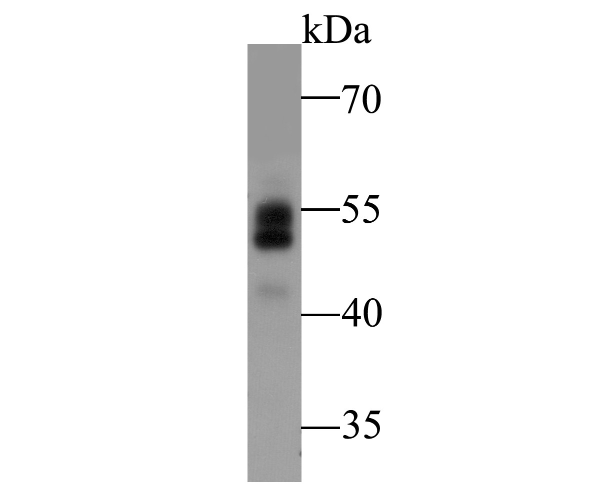
Fig1: Western blot analysis of BIN1 on rat spleen tissue lysate. Proteins were transferred to a PVDF membrane and blocked with 5% BSA in PBS for 1 hour at room temperature. The primary antibody (ER1901-39, 1/500) was used in 5% BSA at room temperature for 2 hours. Goat Anti-Rabbit IgG - HRP Secondary Antibody (HA1001) at 1:5,000 dilution was used for 1 hour at room temperature.Application
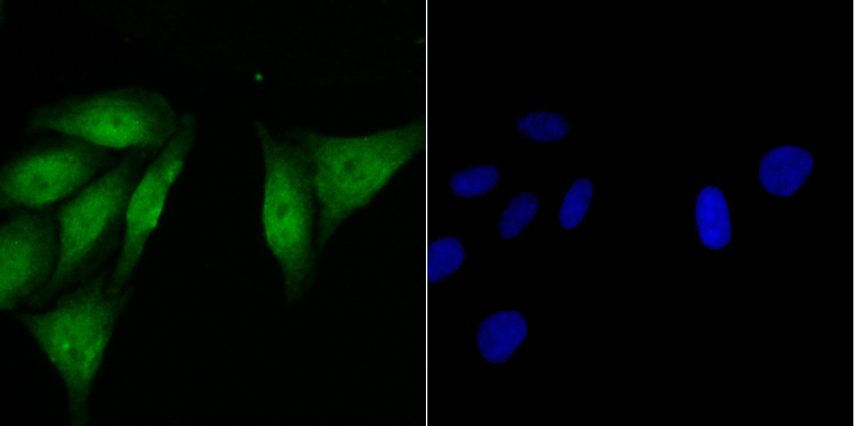
Fig2: ICC staining of BIN1 in SiHa cells (green). Formalin fixed cells were permeabilized with 0.1% Triton X-100 in TBS for 10 minutes at room temperature and blocked with 1% Blocker BSA for 15 minutes at room temperature. Cells were probed with the primary antibody (ER1901-39, 1/50) for 1 hour at room temperature, washed with PBS. Alexa Fluor®488 Goat anti-Rabbit IgG was used as the secondary antibody at 1/100 dilution. The nuclear counter stain is DAPI (blue).Application
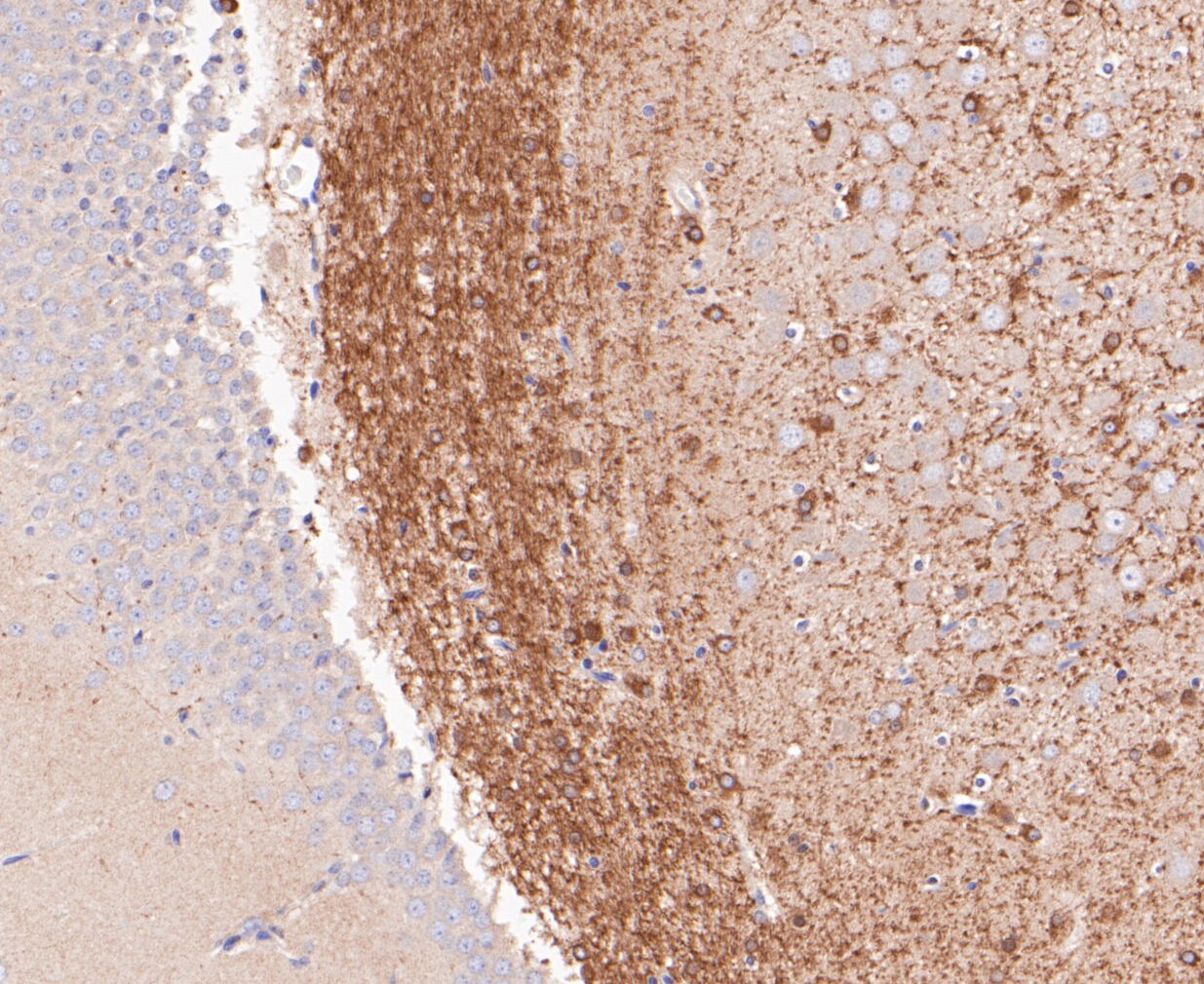
Fig3: Immunohistochemical analysis of paraffin-embedded rat brain tissue using anti-BIN1 antibody. The section was pre-treated using heat mediated antigen retrieval with Tris-EDTA buffer (pH 8.0-8.4) for 20 minutes.The tissues were blocked in 5% BSA for 30 minutes at room temperature, washed with ddH2O and PBS, and then probed with the primary antibody (ER1901-39, 1/200) for 30 minutes at room temperature. The detection was performed using an HRP conjugated compact polymer system. DAB was used as the chromogen. Tissues were counterstained with hematoxylin and mounted with DPX.Application
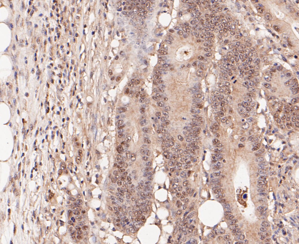
Fig4: Immunohistochemical analysis of paraffin-embedded human colon cancer tissue using anti-BIN1 antibody. The section was pre-treated using heat mediated antigen retrieval with Tris-EDTA buffer (pH 8.0-8.4) for 20 minutes.The tissues were blocked in 5% BSA for 30 minutes at room temperature, washed with ddH2O and PBS, and then probed with the primary antibody (ER1901-39, 1/50) for 30 minutes at room temperature. The detection was performed using an HRP conjugated compact polymer system. DAB was used as the chromogen. Tissues were counterstained with hematoxylin and mounted with DPX.Application
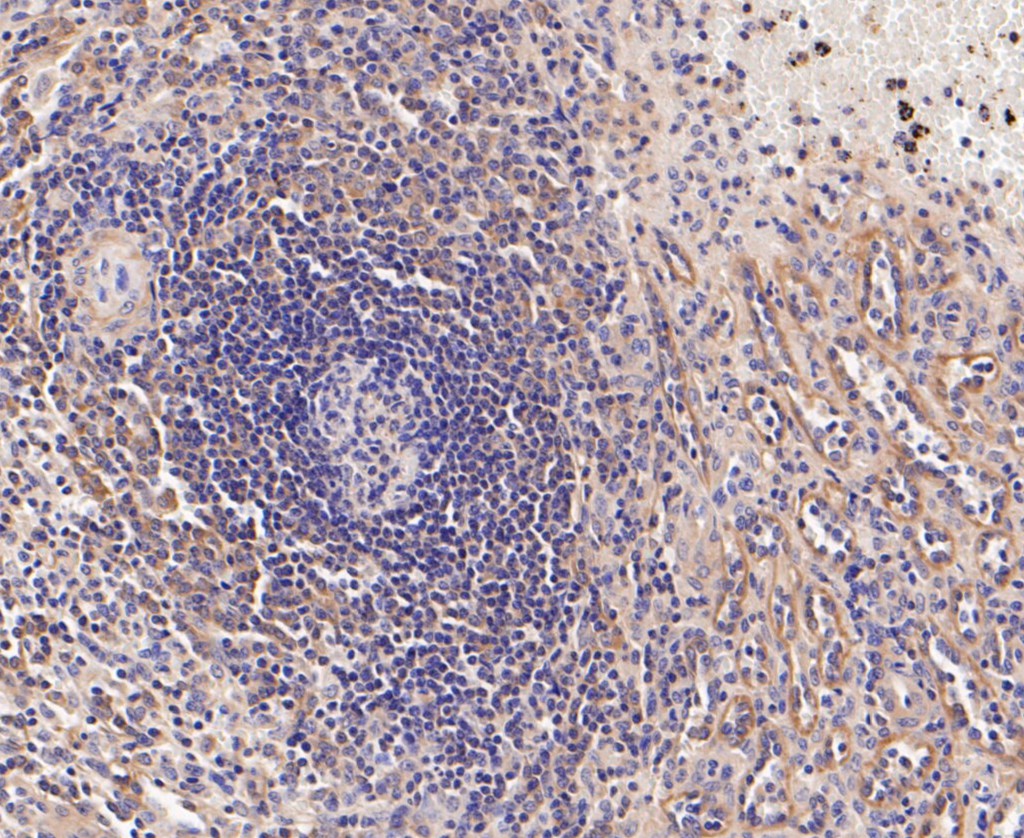
Fig5: Immunohistochemical analysis of paraffin-embedded human spleen tissue using anti-BIN1 antibody. The section was pre-treated using heat mediated antigen retrieval with Tris-EDTA buffer (pH 8.0-8.4) for 20 minutes.The tissues were blocked in 5% BSA for 30 minutes at room temperature, washed with ddH2O and PBS, and then probed with the primary antibody (ER1901-39, 1/200) for 30 minutes at room temperature. The detection was performed using an HRP conjugated compact polymer system. DAB was used as the chromogen. Tissues were counterstained with hematoxylin and mounted with DPX.Application
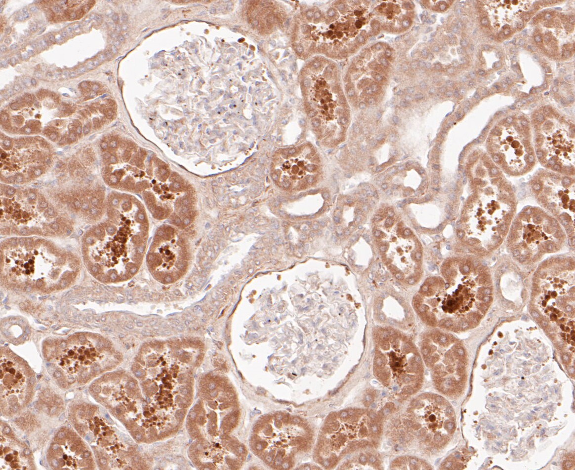
Fig6: Immunohistochemical analysis of paraffin-embedded human kidney tissue using anti-BIN1 antibody. The section was pre-treated using heat mediated antigen retrieval with Tris-EDTA buffer (pH 8.0-8.4) for 20 minutes.The tissues were blocked in 5% BSA for 30 minutes at room temperature, washed with ddH2O and PBS, and then probed with the primary antibody (ER1901-39, 1/200) for 30 minutes at room temperature. The detection was performed using an HRP conjugated compact polymer system. DAB was used as the chromogen. Tissues were counterstained with hematoxylin and mounted with DPX.Application
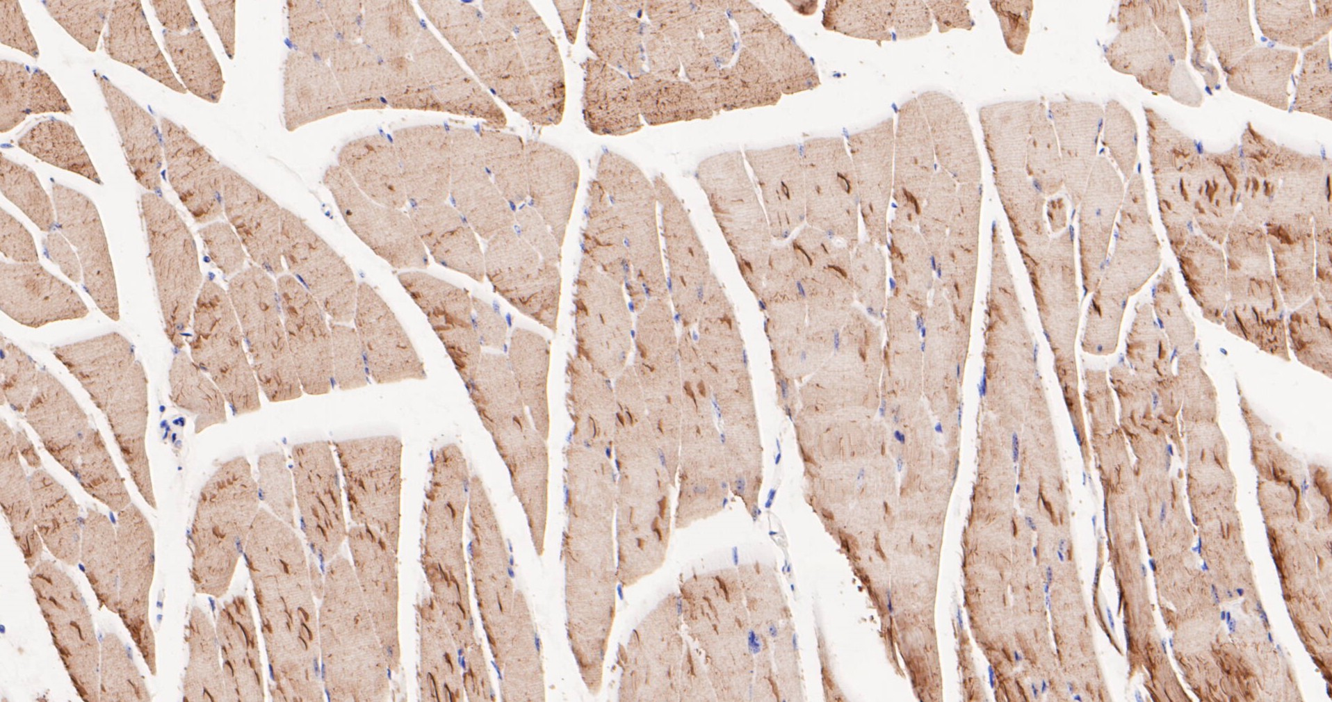
Fig7: Immunohistochemical analysis of paraffin-embedded mouse skeletal muscle tissue using anti-BIN1 antibody. The section was pre-treated using heat mediated antigen retrieval with Tris-EDTA buffer (pH 8.0-8.4) for 20 minutes.The tissues were blocked in 5% BSA for 30 minutes at room temperature, washed with ddH2O and PBS, and then probed with the primary antibody (ER1901-39, 1/200) for 30 minutes at room temperature. The detection was performed using an HRP conjugated compact polymer system. DAB was used as the chromogen. Tissues were counterstained with hematoxylin and mounted with DPX.| Product Name | BIN1 |
|---|---|
| Antibody Type | Primary Antibodies |
| Antigen Alias | AMPH 2 antibody AMPH2 antibody Amphiphysin 2 antibody Amphiphysin II antibody Amphiphysin like protein antibody amphiphysin-like antibody Amphiphysin-like protein antibody AMPHL antibody Bin1 antibody BIN1_HUMAN antibody Box Dependant MYC Interacting Protein 1 antibody Box-dependent myc-interacting protein 1 antibody Bridging integrator 1 antibody DKFZp547F068 antibody MGC10367 antibody MGC105358 antibody Myc box dependent interacting protein 1 antibody Myc box-dependent-interacting protein 1 antibody SH3P9 antibody |
| Product description | Is a key player in the control of plasma membrane curvature, membrane shaping and membrane remodeling. Required in muscle cells for the formation of T-tubules, tubular invaginations of the plasma membrane that function in depolarization-contraction coupling. Is a negative regulator of endocytosis (By similarity). Is also involved in the regulation of intracellular vesicles sorting, modulation of BACE1 trafficking and the control of amyloid-beta production. In neuronal circuits, endocytosis regulation may influence the internalization of PHF-tau aggregates (By similarity). May be involved in the regulation of MYC activity and the control cell proliferation. Has actin bundling activity and stabilizes actin filaments against depolymerization in vitro. |
| Immunogen | Recombinant protein within human BIN1 aa 56-257. |
| Clonality | Polyclonal |
|---|---|
| Isotype | IgG |
| Host Species | Rabbit |
| Tested Applications | WBICCIHC |
| WB:1:500-1:1000: ICC:1:50-1:200: IHC:1:50-1:200: |
|
| Species Reactivity | HumanMouseRat |
| Concentration | 1mg/ml |
| Alternative Names | AMPH 2 antibody
AMPH2 antibody
Amphiphysin 2 antibody Amphiphysin II antibody Amphiphysin like protein antibody amphiphysin-like antibody Amphiphysin-like protein antibody AMPHL antibody Bin1 antibody BIN1_HUMAN antibody Box Dependant MYC Interacting Protein 1 antibody Box-dependent myc-interacting protein 1 antibody Bridging integrator 1 antibody DKFZp547F068 antibody MGC10367 antibody MGC105358 antibody Myc box dependent interacting protein 1 antibody Myc box-dependent-interacting protein 1 antibody SH3P9 antibody |
|---|---|
| Molecular Weight(MW) | 55 kDa |
| Cellular Localization | Endosome. Nucleus. Plasma membrane. |
Application
Fig1: Western blot analysis of BIN1 on rat spleen tissue lysate. Proteins were transferred to a PVDF membrane and blocked with 5% BSA in PBS for 1 hour at room temperature. The primary antibody (ER1901-39, 1/500) was used in 5% BSA at room temperature for 2 hours. Goat Anti-Rabbit IgG - HRP Secondary Antibody (HA1001) at 1:5,000 dilution was used for 1 hour at room temperature.
Application
Fig2: ICC staining of BIN1 in SiHa cells (green). Formalin fixed cells were permeabilized with 0.1% Triton X-100 in TBS for 10 minutes at room temperature and blocked with 1% Blocker BSA for 15 minutes at room temperature. Cells were probed with the primary antibody (ER1901-39, 1/50) for 1 hour at room temperature, washed with PBS. Alexa Fluor®488 Goat anti-Rabbit IgG was used as the secondary antibody at 1/100 dilution. The nuclear counter stain is DAPI (blue).
Application
Fig3: Immunohistochemical analysis of paraffin-embedded rat brain tissue using anti-BIN1 antibody. The section was pre-treated using heat mediated antigen retrieval with Tris-EDTA buffer (pH 8.0-8.4) for 20 minutes.The tissues were blocked in 5% BSA for 30 minutes at room temperature, washed with ddH2O and PBS, and then probed with the primary antibody (ER1901-39, 1/200) for 30 minutes at room temperature. The detection was performed using an HRP conjugated compact polymer system. DAB was used as the chromogen. Tissues were counterstained with hematoxylin and mounted with DPX.
Application
Fig4: Immunohistochemical analysis of paraffin-embedded human colon cancer tissue using anti-BIN1 antibody. The section was pre-treated using heat mediated antigen retrieval with Tris-EDTA buffer (pH 8.0-8.4) for 20 minutes.The tissues were blocked in 5% BSA for 30 minutes at room temperature, washed with ddH2O and PBS, and then probed with the primary antibody (ER1901-39, 1/50) for 30 minutes at room temperature. The detection was performed using an HRP conjugated compact polymer system. DAB was used as the chromogen. Tissues were counterstained with hematoxylin and mounted with DPX.
Application
Fig5: Immunohistochemical analysis of paraffin-embedded human spleen tissue using anti-BIN1 antibody. The section was pre-treated using heat mediated antigen retrieval with Tris-EDTA buffer (pH 8.0-8.4) for 20 minutes.The tissues were blocked in 5% BSA for 30 minutes at room temperature, washed with ddH2O and PBS, and then probed with the primary antibody (ER1901-39, 1/200) for 30 minutes at room temperature. The detection was performed using an HRP conjugated compact polymer system. DAB was used as the chromogen. Tissues were counterstained with hematoxylin and mounted with DPX.
Application
Fig6: Immunohistochemical analysis of paraffin-embedded human kidney tissue using anti-BIN1 antibody. The section was pre-treated using heat mediated antigen retrieval with Tris-EDTA buffer (pH 8.0-8.4) for 20 minutes.The tissues were blocked in 5% BSA for 30 minutes at room temperature, washed with ddH2O and PBS, and then probed with the primary antibody (ER1901-39, 1/200) for 30 minutes at room temperature. The detection was performed using an HRP conjugated compact polymer system. DAB was used as the chromogen. Tissues were counterstained with hematoxylin and mounted with DPX.
Application
Fig7: Immunohistochemical analysis of paraffin-embedded mouse skeletal muscle tissue using anti-BIN1 antibody. The section was pre-treated using heat mediated antigen retrieval with Tris-EDTA buffer (pH 8.0-8.4) for 20 minutes.The tissues were blocked in 5% BSA for 30 minutes at room temperature, washed with ddH2O and PBS, and then probed with the primary antibody (ER1901-39, 1/200) for 30 minutes at room temperature. The detection was performed using an HRP conjugated compact polymer system. DAB was used as the chromogen. Tissues were counterstained with hematoxylin and mounted with DPX.| Positive Control | Rat spleen tissue, SiHa, rat brain tissue, human colon cancer tissue, human spleen tissue, human kidney tissue, mouse skeletal muscle tissue. |
|---|---|
| Application Notes | WB:1:500-1:1000: ICC:1:50-1:200: IHC:1:50-1:200: |
| Form | Liquid |
|---|---|
| Storage Instructions | Store at +4℃ after thawing. Aliquot store at -20℃. Avoid repeated freeze / thaw cycles. |
| Storage Buffer | 1*TBS (pH7.4), 1%BSA, 50%Glycerol. Preservative: 0.05% Sodium Azide. |


