Application
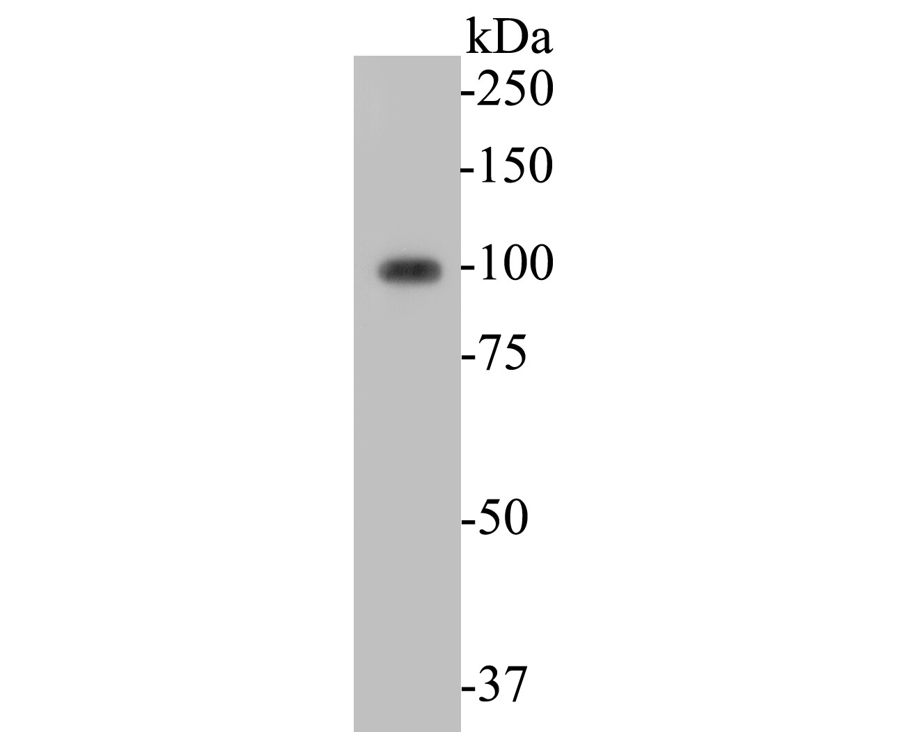
Fig1: Western blot analysis of KCNB1 on rat cerebellum tissue lysates. Proteins were transferred to a PVDF membrane and blocked with 5% BSA in PBS for 1 hour at room temperature. The primary antibody (ER1901-41, 1/500) was used in 5% BSA at room temperature for 2 hours. Goat Anti-Rabbit IgG - HRP Secondary Antibody (HA1001) at 1:5,000 dilution was used for 1 hour at room temperature.Application
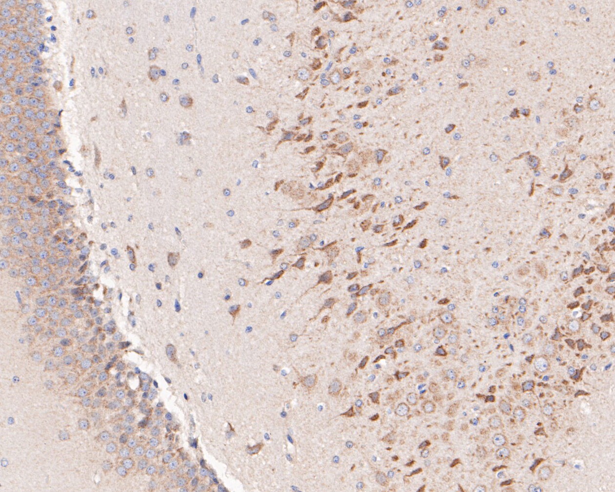
Fig2: Immunohistochemical analysis of paraffin-embedded rat brain tissue using anti-KCNB1 antibody. The section was pre-treated using heat mediated antigen retrieval with Tris-EDTA buffer (pH 8.0-8.4) for 20 minutes.The tissues were blocked in 5% BSA for 30 minutes at room temperature, washed with ddH2O and PBS, and then probed with the primary antibody (ER1901-41, 1/50) for 30 minutes at room temperature. The detection was performed using an HRP conjugated compact polymer system. DAB was used as the chromogen. Tissues were counterstained with hematoxylin and mounted with DPX.Application
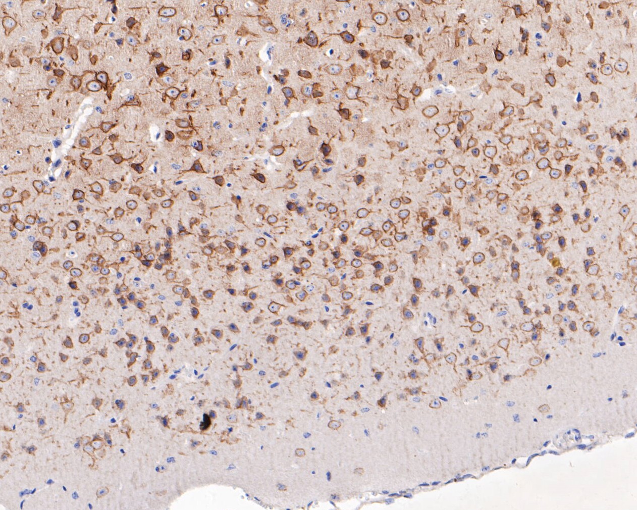
Fig3: Immunohistochemical analysis of paraffin-embedded mouse brain tissue using anti-KCNB1 antibody. The section was pre-treated using heat mediated antigen retrieval with Tris-EDTA buffer (pH 8.0-8.4) for 20 minutes.The tissues were blocked in 5% BSA for 30 minutes at room temperature, washed with ddH2O and PBS, and then probed with the primary antibody (ER1901-41, 1/200) for 30 minutes at room temperature. The detection was performed using an HRP conjugated compact polymer system. DAB was used as the chromogen. Tissues were counterstained with hematoxylin and mounted with DPX.Application
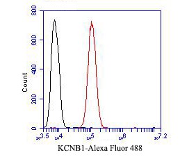
Fig4: Flow cytometric analysis of KCNB1 was done on SH-SY5Y cells. The cells were fixed, permeabilized and stained with the primary antibody (ER1901-41, 1/50) (red). After incubation of the primary antibody at room temperature for an hour, the cells were stained with a Alexa Fluor 488-conjugated Goat anti-Rabbit IgG Secondary antibody at 1/1000 dilution for 30 minutes.Unlabelled sample was used as a control (cells without incubation with primary antibody; black).| Product Name | KCNB1 |
|---|---|
| Antibody Type | Primary Antibodies |
| Antigen Alias | Delayed rectifier potassium channel 1 antibody Delayed rectifier potassium channel Kv2.1 antibody DRK 1 antibody DRK1 antibody h DRK1 K(+) channel antibody h-DRK1 antibody hDRK 1 antibody hDRK1 antibody KCB 1 antibody KCB1 antibody KCNB1 antibody KCNB1_HUMAN antibody KV2.1 antibody Potassium channel protein DRK1 antibody Potassium voltage gated channel shab related subfamily member 1 antibody Potassium voltage-gated channel subfamily B member 1 antibody Voltage-gated potassium channel subunit Kv2.1 antibody |
| Product description | Potassium voltage-gated channel, Shab-related subfamily, member 1, also known as KCNB1 or Kv2.1, is a protein that, in humans, is encoded by the KCNB1 gene. Potassium voltage-gated channel subfamily B member one, or simply known as KCNB1, is a delayed rectifier and voltage-gated potassium channel found throughout the body. The channel has a diverse number of functions. However, its main function, as a delayed rectifier, is to propagate current in its respective location. It is commonly expressed in the central nervous system, but may also be found in pulmonary arteries, auditory outer hair cells, stem cells, the retina, and organs such as the heart and pancreas. Modulation of K+ channel activity and expression has been found to be at the crux of many profound pathophysiological disorders in several cell types. Potassium channels are among the most diverse of all ion channels in eukaryotes. With over 100 genes coding numerous functions, many isoforms of potassium channels are present in the body, but most are divided up into two main groups: inactivating transient channels and non-inactivating delayed rectifiers. Due to the multiple varied forms, potassium delayed rectifier channels open or close in response to a myriad of signals. These include: cell depolarization or hyperpolarization, increases in intracellular calcium concentrations, neurotransmitter binding, or second messenger activity such as G-proteins or kinases. |
| Immunogen | Synthetic peptide within C-terminal rat KCNB1. |
| Clonality | Polyclonal |
|---|---|
| Isotype | IgG |
| Host Species | Rabbit |
| Tested Applications | WBIHC-PFC |
| WB:1:500-1:2,000: IHC-P:1:50-1:200: FC:1:50-1:100: |
|
| Species Reactivity | HumanMouseRat |
| Concentration | 1mg/ml |
| Alternative Names | Delayed rectifier potassium channel 1 antibody
Delayed rectifier potassium channel Kv2.1 antibody
DRK 1 antibody DRK1 antibody h DRK1 K(+) channel antibody h-DRK1 antibody hDRK 1 antibody hDRK1 antibody KCB 1 antibody KCB1 antibody KCNB1 antibody KCNB1_HUMAN antibody KV2.1 antibody Potassium channel protein DRK1 antibody Potassium voltage gated channel shab related subfamily member 1 antibody Potassium voltage-gated channel subfamily B member 1 antibody Voltage-gated potassium channel subunit Kv2.1 antibody |
|---|---|
| Molecular Weight(MW) | 96 kDa |
| Cellular Localization | Cell membrane, postsynaptic cell membrane, lateral cell membrane, sarcolemma. |
Application
Fig1: Western blot analysis of KCNB1 on rat cerebellum tissue lysates. Proteins were transferred to a PVDF membrane and blocked with 5% BSA in PBS for 1 hour at room temperature. The primary antibody (ER1901-41, 1/500) was used in 5% BSA at room temperature for 2 hours. Goat Anti-Rabbit IgG - HRP Secondary Antibody (HA1001) at 1:5,000 dilution was used for 1 hour at room temperature.
Application
Fig2: Immunohistochemical analysis of paraffin-embedded rat brain tissue using anti-KCNB1 antibody. The section was pre-treated using heat mediated antigen retrieval with Tris-EDTA buffer (pH 8.0-8.4) for 20 minutes.The tissues were blocked in 5% BSA for 30 minutes at room temperature, washed with ddH2O and PBS, and then probed with the primary antibody (ER1901-41, 1/50) for 30 minutes at room temperature. The detection was performed using an HRP conjugated compact polymer system. DAB was used as the chromogen. Tissues were counterstained with hematoxylin and mounted with DPX.
Application
Fig3: Immunohistochemical analysis of paraffin-embedded mouse brain tissue using anti-KCNB1 antibody. The section was pre-treated using heat mediated antigen retrieval with Tris-EDTA buffer (pH 8.0-8.4) for 20 minutes.The tissues were blocked in 5% BSA for 30 minutes at room temperature, washed with ddH2O and PBS, and then probed with the primary antibody (ER1901-41, 1/200) for 30 minutes at room temperature. The detection was performed using an HRP conjugated compact polymer system. DAB was used as the chromogen. Tissues were counterstained with hematoxylin and mounted with DPX.
Application
Fig4: Flow cytometric analysis of KCNB1 was done on SH-SY5Y cells. The cells were fixed, permeabilized and stained with the primary antibody (ER1901-41, 1/50) (red). After incubation of the primary antibody at room temperature for an hour, the cells were stained with a Alexa Fluor 488-conjugated Goat anti-Rabbit IgG Secondary antibody at 1/1000 dilution for 30 minutes.Unlabelled sample was used as a control (cells without incubation with primary antibody; black).| Positive Control | Rat cerebellum tissue lysates, rat brain tissue, mouse brain tissue, SH-SY5Y. |
|---|---|
| Application Notes | WB:1:500-1:2,000: IHC-P:1:50-1:200: FC:1:50-1:100: |
| Form | Liquid |
|---|---|
| Storage Instructions | Store at +4℃ after thawing. Aliquot store at -20℃. Avoid repeated freeze / thaw cycles. |
| Storage Buffer | 1*TBS (pH7.4), 1%BSA, 50%Glycerol. Preservative: 0.05% Sodium Azide. |


