Application
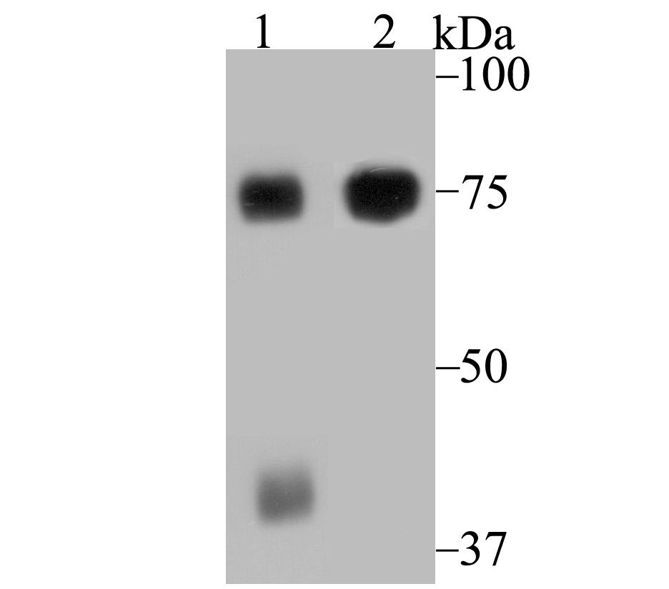
Fig1: Western blot analysis of ELMO1 on mouse spleen tissue and Jurkat cell lysates using anti-ELMO1 antibody at 1/1,000 dilution.Application
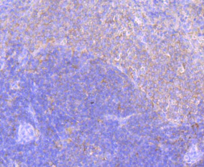
Fig2: Immunohistochemical analysis of paraffin-embedded rat spleen tissue using anti-ELMO1 antibody. Counter stained with hematoxylin. The section was pre-treated using heat mediated antigen retrieval with sodium citrate buffer (pH6) for 20 mins.Application
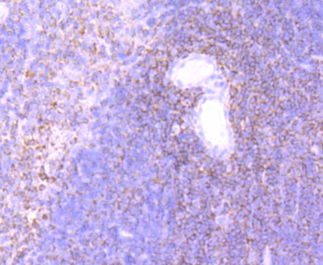
Fig3: Immunohistochemical analysis of paraffin-embedded mouse spleen tissue using anti-ELMO1 antibody. Counter stained with hematoxylin. The section was pre-treated using heat mediated antigen retrieval with sodium citrate buffer (pH6) for 20 mins.Application
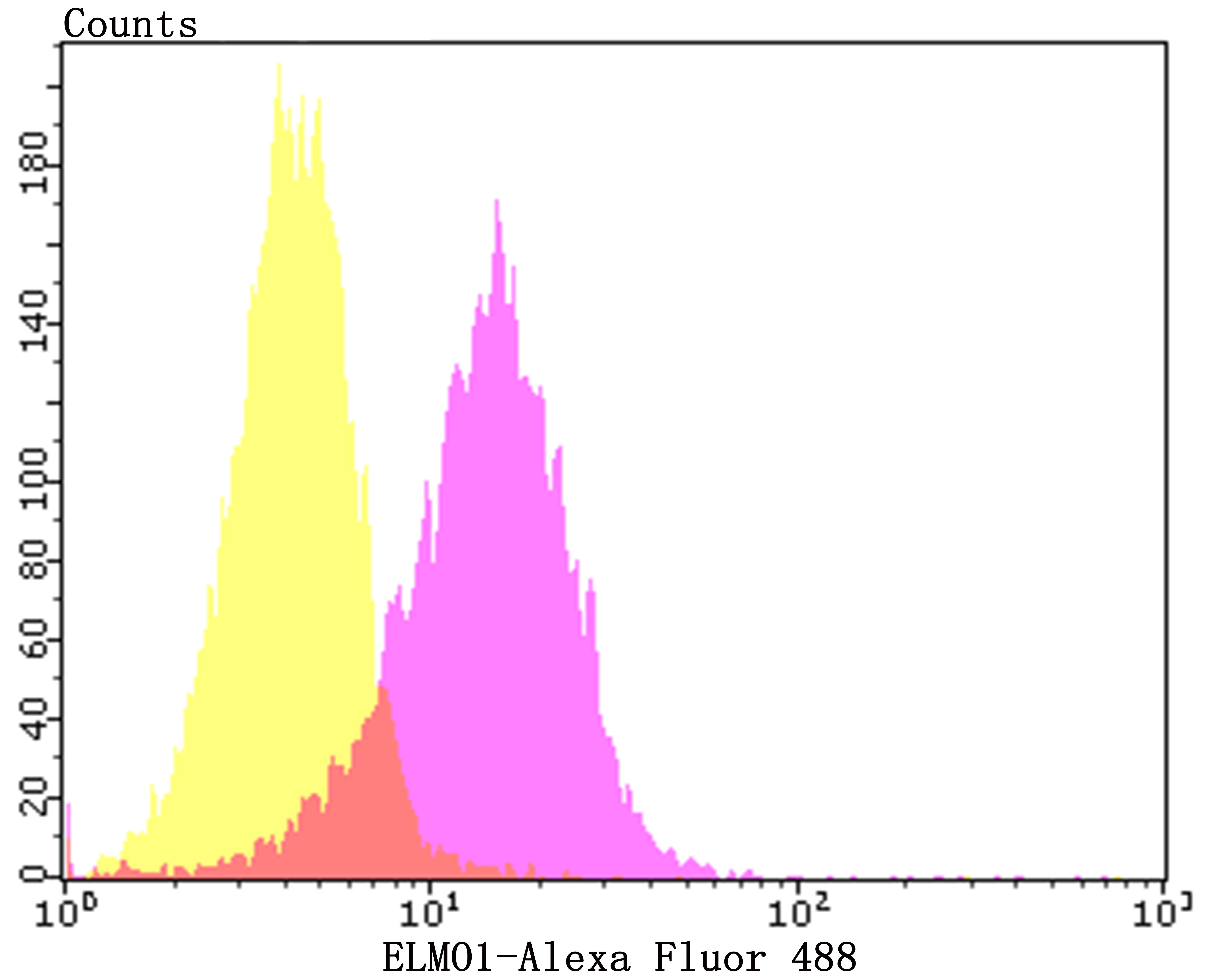
Fig4: Flow cytometric analysis of SH-SY-5Y cells with ELMO1 antibody at 1/50 dilution (purple) compared with an unlabelled control (cells without incubation with primary antibody; yellow). Alexa Fluor 488-conjugated goat anti-rabbit IgG was used as the secondary antibody.| Product Name | ELMO1 [JG34-59] |
|---|---|
| Antibody Type | Primary Antibodies |
| Antigen Alias | CED 12 antibody Ced 12 homolog 1 antibody Ced 12 homolog antibody CED-12 antibody CED12 antibody Ced12 homolog 1 antibody Ced12 homolog antibody ELMO 1 antibody ELMO-1 antibody Elmo1 antibody ELMO1_HUMAN antibody Engulfment and cell motility 1 antibody Engulfment and cell motility protein 1 antibody KIAA0281 antibody MGC126406 antibody Protein ced-12 homolog antibody |
| Product description | Elmo (engulfment and cell motility) proteins share similarity to C. elegans CED-12. The C. elegans genes ced-2, ced-5, ced-10 and ced-12, and their mammalian homologs, CRKII, DOCK1, RAC1 and ELMO, mediate cytoskeletal rear-rangements during phagocytosis of apoptotic cells as well as cell motility. Elmo1 associates with DOCK 180 and may influence phagocytosis and effect cell shape changes. Src family kinase-mediated tyrosine phosphorylation of Elmo1 influences signaling through Elmo1/Crk/DOCK 180 pathways. Elmo2 interacts directly with Rho G in a GTP-dependent manner and forms a ternary complex with DOCK 180 to induce activation of Rac 1. The Rho G-Elmo2-DOCK 180 pathway is required for activation of Rac 1 and cell spreading mediated by integrin, as well as for neurite outgrowth induced by nerve growth factor. Elmo3 acts in assocation with DOCK 180 and Crk II and may be required in complex with DOCK 180 to activate Rac/Rho small GTPases. |
| Immunogen | Recombinant protein within human ELMO1 aa 500 to 700. |
| Clonality | Monoclonal |
|---|---|
| Isotype | IgG |
| Host Species | Recombinant rabbit |
| Tested Applications | WBIHCFCIP |
| WB:1:500-1:2,000 IHC:1:50-1:200 FC:1:50-1:100 IP:1:50-1:100 |
|
| Species Reactivity | HumanMouseRat |
| Concentration | 1mg/ml |
| Alternative Names | CED 12 antibody Ced 12 homolog 1 antibody Ced 12 homolog antibody CED-12 antibody CED12 antibody Ced12 homolog 1 antibody Ced12 homolog antibody ELMO 1 antibody ELMO-1 antibody Elmo1 antibody ELMO1_HUMAN antibody Engulfment and cell motility 1 antibody Engulfment and cell motility protein 1 antibody KIAA0281 antibody MGC126406 antibody Protein ced-12 homolog antibody |
|---|---|
| Molecular Weight(MW) | 84 kDa |
| Cellular Localization | Cell membrane. Cytoplasm. |
Application
Fig1: Western blot analysis of ELMO1 on mouse spleen tissue and Jurkat cell lysates using anti-ELMO1 antibody at 1/1,000 dilution.
Application
Fig2: Immunohistochemical analysis of paraffin-embedded rat spleen tissue using anti-ELMO1 antibody. Counter stained with hematoxylin. The section was pre-treated using heat mediated antigen retrieval with sodium citrate buffer (pH6) for 20 mins.
Application
Fig3: Immunohistochemical analysis of paraffin-embedded mouse spleen tissue using anti-ELMO1 antibody. Counter stained with hematoxylin. The section was pre-treated using heat mediated antigen retrieval with sodium citrate buffer (pH6) for 20 mins.
Application
Fig4: Flow cytometric analysis of SH-SY-5Y cells with ELMO1 antibody at 1/50 dilution (purple) compared with an unlabelled control (cells without incubation with primary antibody; yellow). Alexa Fluor 488-conjugated goat anti-rabbit IgG was used as the secondary antibody.| Positive Control | Rat spleen tissue, mouse spleen tissue, Jurkat, SH-SY-5Y. |
|---|---|
| Application Notes | WB:1:500-1:2,000 IHC:1:50-1:200 FC:1:50-1:100 IP:1:50-1:100 |
| Form | Liquid |
|---|---|
| Storage Instructions | Store at +4℃ after thawing. Aliquot store at -20℃. Avoid repeated freeze / thaw cycles. |
| Storage Buffer | 1*TBS (pH7.4), 1%BSA, 40%Glycerol. Preservative: 0.05% Sodium Azide. |


