Application
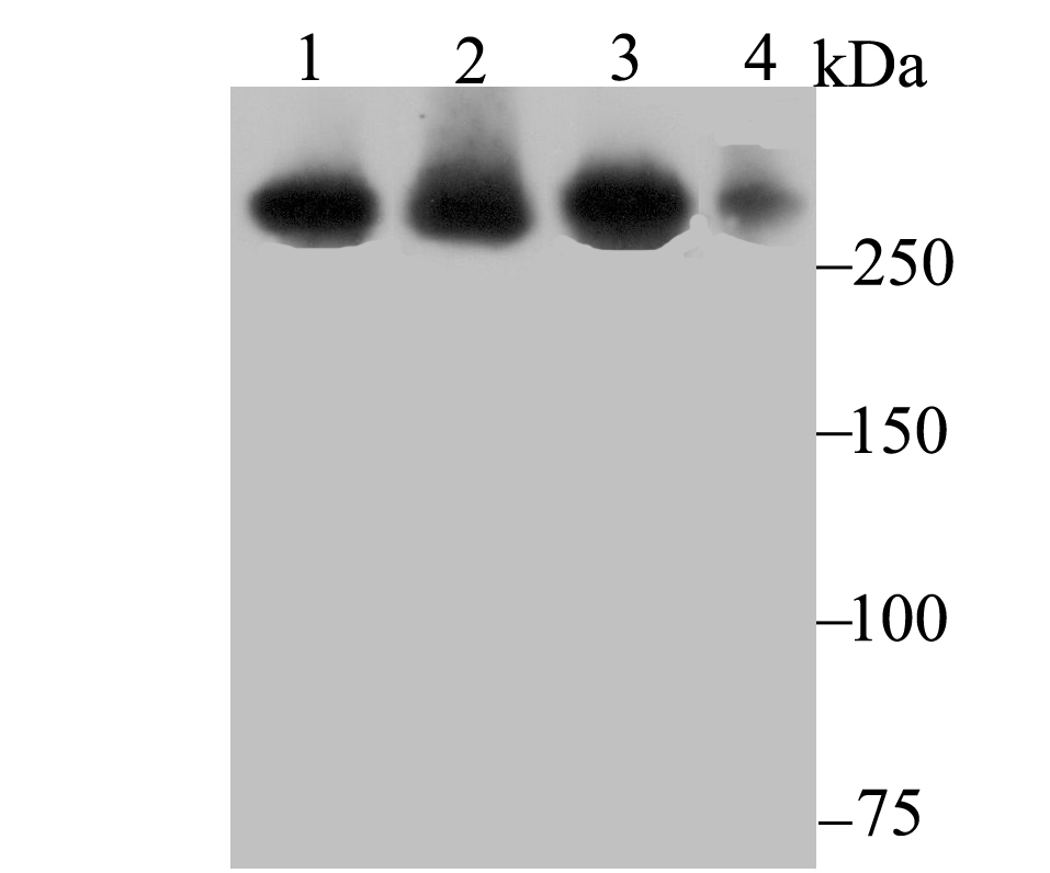
Fig1: Western blot analysis of USP9X on different lysates using anti-USP9X antibody at 1/1,000 dilution. Positive control: Lane 1: SiHa Lane 2: A549 Lane 3: 293 Lane 4: Mouse colonApplication
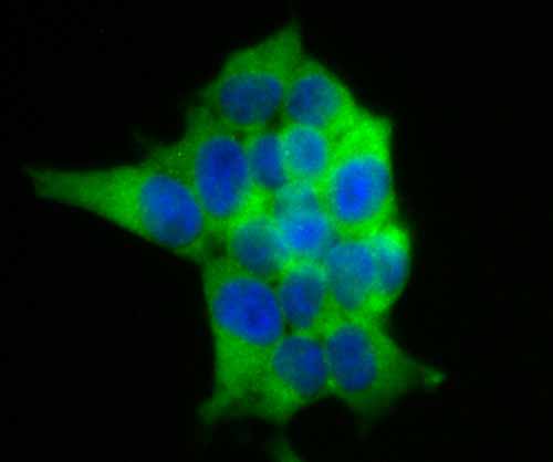
Fig2: ICC staining USP9X in 293T cells (green). The nuclear counter stain is DAPI (blue). Cells were fixed in paraformaldehyde, permeabilised with 0.25% Triton X100/PBS.Application
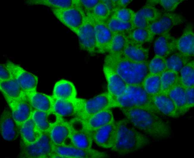
Fig3: ICC staining USP9X in F9 cells (green). The nuclear counter stain is DAPI (blue). Cells were fixed in paraformaldehyde, permeabilised with 0.25% Triton X100/PBS.Application
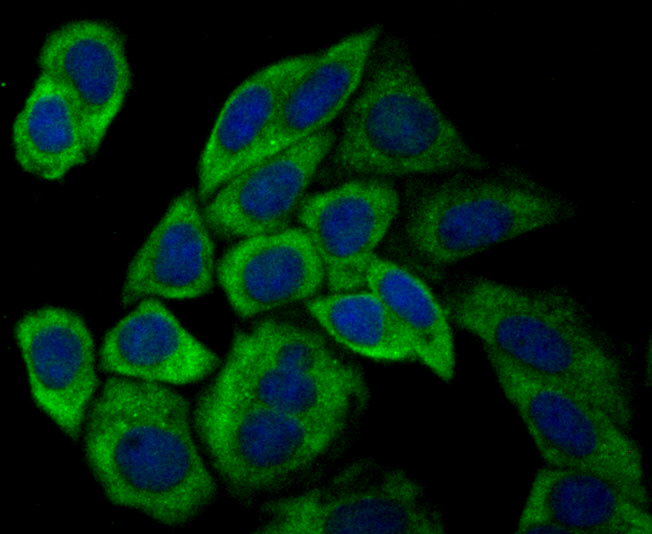
Fig4: ICC staining USP9X in SiHa cells (green). The nuclear counter stain is DAPI (blue). Cells were fixed in paraformaldehyde, permeabilised with 0.25% Triton X100/PBS.Application
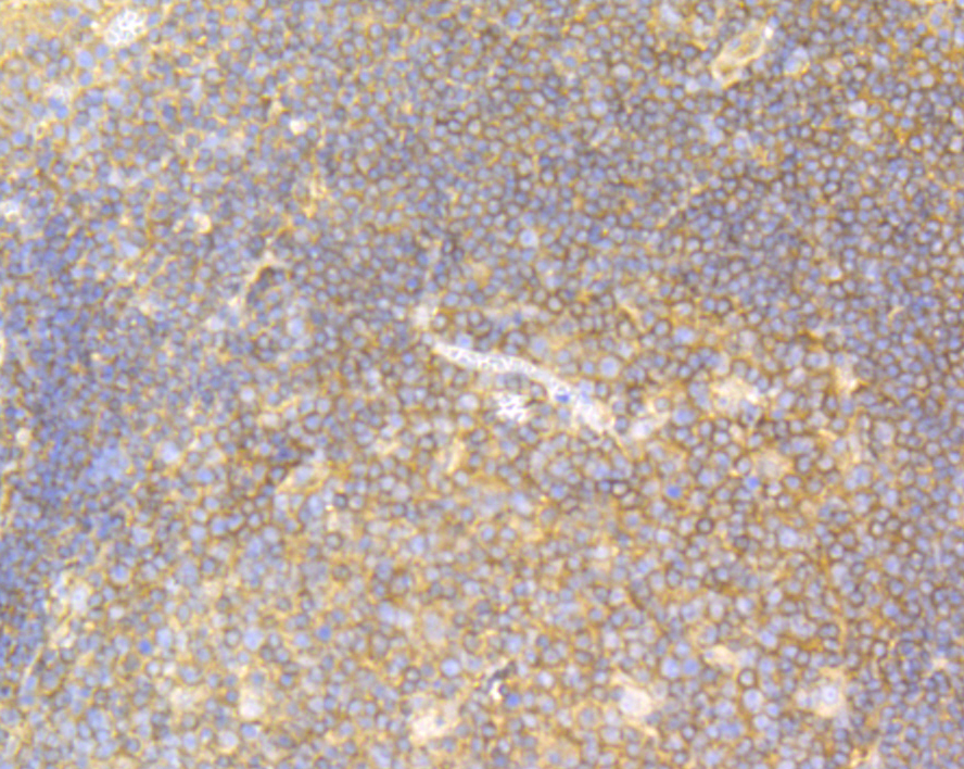
Fig5: Immunohistochemical analysis of paraffin-embedded human tonsil tissue using anti-USP9X antibody. Counter stained with hematoxylin. The section was pre-treated using heat mediated antigen retrieval with sodium citrate buffer (pH6) for 20 mins.Application
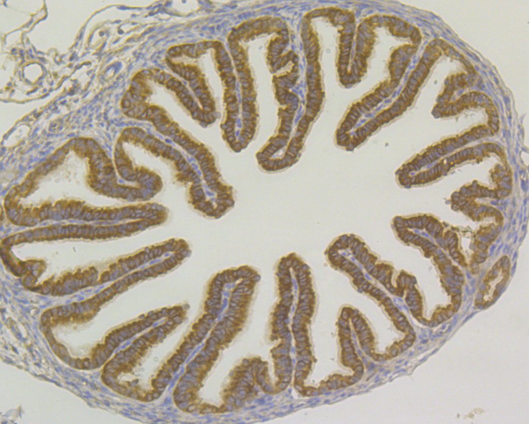
Fig6: Immunohistochemical analysis of paraffin-embedded mouse fallopian tube tissue using anti-USP9X antibody. Counter stained with hematoxylin. The section was pre-treated using heat mediated antigen retrieval with sodium citrate buffer (pH6) for 20 mins.Application
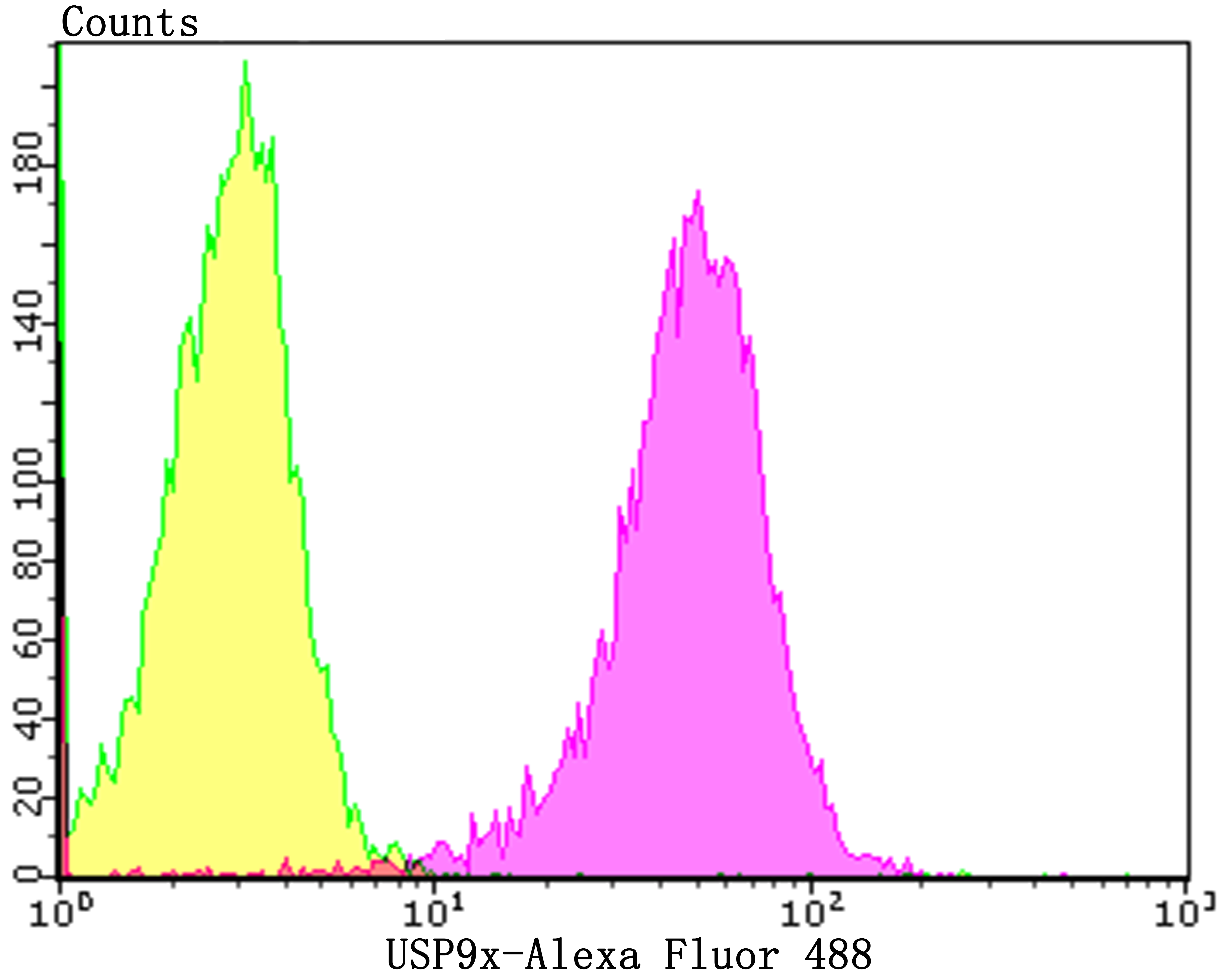
Fig7: Flow cytometric analysis of 293T cells with USP9X antibody at 1/100 dilution (purple) compared with an unlabelled control (cells without incubation with primary antibody; yellow).Alexa Fluor 488-conjugated goat anti-rabbit IgG was used as the secondary antibody.| Product Name | USP9x [JG35-11] |
|---|---|
| Antibody Type | Primary Antibodies |
| Antigen Alias | Deubiquitinating enzyme FAF X antibody Deubiquitinating enzyme FAF-X antibody DFFRX antibody Drosophila fat facets related X linked antibody FAF antibody Fafl antibody Fam antibody Fat facets homolog antibody Fat facets in mammals antibody Fat facets protein related X linked antibody Fat facets protein related, X-linked antibody Fat facets protein-related antibody hFAM antibody MRX99 antibody Probable ubiquitin carboxyl terminal hydrolase FAF X antibody Probable ubiquitin carboxyl-terminal hydrolase FAF-X antibody Ubiquitin carboxyl-terminal hydrolase FAM antibody Ubiquitin specific peptidase 9 X linked antibody Ubiquitin specific peptidase 9, X-linked antibody Ubiquitin specific processing protease FAF X antibody Ubiquitin specific protease 9 X chromosome antibody Ubiquitin thioesterase FAF X antibody Ubiquitin thiolesterase FAF X antibody Ubiquitin thiolesterase FAF-X antibody Ubiquitin-specific protease 9 antibody Ubiquitin-specific-processing protease FAF-X antibody USP9 (gene name) antibody Usp9x antibody USP9X_HUMAN antibody Uubiquitin specific protease 9, X chromosome (fat facets like Drosophila) antibody X chromosome antibody X-linked antibody |
| Product description | Deubiquitinase involved both in the processing of ubiquitin precursors and of ubiquitinated proteins. May therefore play an important regulatory role at the level of protein turnover by preventing degradation of proteins through the removal of conjugated ubiquitin. Specifically hydrolyzes 'Lys-48'-, 'Lys-29'- and 'Lys-33'-linked polyubiquitins chains. Essential component of TGF-beta/BMP signaling cascade. Specifically deubiquitinates monoubiquitinated SMAD4, opposing the activity of E3 ubiquitin-protein ligase TRIM33. Deubiquitinates alkylation repair enzyme ALKBH3. OTUD4 recruits USP7 and USP9X to stabilize ALKBH3, thereby promoting the repair of alkylated DNA lesions. Regulates chromosome alignment and segregation in mitosis by regulating the localization of BIRC5/survivin to mitotic centromeres. Involved in axonal growth and neuronal cell migration. |
| Immunogen | Recombinant protein within human USP9X aa 1-200. |
| Clonality | Monoclonal |
|---|---|
| Isotype | IgG |
| Host Species | Recombinant rabbit |
| Tested Applications | WBICCIFIHCFC |
| WB:1:1,000-1:2,000 ICC:1:50-1:200 IHC:1:50-1:200 FC:1:50-1:100 |
|
| Species Reactivity | HumanMouseRat |
| Concentration | 1mg/ml |
| Alternative Names | Deubiquitinating enzyme FAF X antibody
Deubiquitinating enzyme FAF-X antibody
DFFRX antibody
Drosophila fat facets related X linked antibody
FAF antibody
Fafl antibody
Fam antibody
Fat facets homolog antibody
Fat facets in mammals antibody
Fat facets protein related X linked antibody
Fat facets protein related X-linked antibody Fat facets protein-related antibody hFAM antibody MRX99 antibody Probable ubiquitin carboxyl terminal hydrolase FAF X antibody Probable ubiquitin carboxyl-terminal hydrolase FAF-X antibody Ubiquitin carboxyl-terminal hydrolase FAM antibody Ubiquitin specific peptidase 9 X linked antibody Ubiquitin specific peptidase 9 X-linked antibody Ubiquitin specific processing protease FAF X antibody Ubiquitin specific protease 9 X chromosome antibody Ubiquitin thioesterase FAF X antibody Ubiquitin thiolesterase FAF X antibody Ubiquitin thiolesterase FAF-X antibody Ubiquitin-specific protease 9 antibody Ubiquitin-specific-processing protease FAF-X antibody USP9 (gene name) antibody Usp9x antibody USP9X_HUMAN antibody Uubiquitin specific protease 9 X chromosome (fat facets like Drosophila) antibody X chromosome antibody X-linked antibody |
|---|---|
| Molecular Weight(MW) | 292 kDa |
| Cellular Localization | Cytoplasm. |
Application
Fig1: Western blot analysis of USP9X on different lysates using anti-USP9X antibody at 1/1,000 dilution. Positive control: Lane 1: SiHa Lane 2: A549 Lane 3: 293 Lane 4: Mouse colon
Application
Fig2: ICC staining USP9X in 293T cells (green). The nuclear counter stain is DAPI (blue). Cells were fixed in paraformaldehyde, permeabilised with 0.25% Triton X100/PBS.
Application
Fig3: ICC staining USP9X in F9 cells (green). The nuclear counter stain is DAPI (blue). Cells were fixed in paraformaldehyde, permeabilised with 0.25% Triton X100/PBS.
Application
Fig4: ICC staining USP9X in SiHa cells (green). The nuclear counter stain is DAPI (blue). Cells were fixed in paraformaldehyde, permeabilised with 0.25% Triton X100/PBS.
Application
Fig5: Immunohistochemical analysis of paraffin-embedded human tonsil tissue using anti-USP9X antibody. Counter stained with hematoxylin. The section was pre-treated using heat mediated antigen retrieval with sodium citrate buffer (pH6) for 20 mins.
Application
Fig6: Immunohistochemical analysis of paraffin-embedded mouse fallopian tube tissue using anti-USP9X antibody. Counter stained with hematoxylin. The section was pre-treated using heat mediated antigen retrieval with sodium citrate buffer (pH6) for 20 mins.
Application
Fig7: Flow cytometric analysis of 293T cells with USP9X antibody at 1/100 dilution (purple) compared with an unlabelled control (cells without incubation with primary antibody; yellow).Alexa Fluor 488-conjugated goat anti-rabbit IgG was used as the secondary antibody.| Positive Control | SiHa, A549, 293, mouse colon tissue, 293T, F9, human tonsil tissue, mouse fallopian tube tissue. |
|---|---|
| Application Notes | WB:1:1,000-1:2,000 ICC:1:50-1:200 IHC:1:50-1:200 FC:1:50-1:100 |
| Form | Liquid |
|---|---|
| Storage Instructions | Store at +4℃ after thawing. Aliquot store at -20℃. Avoid repeated freeze / thaw cycles. |
| Storage Buffer | 1*TBS (pH7.4), 1%BSA, 40%Glycerol. Preservative: 0.05% Sodium Azide. |


