Application
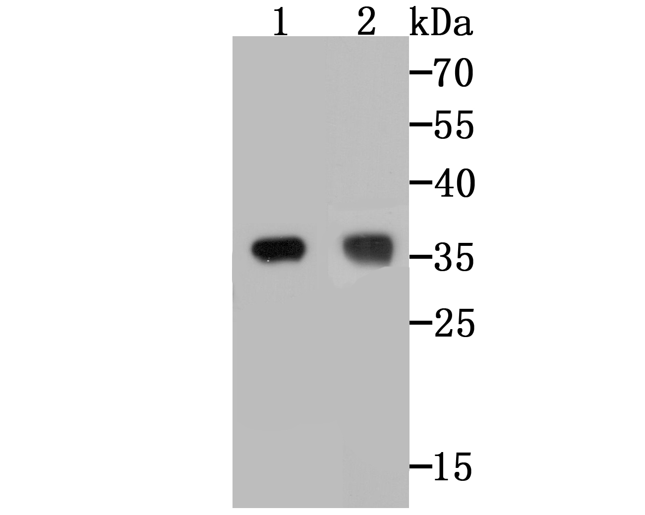
Fig1: Western blot analysis of TREX1 on different cell lysates using anti-TREX1 at 1/500 dilution. Positive control: Line 1: A431 Line 2: SK-Br-3Application
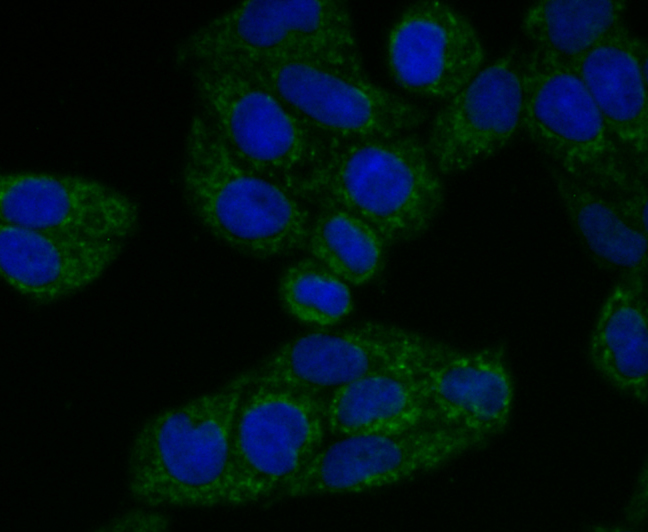
Fig2: ICC staining TREX1 in Hela cells (green). The nuclear counter stain is DAPI (blue). Cells were fixed in paraformaldehyde, permeabilised with 0.25% Triton X100/PBS.Application
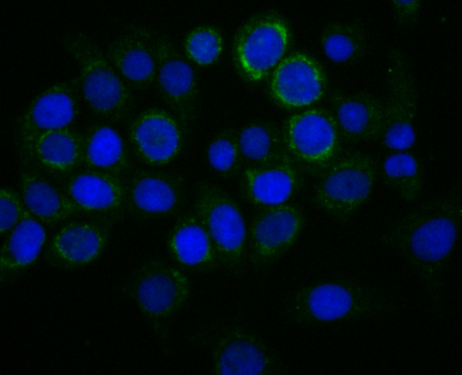
Fig3: ICC staining TREX1 in LOVO cells (green). The nuclear counter stain is DAPI (blue). Cells were fixed in paraformaldehyde, permeabilised with 0.25% Triton X100/PBS.Application
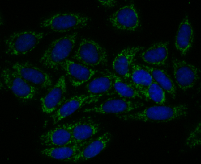
Fig4: ICC staining TREX1 in SiHa cells (green). The nuclear counter stain is DAPI (blue). Cells were fixed in paraformaldehyde, permeabilised with 0.25% Triton X100/PBS.Application
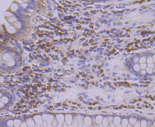
Fig5: Immunohistochemical analysis of paraffin-embedded human colon tissue using anti-TREX1 antibody. Counter stained with hematoxylin.Application
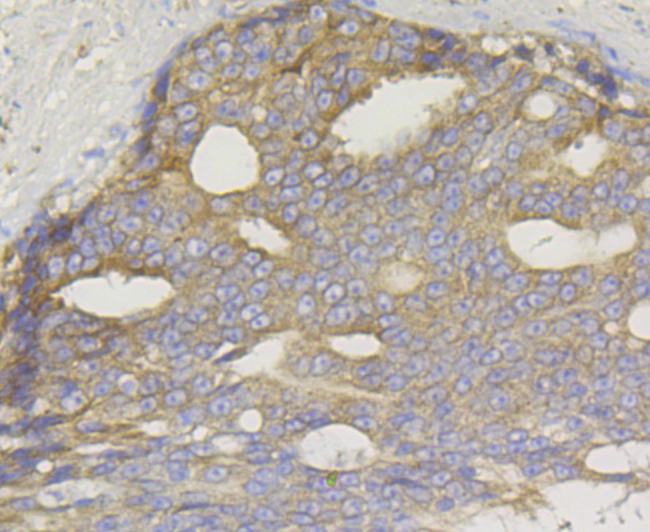
Fig6: Immunohistochemical analysis of paraffin-embedded human prostate cancer tissue using anti-TREX1 antibody. Counter stained with hematoxylin.| Product Name | TREX1 [JG35-71] |
|---|---|
| Antibody Type | Primary Antibodies |
| Antigen Alias | 3' 5' exonuclease TREX1 antibody 3' repair exonuclease 1 antibody AGS1 antibody AGS5 antibody CRV antibody Deoxyribonuclease III, dnaQ/mutD (E. coli) like antibody DKFZp434J0310 antibody DNase III antibody DRN3 antibody HERNS antibody Three prime repair exonuclease 1 antibody TREX1 antibody |
| Product description | Major cellular 3'-to-5' DNA exonuclease which digests single-stranded DNA (ssDNA) and double-stranded DNA (dsDNA) with mismatched 3' termini. Prevents cell-intrinsic initiation of autoimmunity. Acts by metabolizing DNA fragments from endogenous retroelements, including L1, LTR and SINE elements. Unless degraded, these DNA fragments accumulate in the cytosol and activate the IFN-stimulatory DNA (ISD) response and innate immune signaling. Prevents chronic ATM-dependent checkpoint activation, by processing ssDNA polynucleotide species arising from the processing of aberrant DNA replication intermediates. Inefficiently degrades oxidized DNA, such as that generated upon antimicrobial reactive oxygen production or upon absorption of UV light. During GZMA-mediated cell death, contributes to DNA damage in concert with NME1. NME1 nicks one strand of DNA and TREX1 removes bases from the free 3' end to enhance DNA damage and prevent DNA end reannealing and rapid repair. |
| Immunogen | Recombinant protein within human TREX1 aa 100-300. |
| Clonality | Monoclonal |
|---|---|
| Isotype | IgG |
| Host Species | Recombinant rabbit |
| Tested Applications | WBICCIFIHC |
| WB:1:500-1:2,000 ICC:1:50-1:200 IHC:1:50-1:200 |
|
| Species Reactivity | Human |
| Concentration | 1mg/ml |
| Alternative Names | 3' 5' exonuclease TREX1 antibody
3' repair exonuclease 1 antibody
AGS1 antibody
AGS5 antibody
CRV antibody
Deoxyribonuclease III dnaQ/mutD (E. coli) like antibody DKFZp434J0310 antibody DNase III antibody DRN3 antibody HERNS antibody Three prime repair exonuclease 1 antibody TREX1 antibody |
|---|---|
| Molecular Weight(MW) | 34 kDa |
| Cellular Localization | Cytoplasm. Nucleus. Endoplasmic reticulum. |
| SwissProt ID | Q9NSU2 |
|---|
Application
Fig1: Western blot analysis of TREX1 on different cell lysates using anti-TREX1 at 1/500 dilution. Positive control: Line 1: A431 Line 2: SK-Br-3
Application
Fig2: ICC staining TREX1 in Hela cells (green). The nuclear counter stain is DAPI (blue). Cells were fixed in paraformaldehyde, permeabilised with 0.25% Triton X100/PBS.
Application
Fig3: ICC staining TREX1 in LOVO cells (green). The nuclear counter stain is DAPI (blue). Cells were fixed in paraformaldehyde, permeabilised with 0.25% Triton X100/PBS.
Application
Fig4: ICC staining TREX1 in SiHa cells (green). The nuclear counter stain is DAPI (blue). Cells were fixed in paraformaldehyde, permeabilised with 0.25% Triton X100/PBS.
Application
Fig5: Immunohistochemical analysis of paraffin-embedded human colon tissue using anti-TREX1 antibody. Counter stained with hematoxylin.
Application
Fig6: Immunohistochemical analysis of paraffin-embedded human prostate cancer tissue using anti-TREX1 antibody. Counter stained with hematoxylin.| Positive Control | A431, SK-Br-3, Hela, LOVO, SiHa, human colon tissue, human prostate cancer tissue. |
|---|---|
| Application Notes | WB:1:500-1:2,000 ICC:1:50-1:200 IHC:1:50-1:200 |
| Form | Liquid |
|---|---|
| Storage Instructions | Store at +4℃ after thawing. Aliquot store at -20℃. Avoid repeated freeze / thaw cycles. |
| Storage Buffer | 1*TBS (pH7.4), 1%BSA, 40%Glycerol. Preservative: 0.05% Sodium Azide. |


