Application
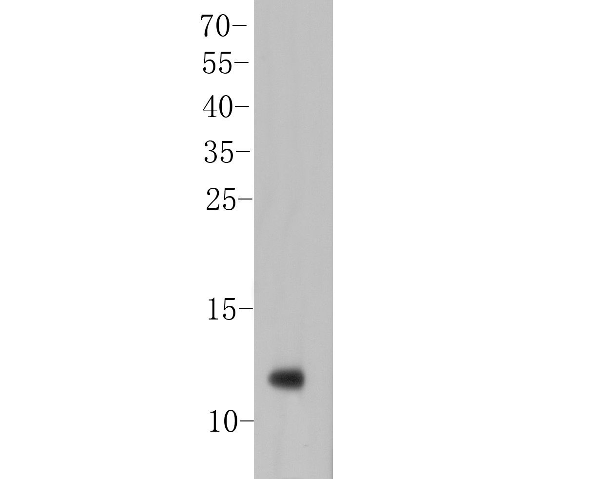
Fig1: Western blot analysis of IL8 on Mouse stomach tissue lysate. Proteins were transferred to a PVDF membrane and blocked with 5% BSA in PBS for 1 hour at room temperature. The primary antibody (ER1901-61, 1/100) was used in 5% BSA at room temperature for 2 hours. Goat Anti-Rabbit IgG - HRP Secondary Antibody (HA1001) at 1:5,000 dilution was used for 1 hour at room temperature.Application
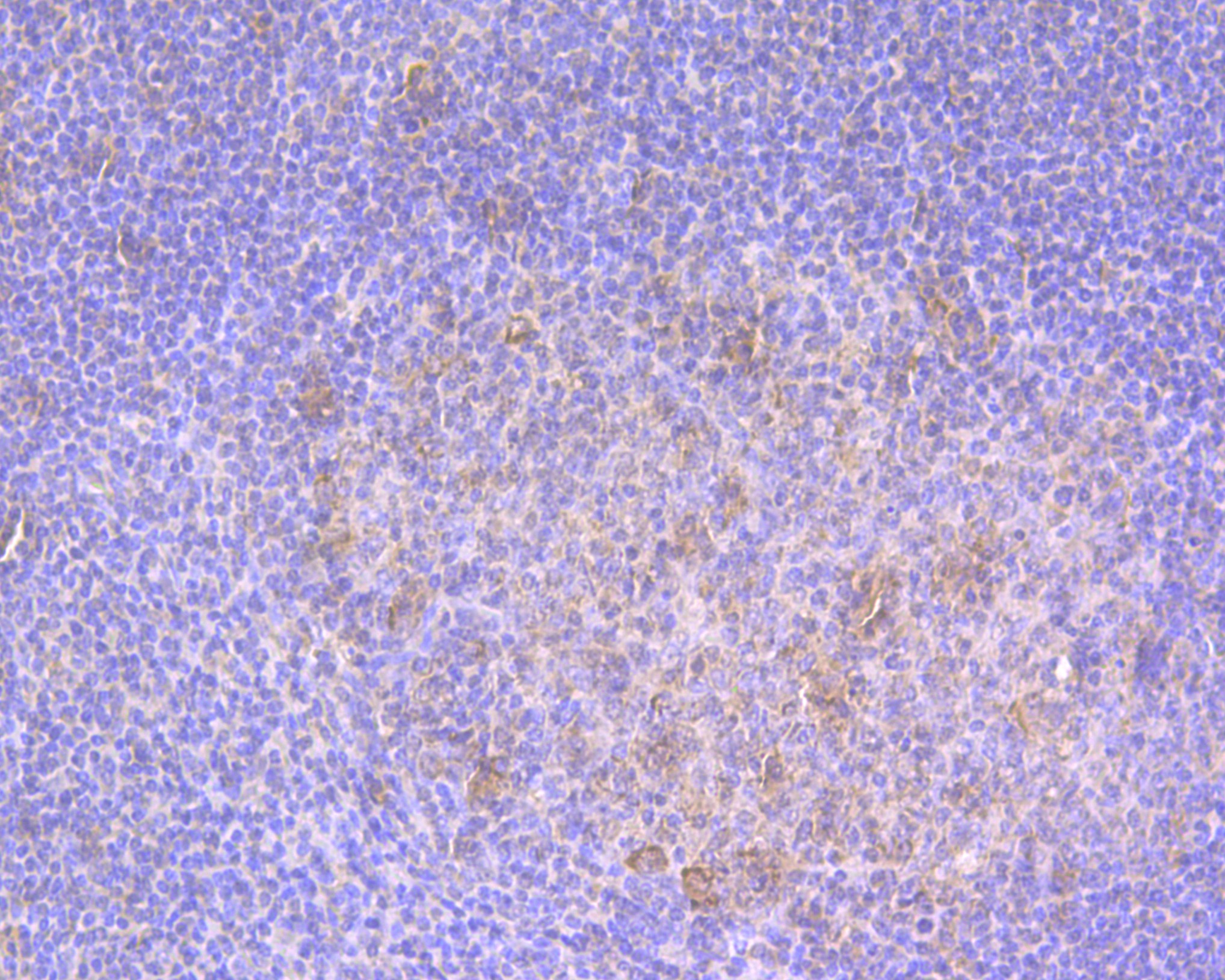
Fig2: Immunohistochemical analysis of paraffin-embedded human tonsil tissue using anti-IL8 antibody. The section was pre-treated using heat mediated antigen retrieval with Tris-EDTA buffer (pH 8.0-8.4) for 20 minutes.The tissues were blocked in 5% BSA for 30 minutes at room temperature, washed with ddH2O and PBS, and then probed with the primary antibody (ER1901-61, 1/100) for 30 minutes at room temperature. The detection was performed using an HRP conjugated compact polymer system. DAB was used as the chromogen. Tissues were counterstained with hematoxylin and mounted with DPX.Application
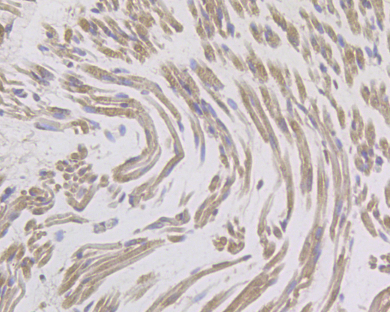
Fig3: Immunohistochemical analysis of paraffin-embedded human skeletal muscle tissue using anti-IL8 antibody. The section was pre-treated using heat mediated antigen retrieval with Tris-EDTA buffer (pH 8.0-8.4) for 20 minutes.The tissues were blocked in 5% BSA for 30 minutes at room temperature, washed with ddH2O and PBS, and then probed with the primary antibody (ER1901-61, 1/100) for 30 minutes at room temperature. The detection was performed using an HRP conjugated compact polymer system. DAB was used as the chromogen. Tissues were counterstained with hematoxylin and mounted with DPX.Application
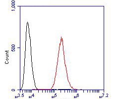
Fig4: Flow cytometric analysis of IL8 was done on AGS cells. The cells were fixed, permeabilized and stained with the primary antibody (ER1901-61, 1/100) (red). After incubation of the primary antibody at room temperature for an hour, the cells were stained with a Alexa Fluor 488-conjugated goat anti-rabbit IgG Secondary antibody at 1/500 dilution for 30 minutes.Unlabelled sample was used as a control (cells without incubation with primary antibody; black).| Product Name | IL8 |
|---|---|
| Antibody Type | Primary Antibodies |
| Antigen Alias | (Ala-IL-8)77 antibody (Ser-IL-8)72 antibody 9E3 antibody Beta thromboglobulin like protein antibody C-X-C motif chemokine 8 antibody CEF-4 antibody chemokine, CXC motif, ligand 8 antibody CXCL8 antibody Emoctakin antibody GCP-1 antibody GCP/IL-8 protein I antibody GCP/IL-8 protein II antibody GCP/IL-8 protein III antibody GCP/IL-8 protein IV antibody GCP/IL-8 protein V antibody GCP/IL-8 protein VI antibody GCP1 antibody Granulocyte chemotactic protein 1 antibody IL-8 antibody IL-8(1-77) antibody IL-8(9-77) antibody IL8 antibody IL8/NAP1 form I antibody IL8/NAP1 form II antibody IL8/NAP1 form III antibody IL8/NAP1 form IV antibody IL8/NAP1 form V antibody IL8/NAP1 form VI antibody IL8_HUMAN antibody Inteleukin 8 antibody LECT antibody LUCT antibody Lymphocyte-derived neutrophil-activating factor antibody LYNAP antibody MDNCF antibody MDNCF-b antibody MDNCF-c antibody MONAP antibody Monocyte derived neutrophil activating peptide antibody Monocyte derived neutrophil chemotactic factor antibody Monocyte-derived neutrophil chemotactic factor antibody Monocyte-derived neutrophil-activating peptide antibody NAF antibody NAP 1 antibody NAP-1 antibody NAP1 antibody Neutrophil activating peptide 1 antibody Neutrophil activating protein 1 antibody Neutrophil-activating factor antibody Neutrophil-activating protein 1 antibody Protein 3 10C antibody Protein 3-10C antibody SCYB 8 antibody SCYB8 antibody Small inducible cytokine subfamily B member 8 antibody T cell chemotactic factor antibody T-cell chemotactic factor antibody TSG 1 antibody TSG1 antibody |
| Product description | IL-8, also known as neutrophil chemotactic factor, has two primary functions. It induces chemotaxis in target cells, primarily neutrophils but also other granulocytes, causing them to migrate toward the site of infection. IL-8 also stimulates phagocytosis once they have arrived. IL-8 is also known to be a potent promoter of angiogenesis. In target cells, IL-8 induces a series of physiological responses required for migration and phagocytosis, such as increases in intracellular Ca2+, exocytosis (e.g. histamine release), and the respiratory burst.IL-8 can be secreted by any cells with toll-like receptors that are involved in the innate immune response. Usually, it is the macrophages that see an antigen first, and thus are the first cells to release IL-8 to recruit other cells. Both monomer and homodimer forms of IL-8 have been reported to be potent inducers of the chemokine receptors CXCR1 and CXCR2. The homodimer is more potent, but methylation of Leu25 can block the activity of homodimers. |
| Immunogen | Recombinant protein within Human IL8 aa 1-99. |
| Clonality | Polyclonal |
|---|---|
| Isotype | IgG |
| Host Species | Rabbit |
| Tested Applications | WBIHCICCFC |
| WB:1:500-1:1000 IHC:1:50-1:100 ICC:1:50-1:100 FC:1:50-1:100 |
|
| Species Reactivity | HumanMouse |
| Concentration | 1mg/ml |
| Alternative Names | (Ala-IL-8)77 antibody
(Ser-IL-8)72 antibody
9E3 antibody Beta thromboglobulin like protein antibody C-X-C motif chemokine 8 antibody CEF-4 antibody chemokine CXC motif ligand 8 antibody CXCL8 antibody Emoctakin antibody GCP-1 antibody GCP/IL-8 protein I antibody GCP/IL-8 protein II antibody GCP/IL-8 protein III antibody GCP/IL-8 protein IV antibody GCP/IL-8 protein V antibody GCP/IL-8 protein VI antibody GCP1 antibody Granulocyte chemotactic protein 1 antibody IL-8 antibody IL-8(1-77) antibody IL-8(9-77) antibody IL8 antibody IL8/NAP1 form I antibody IL8/NAP1 form II antibody IL8/NAP1 form III antibody IL8/NAP1 form IV antibody IL8/NAP1 form V antibody IL8/NAP1 form VI antibody IL8_HUMAN antibody Inteleukin 8 antibody LECT antibody LUCT antibody Lymphocyte-derived neutrophil-activating factor antibody LYNAP antibody MDNCF antibody MDNCF-b antibody MDNCF-c antibody MONAP antibody Monocyte derived neutrophil activating peptide antibody Monocyte derived neutrophil chemotactic factor antibody Monocyte-derived neutrophil chemotactic factor antibody Monocyte-derived neutrophil-activating peptide antibody NAF antibody NAP 1 antibody NAP-1 antibody NAP1 antibody Neutrophil activating peptide 1 antibody Neutrophil activating protein 1 antibody Neutrophil-activating factor antibody Neutrophil-activating protein 1 antibody Protein 3 10C antibody Protein 3-10C antibody SCYB 8 antibody SCYB8 antibody Small inducible cytokine subfamily B member 8 antibody T cell chemotactic factor antibody T-cell chemotactic factor antibody TSG 1 antibody TSG1 antibody |
|---|---|
| Molecular Weight(MW) | 11 kDa |
| Cellular Localization | Secreted. |
| SwissProt ID | P10145 |
|---|
Application
Fig1: Western blot analysis of IL8 on Mouse stomach tissue lysate. Proteins were transferred to a PVDF membrane and blocked with 5% BSA in PBS for 1 hour at room temperature. The primary antibody (ER1901-61, 1/100) was used in 5% BSA at room temperature for 2 hours. Goat Anti-Rabbit IgG - HRP Secondary Antibody (HA1001) at 1:5,000 dilution was used for 1 hour at room temperature.
Application
Fig2: Immunohistochemical analysis of paraffin-embedded human tonsil tissue using anti-IL8 antibody. The section was pre-treated using heat mediated antigen retrieval with Tris-EDTA buffer (pH 8.0-8.4) for 20 minutes.The tissues were blocked in 5% BSA for 30 minutes at room temperature, washed with ddH2O and PBS, and then probed with the primary antibody (ER1901-61, 1/100) for 30 minutes at room temperature. The detection was performed using an HRP conjugated compact polymer system. DAB was used as the chromogen. Tissues were counterstained with hematoxylin and mounted with DPX.
Application
Fig3: Immunohistochemical analysis of paraffin-embedded human skeletal muscle tissue using anti-IL8 antibody. The section was pre-treated using heat mediated antigen retrieval with Tris-EDTA buffer (pH 8.0-8.4) for 20 minutes.The tissues were blocked in 5% BSA for 30 minutes at room temperature, washed with ddH2O and PBS, and then probed with the primary antibody (ER1901-61, 1/100) for 30 minutes at room temperature. The detection was performed using an HRP conjugated compact polymer system. DAB was used as the chromogen. Tissues were counterstained with hematoxylin and mounted with DPX.
Application
Fig4: Flow cytometric analysis of IL8 was done on AGS cells. The cells were fixed, permeabilized and stained with the primary antibody (ER1901-61, 1/100) (red). After incubation of the primary antibody at room temperature for an hour, the cells were stained with a Alexa Fluor 488-conjugated goat anti-rabbit IgG Secondary antibody at 1/500 dilution for 30 minutes.Unlabelled sample was used as a control (cells without incubation with primary antibody; black).| Positive Control | Mouse stomach tissue lysates, human tonsil tissue, human skeletal muscle tissue, AGS. |
|---|---|
| Application Notes | WB:1:500-1:1000 IHC:1:50-1:100 ICC:1:50-1:100 FC:1:50-1:100 |
| Form | Liquid |
|---|---|
| Storage Instructions | Store at +4℃ after thawing. Aliquot store at -20℃. Avoid repeated freeze / thaw cycles. |
| Storage Buffer | 1*TBS (pH7.4), 1%BSA, 50%Glycerol. Preservative: 0.05% Sodium Azide. |


