Application
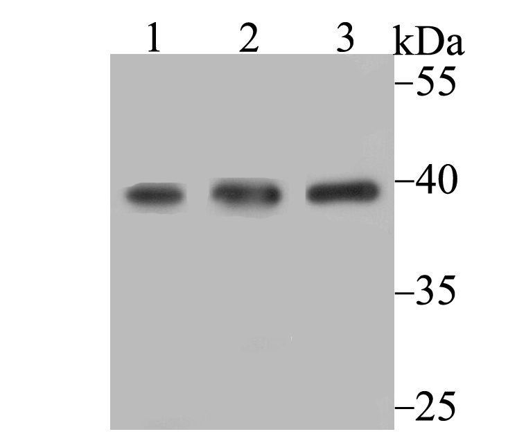
Fig1: Western blot analysis of SAE1 on different cell lysates using anti-SAE1 antibody at 1/1,000 dilution. Positive control: Lane 1: SiHa Lane 2: K562 Lane 3: 293Application
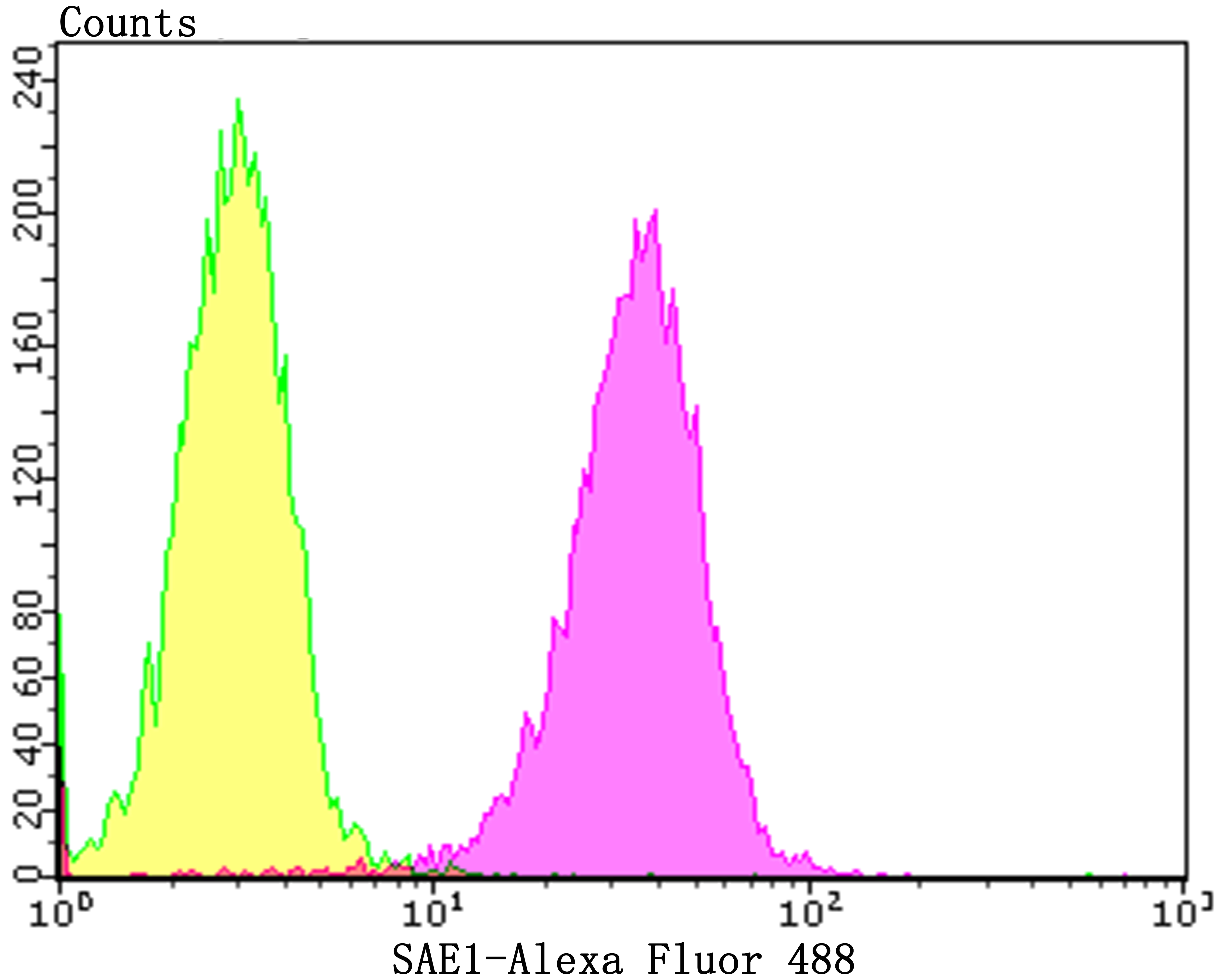
Fig2: Flow cytometric analysis of A549 cells with SAE1 antibody at 1/100 dilution (yellow) compared with an unlabelled control (cells without incubation with primary antibody; purple).Alexa Fluor 488-conjugated goat anti-rabbit IgG was used as the secondary antibody.| Product Name | SAE1 [JG35-88] |
|---|---|
| Antibody Type | Primary Antibodies |
| Antigen Alias | Activator of SUMO1 antibody AOS1 antibody HSPC140 antibody Sae1 antibody SAE1_HUMAN antibody Sentrin/SUMO activating protein AOS1 antibody SUA1 antibody SUMO 1 activating enzyme E1 N subunit antibody SUMO 1 activating enzyme subunit 1 antibody SUMO-activating enzyme subunit 1 antibody Ubiquitin like protein SUMO1 activating enzyme antibody Ubiquitin-like 1-activating enzyme E1A antibody UBL E1A antibody UBLE1A antibody |
| Product description | Proteolytic degradation by the ubiquitin (Ub) system is essential for normal cell cycle progression, cellular differentiation and stress responses. Proteins conjugated to Ub are marked for progressive degradation by the 26S Proteasome. AOS-1, also designated SUMO-1-activating enzyme or ubiquitin-like 1-activating enzyme E1A, belongs to the ubiquitin-activating E1 family of proteins and plays an important role in the first step of the UBL1 conjugation pathway. AOS-1, which is a dimeric enzyme, functions as a UBLI E1 ligase, mediating the ATP-dependent activation of UBL1. AOS-1 can bind with UBLE1A and UBLE1B to form a heterodimer which can bind UBL1. |
| Immunogen | Recombinant protein within human SAE1 aa 150-300. |
| Clonality | Monoclonal |
|---|---|
| Isotype | IgG |
| Host Species | Recombinant rabbit |
| Tested Applications | WBFC |
| WB:1:500-1:2,000 FC:1:50-1:100 |
|
| Species Reactivity | Human |
| Concentration | 1mg/ml |
| Alternative Names | Activator of SUMO1 antibody AOS1 antibody HSPC140 antibody Sae1 antibody SAE1_HUMAN antibody Sentrin/SUMO activating protein AOS1 antibody SUA1 antibody SUMO 1 activating enzyme E1 N subunit antibody SUMO 1 activating enzyme subunit 1 antibody SUMO-activating enzyme subunit 1 antibody Ubiquitin like protein SUMO1 activating enzyme antibody Ubiquitin-like 1-activating enzyme E1A antibody UBL E1A antibody UBLE1A antibody |
|---|---|
| Molecular Weight(MW) | 38 kDa |
| Cellular Localization | Nucleus. |
| SwissProt ID | Q9UBE0 |
|---|
Application
Fig1: Western blot analysis of SAE1 on different cell lysates using anti-SAE1 antibody at 1/1,000 dilution. Positive control: Lane 1: SiHa Lane 2: K562 Lane 3: 293
Application
Fig2: Flow cytometric analysis of A549 cells with SAE1 antibody at 1/100 dilution (yellow) compared with an unlabelled control (cells without incubation with primary antibody; purple).Alexa Fluor 488-conjugated goat anti-rabbit IgG was used as the secondary antibody.| Positive Control | SiHa, K562, 293, A549. |
|---|---|
| Application Notes | WB:1:500-1:2,000 FC:1:50-1:100 |
| Form | Liquid |
|---|---|
| Storage Instructions | Store at +4℃ after thawing. Aliquot store at -20℃. Avoid repeated freeze / thaw cycles. |
| Storage Buffer | 1*TBS (pH7.4), 1%BSA, 40%Glycerol. Preservative: 0.05% Sodium Azide. |


