Application
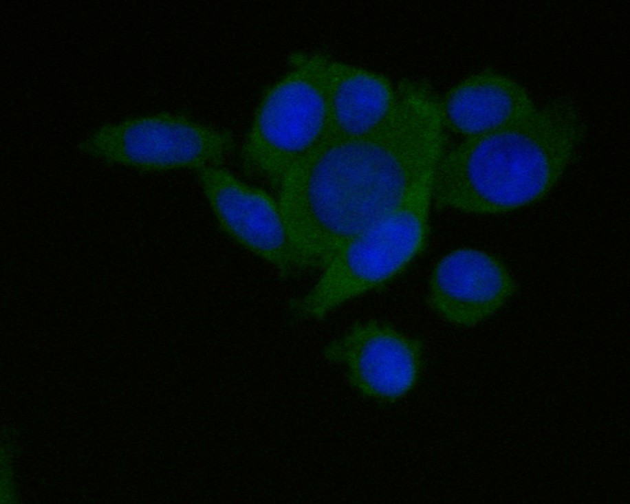
Fig1: ICC staining of CD133 in SW620 cells (green). Formalin fixed cells were permeabilized with 0.1% Triton X-100 in TBS for 10 minutes at room temperature and blocked with 1% Blocker BSA for 15 minutes at room temperature. Cells were probed with the primary antibody (ER1901-63, 1/100) for 1 hour at room temperature, washed with PBS. Alexa Fluor®488 Goat anti-Rabbit IgG was used as the secondary antibody at 1/100 dilution. The nuclear counter stain is DAPI (blue).Application
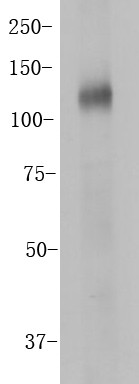
Fig2: Western blot analysis of CD133 on HT-29 cell lysates. Proteins were transferred to a PVDF membrane and blocked with 5% BSA in PBS for 1 hour at room temperature. The primary antibody (ER1901-63, 1/100) was used in 5% BSA at room temperature for 2 hours. Goat Anti-Rabbit IgG - HRP Secondary Antibody (HA1001) at 1:5,000 dilution was used for 1 hour at room temperature.Application
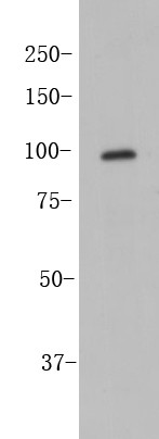
Fig3: Western blot analysis of CD133 on SW480 cell lysates. Proteins were transferred to a PVDF membrane and blocked with 5% BSA in PBS for 1 hour at room temperature. The primary antibody (ER1901-63, 1/100) was used in 5% BSA at room temperature for 2 hours. Goat Anti-Rabbit IgG - HRP Secondary Antibody (HA1001) at 1:5,000 dilution was used for 1 hour at room temperature.Application
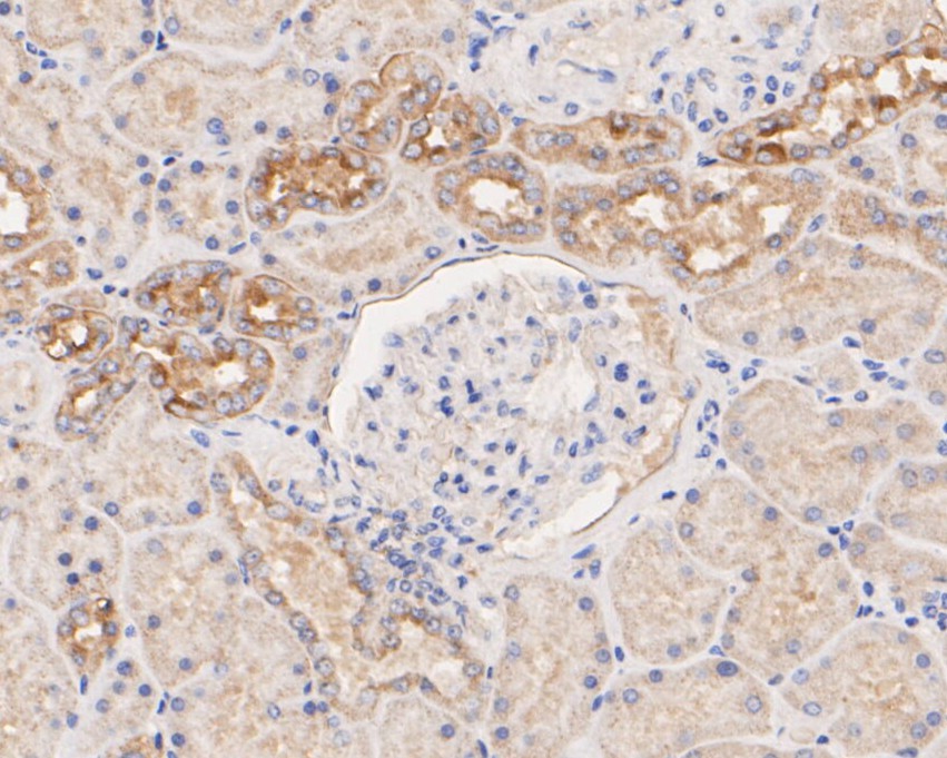
Fig4: Immunohistochemical analysis of paraffin-embedded human kidney tissue using anti-CD133 antibody. The section was pre-treated using heat mediated antigen retrieval with Tris-EDTA buffer (pH 8.0-8.4) for 20 minutes.The tissues were blocked in 5% BSA for 30 minutes at room temperature, washed with ddH2O and PBS, and then probed with the primary antibody (ER1901-63, 1/100) for 30 minutes at room temperature. The detection was performed using an HRP conjugated compact polymer system. DAB was used as the chromogen. Tissues were counterstained with hematoxylin and mounted with DPX.Application
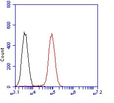
Fig5: Flow cytometric analysis of CD133 was done on F9 cells. The cells were fixed, permeabilized and stained with the primary antibody (ER1901-63, 1/100) (red). After incubation of the primary antibody at room temperature for an hour, the cells were stained with a Alexa Fluor 488-conjugated goat anti-rabbit IgG Secondary antibody at 1/500 dilution for 30 minutes.Unlabelled sample was used as a control (cells without incubation with primary antibody; black).| Product Name | CD133 |
|---|---|
| Antibody Type | Primary Antibodies |
| Antigen Alias | AC133 antibody Antigen AC133 antibody CD133 antibody CORD12 antibody Hematopoietic stem cell antigen antibody hProminin antibody MCDR2 antibody MSTP061 antibody OTTHUMP00000217744 antibody OTTHUMP00000217745 antibody OTTHUMP00000217746 antibody PROM1 antibody PROM1_HUMAN antibody Prominin I antibody Prominin like 1 antibody Prominin like protein 1 precursor antibody Prominin mouse like 1 antibody Prominin-1 antibody Prominin-like protein 1 antibody Prominin1 antibody PROML1 antibody RP41 antibody STGD4 antibody |
| Product description | CD133 antigen, also known as prominin-1, is a glycoprotein that in humans is encoded by the PROM1 gene. It is a member of pentaspan transmembrane glycoproteins, which specifically localize to cellular protrusions. When embedded in the cell membrane, the membrane topology of prominin-1 is such that the N-terminus extends into the extracellular space and the C-terminus resides in the intracellular compartment. The protein consists of five transmembrane segments, with the first and second segments and the third and fourth segments connected by intracellular loops while the second and third as well as fourth and fifth transmembrane segments are connected by extracellular loops. While the precise function of CD133 remains unknown, it has been proposed that it acts as an organizer of cell membrane topology. CD133 is expressed in hematopoietic stem cells, endothelial progenitor cells, glioblastoma, neuronal and glial stem cells, various pediatric brain tumors, as well as adult kidney, mammary glands, trachea, salivary glands, uterus, placenta, digestive tract, testes, and some other cell types. |
| Immunogen | Synthetic peptide within C-terminal residues of human CD133. |
| Clonality | Polyclonal |
|---|---|
| Isotype | IgG |
| Host Species | Rabbit |
| Tested Applications | WBIHCICCFC |
| WB:1:500-1:2000: |
|
| Species Reactivity | Human |
| Concentration | 1mg/ml |
| Alternative Names | AC133 antibody
Antigen AC133 antibody
CD133 antibody CORD12 antibody Hematopoietic stem cell antigen antibody hProminin antibody MCDR2 antibody MSTP061 antibody OTTHUMP00000217744 antibody OTTHUMP00000217745 antibody OTTHUMP00000217746 antibody PROM1 antibody PROM1_HUMAN antibody Prominin I antibody Prominin like 1 antibody Prominin like protein 1 precursor antibody Prominin mouse like 1 antibody Prominin-1 antibody Prominin-like protein 1 antibody Prominin1 antibody PROML1 antibody RP41 antibody STGD4 antibody |
|---|---|
| Molecular Weight(MW) | Predicted band size 100 kDa. |
| Cellular Localization | Cell membrane. |
| SwissProt ID | O43490 |
|---|
Application
Fig1: ICC staining of CD133 in SW620 cells (green). Formalin fixed cells were permeabilized with 0.1% Triton X-100 in TBS for 10 minutes at room temperature and blocked with 1% Blocker BSA for 15 minutes at room temperature. Cells were probed with the primary antibody (ER1901-63, 1/100) for 1 hour at room temperature, washed with PBS. Alexa Fluor®488 Goat anti-Rabbit IgG was used as the secondary antibody at 1/100 dilution. The nuclear counter stain is DAPI (blue).
Application
Fig2: Western blot analysis of CD133 on HT-29 cell lysates. Proteins were transferred to a PVDF membrane and blocked with 5% BSA in PBS for 1 hour at room temperature. The primary antibody (ER1901-63, 1/100) was used in 5% BSA at room temperature for 2 hours. Goat Anti-Rabbit IgG - HRP Secondary Antibody (HA1001) at 1:5,000 dilution was used for 1 hour at room temperature.
Application
Fig3: Western blot analysis of CD133 on SW480 cell lysates. Proteins were transferred to a PVDF membrane and blocked with 5% BSA in PBS for 1 hour at room temperature. The primary antibody (ER1901-63, 1/100) was used in 5% BSA at room temperature for 2 hours. Goat Anti-Rabbit IgG - HRP Secondary Antibody (HA1001) at 1:5,000 dilution was used for 1 hour at room temperature.
Application
Fig4: Immunohistochemical analysis of paraffin-embedded human kidney tissue using anti-CD133 antibody. The section was pre-treated using heat mediated antigen retrieval with Tris-EDTA buffer (pH 8.0-8.4) for 20 minutes.The tissues were blocked in 5% BSA for 30 minutes at room temperature, washed with ddH2O and PBS, and then probed with the primary antibody (ER1901-63, 1/100) for 30 minutes at room temperature. The detection was performed using an HRP conjugated compact polymer system. DAB was used as the chromogen. Tissues were counterstained with hematoxylin and mounted with DPX.
Application
Fig5: Flow cytometric analysis of CD133 was done on F9 cells. The cells were fixed, permeabilized and stained with the primary antibody (ER1901-63, 1/100) (red). After incubation of the primary antibody at room temperature for an hour, the cells were stained with a Alexa Fluor 488-conjugated goat anti-rabbit IgG Secondary antibody at 1/500 dilution for 30 minutes.Unlabelled sample was used as a control (cells without incubation with primary antibody; black).| Positive Control | SW620, HT-29 cell lysates, SW480 cell lysates, human kidney tissue, F9. |
|---|---|
| Application Notes | WB:1:500-1:2000: |
| Form | Liquid |
|---|---|
| Storage Instructions | Store at +4℃ after thawing. Aliquot store at -20℃. Avoid repeated freeze / thaw cycles. |
| Storage Buffer | 1*TBS (pH7.4), 1%BSA, 50%Glycerol. Preservative: 0.05% Sodium Azide. |


