Application
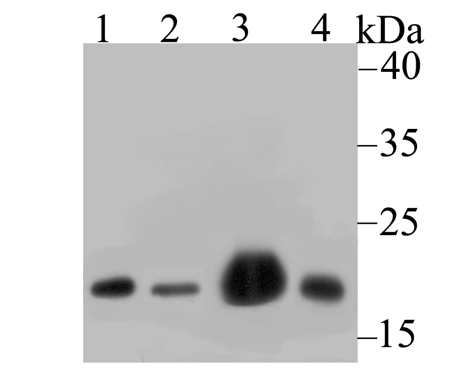
Fig1: Western blot analysis of NDUFB8 on different lysates using anti-NDUFB8 antibody at 1/1,000 dilution. Positive control: Lane 1: 293 Lane 2: A549 Lane 3: Mouse heart Lane 4: Rat spleenApplication
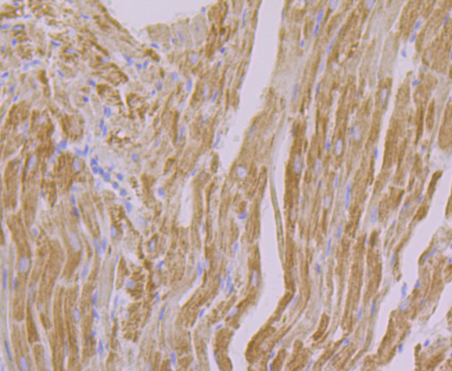
Fig2: Immunohistochemical analysis of paraffin-embedded rat heart tissue using anti-NDUFB8 antibody. Counter stained with hematoxylin.Application
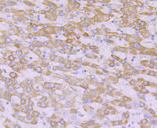
Fig3: Immunohistochemical analysis of paraffin-embedded human liver cancer tissue using anti-NDUFB8 antibody. Counter stained with hematoxylin.Application
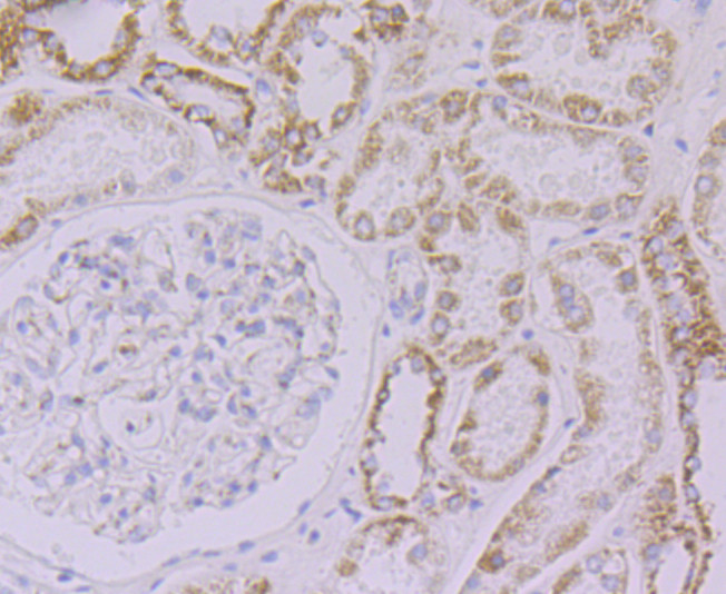
Fig4: Immunohistochemical analysis of paraffin-embedded human kidney tissue using anti-NDUFB8 antibody. Counter stained with hematoxylin.Application
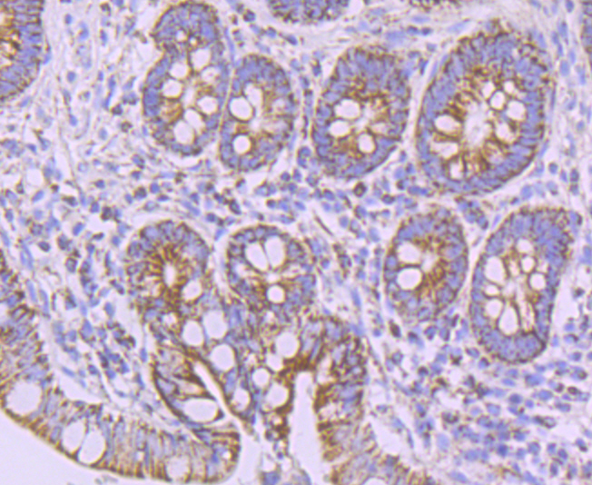
Fig5: Immunohistochemical analysis of paraffin-embedded human small intestine tissue using anti-NDUFB8 antibody. Counter stained with hematoxylin.Application
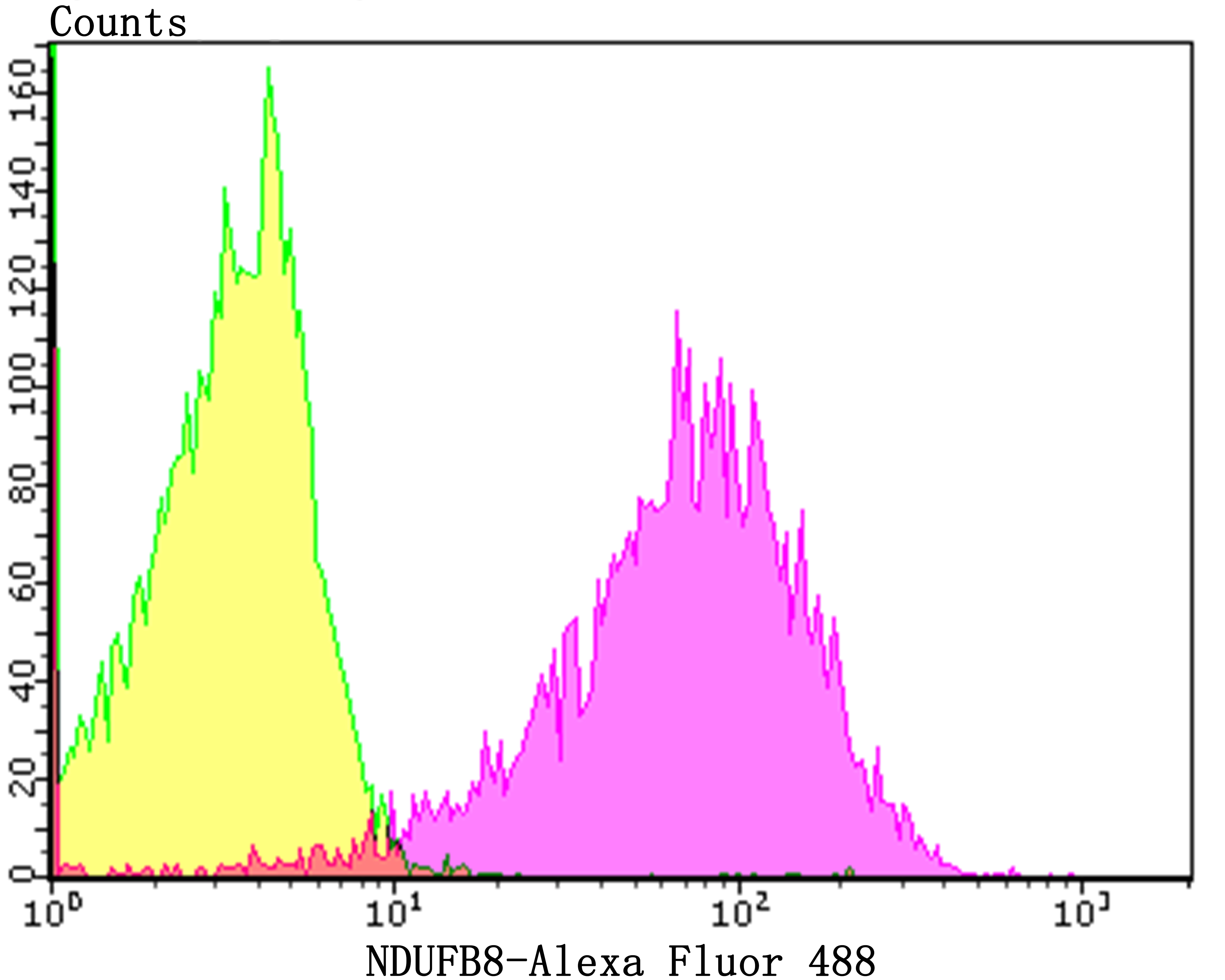
Fig6: Flow cytometric analysis of 293Tcells with NDUFB8 antibody at 1/100 dilution (yellow) compared with an unlabelled control (cells without incubation with primary antibody; purple).Alexa Fluor 488-conjugated goat anti-rabbit IgG was used as the secondary antibody.| Product Name | NDUFB8 [JG61-36] |
|---|---|
| Antibody Type | Primary Antibodies |
| Antigen Alias | ASHI antibody CI-ASHI antibody Complex I ASHI subunit antibody Complex I-ASHI antibody mitochondrial antibody NADH dehydrogenase (ubiquinone) 1 beta subcomplex 8 19kDa antibody NADH dehydrogenase [ubiquinone] 1 beta subcomplex subunit 8 antibody NADH dehydrogenase [ubiquinone] 1 beta subcomplex subunit 8 mitochondrial antibody NADH-ubiquinone oxidoreductase ASHI subunit antibody NDUB8_HUMAN antibody NDUFB8 antibody |
| Product description | Accessory subunit of the mitochondrial membrane respiratory chain NADH dehydrogenase (Complex I), that is believed not to be involved in catalysis. Complex I functions in the transfer of electrons from NADH to the respiratory chain. The immediate electron acceptor for the enzyme is believed to be ubiquinone. |
| Immunogen | Recombinant protein within human NDUFB8 aa 30-130. |
| Clonality | Monoclonal |
|---|---|
| Isotype | IgG |
| Host Species | Recombinant rabbit |
| Tested Applications | WBIPIHCFCICC |
| WB:1:500-1:2,000 IP:1:10-1:50 IHC:1:50-1:200 FC:1:50-1:100 ICC:1:50 |
|
| Species Reactivity | HumanMouseRat |
| Concentration | 1mg/ml |
| Alternative Names | ASHI antibody CI-ASHI antibody Complex I ASHI subunit antibody Complex I-ASHI antibody mitochondrial antibody NADH dehydrogenase (ubiquinone) 1 beta subcomplex 8 19kDa antibody NADH dehydrogenase [ubiquinone] 1 beta subcomplex subunit 8 antibody NADH dehydrogenase [ubiquinone] 1 beta subcomplex subunit 8 mitochondrial antibody NADH-ubiquinone oxidoreductase ASHI subunit antibody NDUB8_HUMAN antibody NDUFB8 antibody |
|---|---|
| Molecular Weight(MW) | 17 kDa |
| Cellular Localization | Mitochondrion. |
Application
Fig1: Western blot analysis of NDUFB8 on different lysates using anti-NDUFB8 antibody at 1/1,000 dilution. Positive control: Lane 1: 293 Lane 2: A549 Lane 3: Mouse heart Lane 4: Rat spleen
Application
Fig2: Immunohistochemical analysis of paraffin-embedded rat heart tissue using anti-NDUFB8 antibody. Counter stained with hematoxylin.
Application
Fig3: Immunohistochemical analysis of paraffin-embedded human liver cancer tissue using anti-NDUFB8 antibody. Counter stained with hematoxylin.
Application
Fig4: Immunohistochemical analysis of paraffin-embedded human kidney tissue using anti-NDUFB8 antibody. Counter stained with hematoxylin.
Application
Fig5: Immunohistochemical analysis of paraffin-embedded human small intestine tissue using anti-NDUFB8 antibody. Counter stained with hematoxylin.
Application
Fig6: Flow cytometric analysis of 293Tcells with NDUFB8 antibody at 1/100 dilution (yellow) compared with an unlabelled control (cells without incubation with primary antibody; purple).Alexa Fluor 488-conjugated goat anti-rabbit IgG was used as the secondary antibody.| Positive Control | 293, A549, 293T, mouse heart tissue lysate, rat spleen tissue lysate, rat heart tissue, human liver cancer tissue, human kidney tissue, human small intestine tissue. |
|---|---|
| Application Notes | WB:1:500-1:2,000 IP:1:10-1:50 IHC:1:50-1:200 FC:1:50-1:100 ICC:1:50 |
| Form | Liquid |
|---|---|
| Storage Instructions | Store at +4℃ after thawing. Aliquot store at -20℃. Avoid repeated freeze / thaw cycles. |
| Storage Buffer | 1*TBS (pH7.4), 1%BSA, 40%Glycerol. Preservative: 0.05% Sodium Azide. |


