Application
Fig1: Western blot analysis of SULT2A1 on mouse kidney tissue lysate using anti-SULT2A1 antibody at 1/1,000 dilution.Application
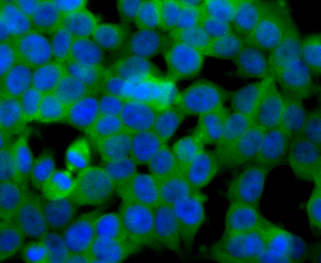
Fig2: ICC staining SULT2A1 in 293T cells (green). The nuclear counter stain is DAPI (blue). Cells were fixed in paraformaldehyde, permeabilised with 0.25% Triton X100/PBS.Application
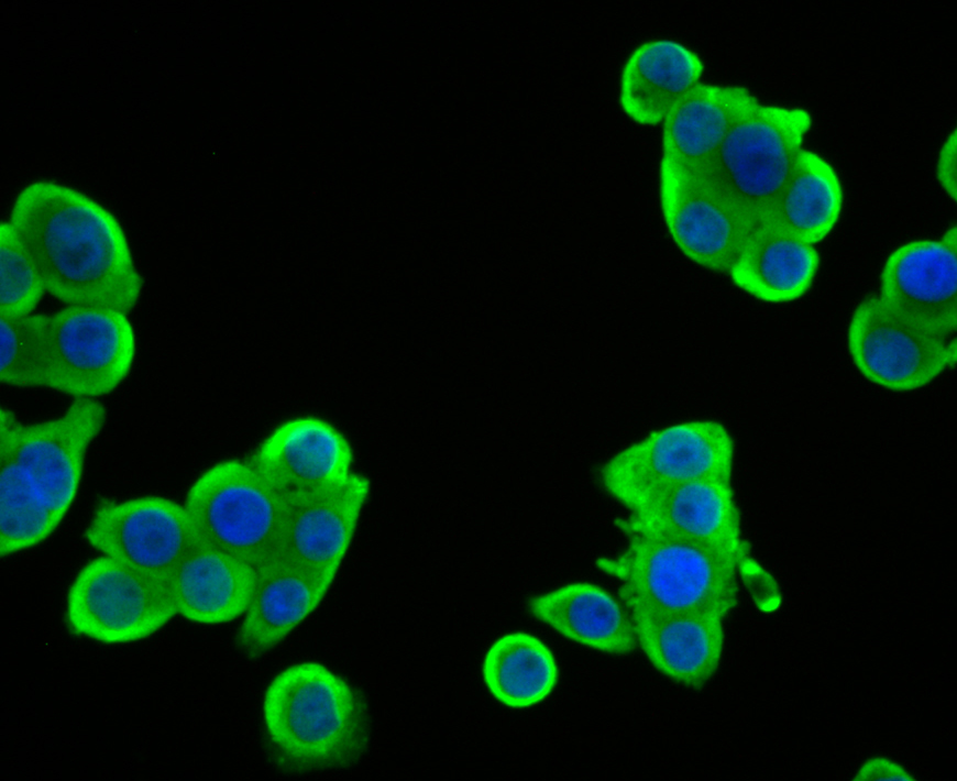
Fig3: ICC staining SULT2A1 in LOVO cells (green). The nuclear counter stain is DAPI (blue). Cells were fixed in paraformaldehyde, permeabilised with 0.25% Triton X100/PBS.Application
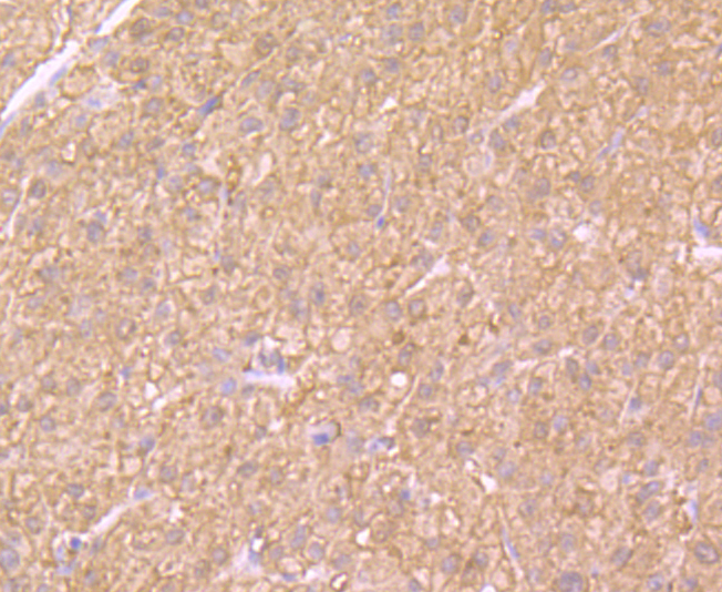
Fig4: Immunohistochemical analysis of paraffin-embedded rat adrenal gland tissue using anti-SULT2A1 antibody. Counter stained with hematoxylin.Application
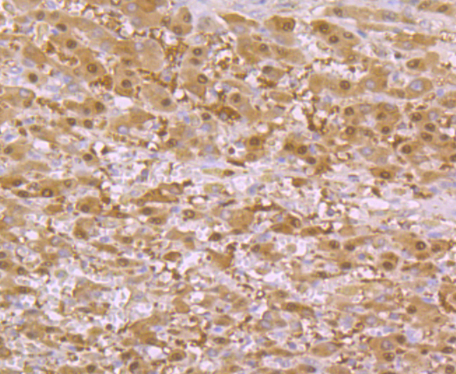
Fig5: Immunohistochemical analysis of paraffin-embedded human liver tissue using anti-SULT2A1 antibody. Counter stained with hematoxylin.Application
Fig6: Immunohistochemical analysis of paraffin-embedded human liver cancer tissue using anti-SULT2A1 antibody. Counter stained with hematoxylin.Application
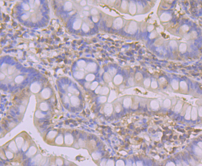
Fig7: Immunohistochemical analysis of paraffin-embedded human small intestine tissue using anti-SULT2A1 antibody. Counter stained with hematoxylin.Application
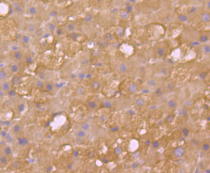
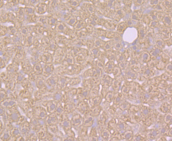
Fig8: Immunohistochemical analysis of paraffin-embedded mouse liver tissue using anti-SULT2A1 antibody. Counter stained with hematoxylin.Application
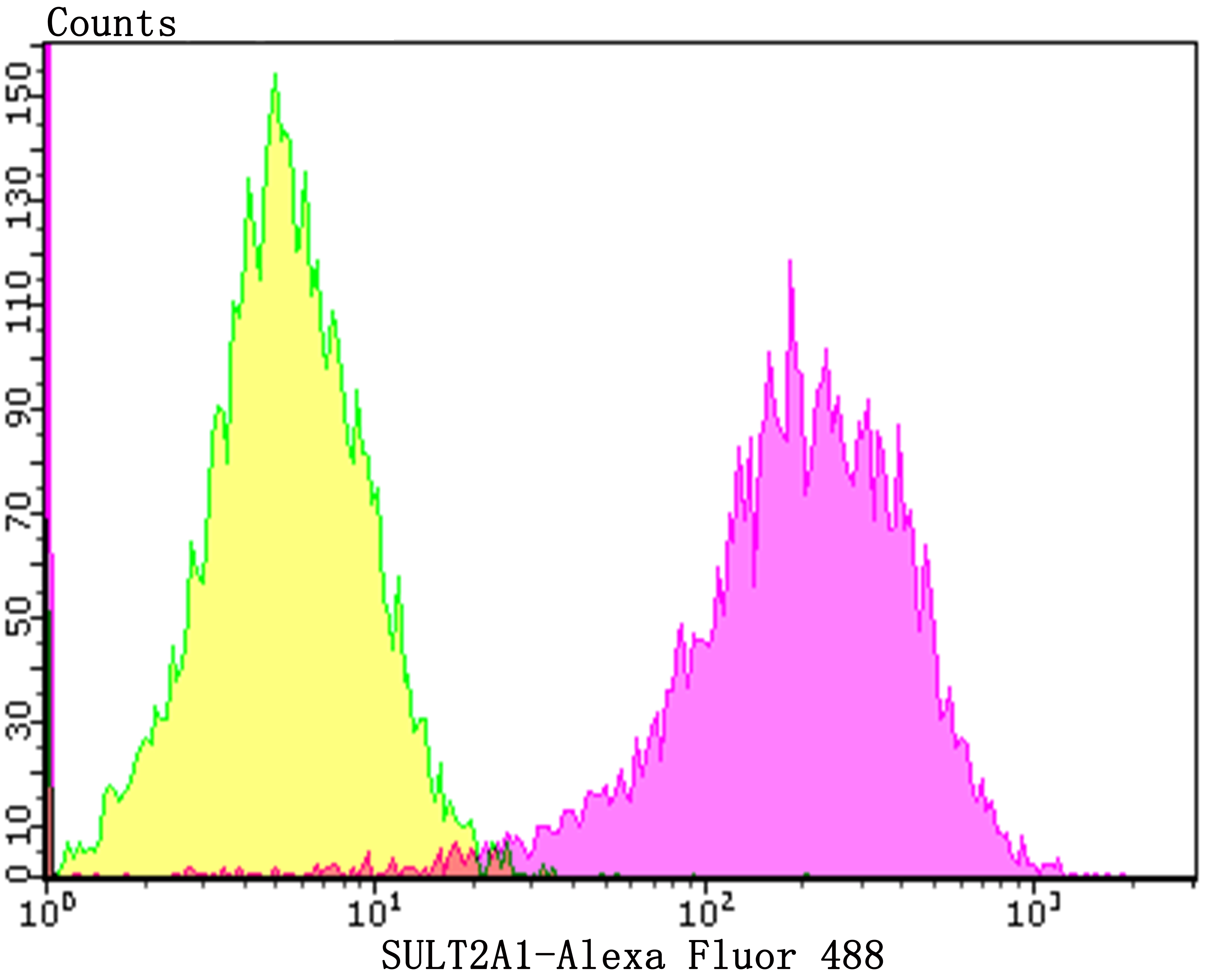
Fig9: Flow cytometric analysis of HepG2 cells with SULT2A1 antibody at 1/100 dilution (yellow) compared with an unlabelled control (cells without incubation with primary antibody; purple).Alexa Fluor 488-conjugated goat anti-rabbit IgG was used as the secondary antibody.| Product Name | SULT2A1 [JG36-18] |
|---|---|
| Antibody Type | Primary Antibodies |
| Antigen Alias | Alcohol/hydroxysteroid sulfotransferase antibody Bile salt sulfotranasferase 2A1 antibody Bile salt sulfotransferase antibody Dehydroepiandrosterone sulfotransferase antibody DHEA ST antibody DHEA sulfotranasferase antibody DHEA-ST antibody DHEAS antibody EC 2.8.2.14 antibody Hst antibody hSTa antibody Hydroxysteroid sulfotransferase antibody ST2 antibody ST2A1 antibody ST2A1_HUMAN antibody ST2A3 antibody STD antibody sulfotranasferase, dehydroepiandrosterone-preferring antibody Sulfotransferase 2A1 antibody Sulfotransferase family 2A, dehydroepiandrosterone-preferring, member 1 antibody Sulfotransferase family cytosolic 2A dehydroepiandrosterone (DHEA) preferring member 1 antibody Sult2a1 antibody |
| Product description | The soluble sulfotransferases contribute to the elimination of xenobiotics, the activation of procarcinogens and the regulation of hormones. Members of the three groups comprising this superfamily show selectivity to certain substrate compounds. SULT1 sulfotransferases exhibit N-sulfating activities of carcinogenic heterocyclic amines, and are selective toward phenols, whereas SULT2 enzymes prefer hydroxysteroids and SULT3 family members are selective for N-substituted aryl and alicyclic compounds. SULT2A1 catalyzes the sulfonation of procarcinogen xenobiotics, hydroxysteroids and bile acids, and is highly expressed in adrenal and liver tissues. SULT2A1 plays a role in hepatic cholesterol homeostasis. SULT2B1 consists of two isoforms, SULT2B1a and SULT2B1b, which are transcribed from the same gene by alternative splicing of their first exons. Both isoforms are highly selective for the sulphation of 3b-hydroxysteroids such as pregnenolone, epiandrosterone, DHEA and androstenediol. SULT2B1b is expressed in prostate, skin, placenta and lung. |
| Immunogen | Recombinant protein within human SULT2A1 aa 100 to the C-terminus. |
| Clonality | Monoclonal |
|---|---|
| Isotype | IgG |
| Host Species | Recombinant rabbit |
| Tested Applications | WBICCIFIHCFC |
| WB:1:500-1:1,000 ICC:1:500-1:2,000 IHC:1:50-1:200 FC:1:50-1:100 |
|
| Species Reactivity | HumanMouseRat |
| Concentration | 1mg/ml |
| Alternative Names | Alcohol/hydroxysteroid sulfotransferase antibody
Bile salt sulfotranasferase 2A1 antibody
Bile salt sulfotransferase antibody
Dehydroepiandrosterone sulfotransferase antibody
DHEA ST antibody
DHEA sulfotranasferase antibody
DHEA-ST antibody
DHEAS antibody
EC 2.8.2.14 antibody
Hst antibody
hSTa antibody
Hydroxysteroid sulfotransferase antibody
ST2 antibody
ST2A1 antibody
ST2A1_HUMAN antibody
ST2A3 antibody
STD antibody
sulfotranasferase dehydroepiandrosterone-preferring antibody Sulfotransferase 2A1 antibody Sulfotransferase family 2A dehydroepiandrosterone-preferring member 1 antibody Sulfotransferase family cytosolic 2A dehydroepiandrosterone (DHEA) preferring member 1 antibody Sult2a1 antibody |
|---|---|
| Molecular Weight(MW) | 34 kDa |
| Cellular Localization | Cytoplasm. |
Application
Fig1: Western blot analysis of SULT2A1 on mouse kidney tissue lysate using anti-SULT2A1 antibody at 1/1,000 dilution.
Application
Fig2: ICC staining SULT2A1 in 293T cells (green). The nuclear counter stain is DAPI (blue). Cells were fixed in paraformaldehyde, permeabilised with 0.25% Triton X100/PBS.
Application
Fig3: ICC staining SULT2A1 in LOVO cells (green). The nuclear counter stain is DAPI (blue). Cells were fixed in paraformaldehyde, permeabilised with 0.25% Triton X100/PBS.
Application
Fig4: Immunohistochemical analysis of paraffin-embedded rat adrenal gland tissue using anti-SULT2A1 antibody. Counter stained with hematoxylin.
Application
Fig5: Immunohistochemical analysis of paraffin-embedded human liver tissue using anti-SULT2A1 antibody. Counter stained with hematoxylin.
Application
Fig6: Immunohistochemical analysis of paraffin-embedded human liver cancer tissue using anti-SULT2A1 antibody. Counter stained with hematoxylin.
Application
Fig7: Immunohistochemical analysis of paraffin-embedded human small intestine tissue using anti-SULT2A1 antibody. Counter stained with hematoxylin.
Application
Fig8: Immunohistochemical analysis of paraffin-embedded mouse liver tissue using anti-SULT2A1 antibody. Counter stained with hematoxylin.
Application
Fig9: Flow cytometric analysis of HepG2 cells with SULT2A1 antibody at 1/100 dilution (yellow) compared with an unlabelled control (cells without incubation with primary antibody; purple).Alexa Fluor 488-conjugated goat anti-rabbit IgG was used as the secondary antibody.| Positive Control | Mouse kidney lysate, 293T, LOVO, rat adrenal gland tissue, human liver tissue, human liver cancer tissue, human small intestine tissue, mouse liver tissue, HepG2. |
|---|---|
| Application Notes | WB:1:500-1:1,000 ICC:1:500-1:2,000 IHC:1:50-1:200 FC:1:50-1:100 |
| Form | Liquid |
|---|---|
| Storage Instructions | Store at +4℃ after thawing. Aliquot store at -20℃. Avoid repeated freeze / thaw cycles. |
| Storage Buffer | 1*TBS (pH7.4), 1%BSA, 40%Glycerol. Preservative: 0.05% Sodium Azide. |


