Application
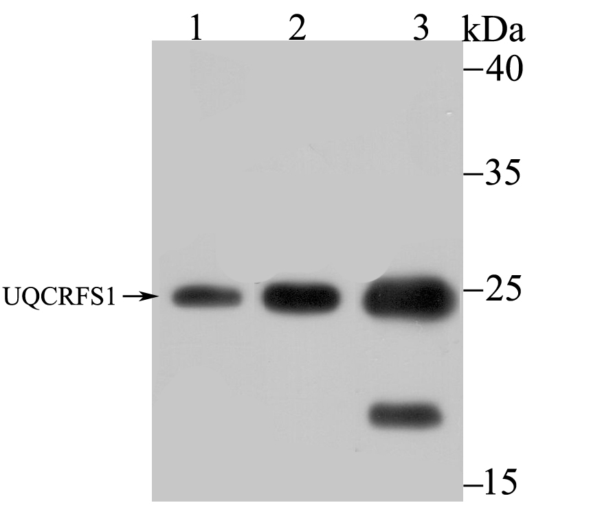
Fig1: Western blot analysis of UQCRFS1 on different lysates using anti-UQCRFS1 antibody at 1/1,000 dilution. Positive control: Lane 1: A549 Lane 2: 293 Lane 3: Mouse kidneyApplication
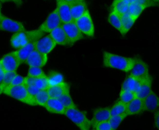
Fig2: ICC staining UQCRFS1 in 293T cells (green). The nuclear counter stain is DAPI (blue). Cells were fixed in paraformaldehyde, permeabilised with 0.25% Triton X100/PBS.Application
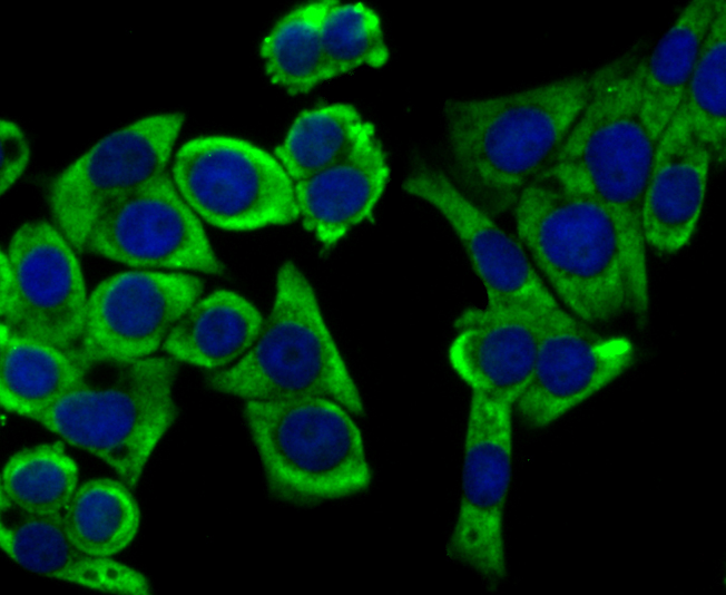
Fig3: ICC staining UQCRFS1 in LOVO cells (green). The nuclear counter stain is DAPI (blue). Cells were fixed in paraformaldehyde, permeabilised with 0.25% Triton X100/PBS.Application
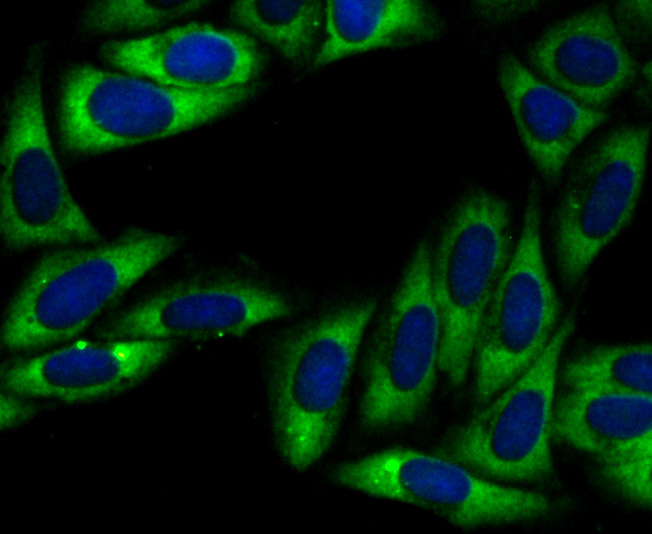
Fig4: ICC staining UQCRFS1 in SiHa cells (green). The nuclear counter stain is DAPI (blue). Cells were fixed in paraformaldehyde, permeabilised with 0.25% Triton X100/PBS.Application
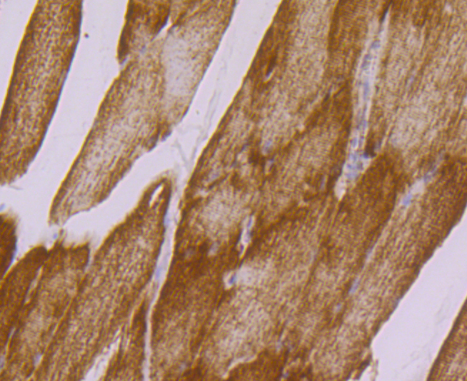
Fig5: Immunohistochemical analysis of paraffin-embedded rat skeletal muscle tissue using anti-UQCRFS1 antibody. Counter stained with hematoxylin.Application
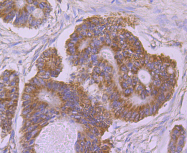
Fig6: Immunohistochemical analysis of paraffin-embedded human colon cancer tissue using anti-UQCRFS1 antibody. Counter stained with hematoxylin.Application
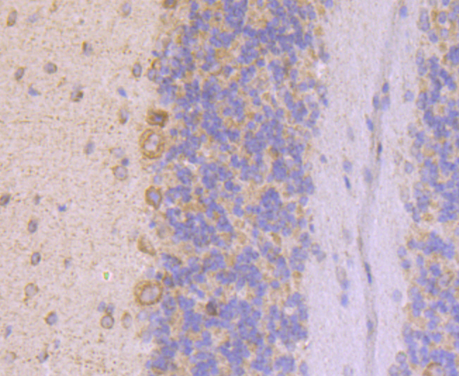
Fig7: Immunohistochemical analysis of paraffin-embedded mouse cerebellum tissue using anti-UQCRFS1 antibody. Counter stained with hematoxylin.Application
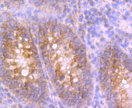
Fig8: Immunohistochemical analysis of paraffin-embedded human appendix tissue using anti-UQCRFS1 antibody. Counter stained with hematoxylin.Application
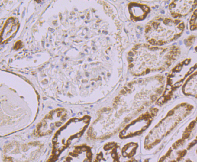
Fig9: Immunohistochemical analysis of paraffin-embedded human kidney tissue using anti-UQCRFS1 antibody. Counter stained with hematoxylin.Application
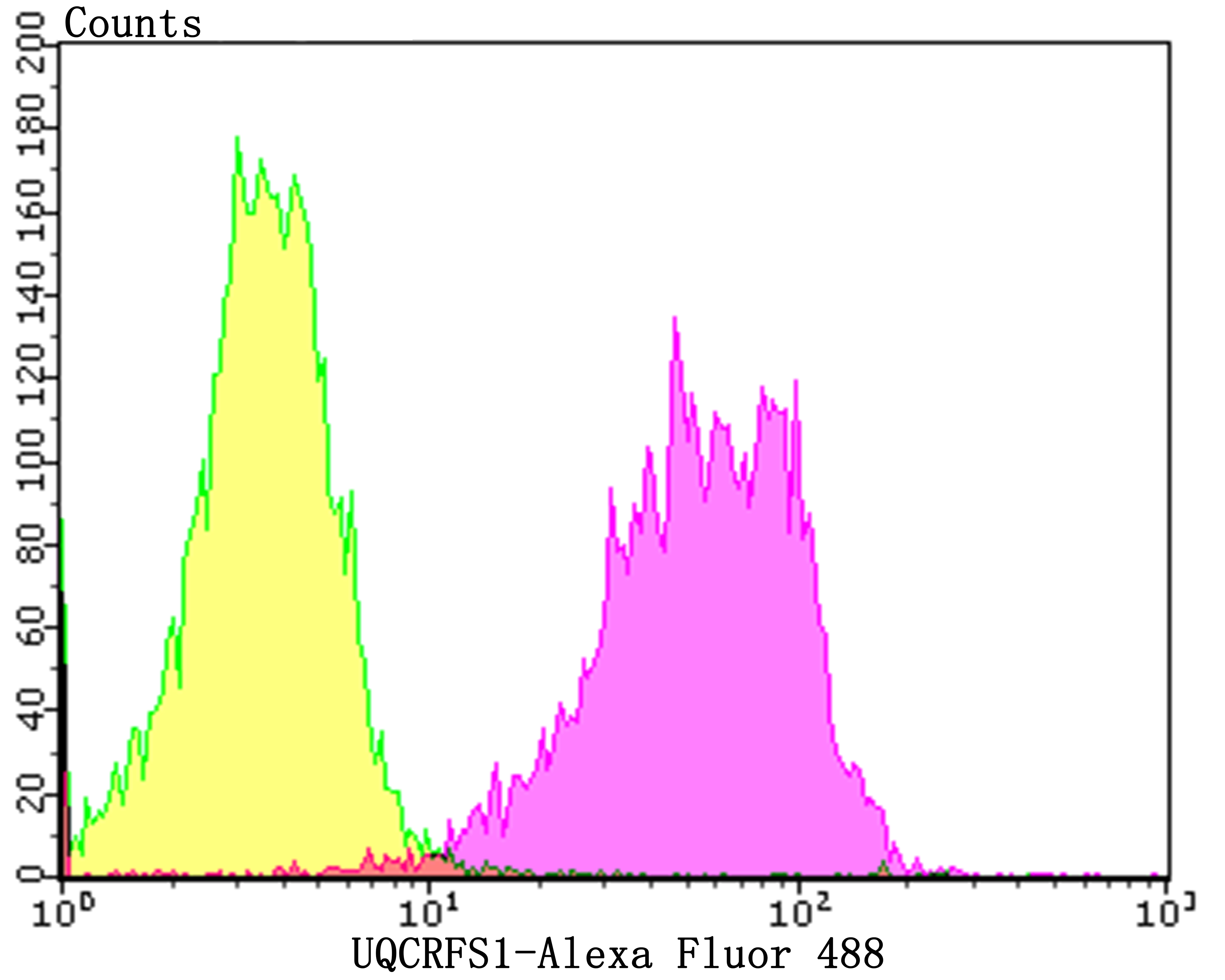
Fig10: Flow cytometric analysis of A549 cells with UQCRFS1 antibody at 1/100 dilution (yellow) compared with an unlabelled control (cells without incubation with primary antibody; purple).Alexa Fluor 488-conjugated goat anti-rabbit IgG was used as the secondary antibody.| Product Name | UQCRFS1 [JG62-31] |
|---|---|
| Antibody Type | Primary Antibodies |
| Antigen Alias | Complex III subunit 5 antibody Complex III subunit IX antibody Cytochrome b c1 complex subunit Rieske mitochondrial antibody Cytochrome b-c1 complex subunit 11 antibody Cytochrome b-c1 complex subunit 5 antibody Cytochrome b6f complex iron sulfur subunit, chloroplastic antibody petC antibody PGR1 antibody Plastohydroquinone:plastocyanin oxidoreductase iron sulfur protein antibody Proton gradient regulation protein 1 antibody Rieske iron sulfur protein antibody Rieske iron-sulfur protein antibody RIP1 antibody RIS1 antibody RISP antibody Ubiquinol cytochrome C reductase rieske iron sulphur antibody Ubiquinol cytochrome c reductase Rieske iron-sulfur polypeptide 1 antibody Ubiquinol-cytochrome c reductase 8 kDa protein antibody Ubiquinol-cytochrome c reductase iron-sulfur subunit antibody UCRI_HUMAN antibody UQCR5 antibody UQCRFS1 antibody |
| Product description | Cytochrome b-c1 complex subunit Rieske, mitochondrial: Component of the mitochondrial ubiquinol-cytochrome c reductase complex dimer (complex III dimer), which is a respiratory chain that generates an electrochemical potential coupled to ATP synthesis. Incorporation of UQCRFS1 is the penultimate step in complex III assembly. |
| Immunogen | Recombinant protein within human UQCRFS1 aa 100 to the C-terminus. |
| Clonality | Monoclonal |
|---|---|
| Isotype | IgG |
| Host Species | Recombinant rabbit |
| Tested Applications | WBICCIFIHCFCIP |
| WB:1:500-1:2,000 ICC:1:500-1:2,000 IHC:1:50-1:200 FC:1:50-1:100 FC:1:10-1:50 |
|
| Species Reactivity | HumanMouseRat |
| Concentration | 1mg/ml |
| Alternative Names | Complex III subunit 5 antibody
Complex III subunit IX antibody
Cytochrome b c1 complex subunit Rieske mitochondrial antibody
Cytochrome b-c1 complex subunit 11 antibody
Cytochrome b-c1 complex subunit 5 antibody
Cytochrome b6f complex iron sulfur subunit chloroplastic antibody petC antibody PGR1 antibody Plastohydroquinone:plastocyanin oxidoreductase iron sulfur protein antibody Proton gradient regulation protein 1 antibody Rieske iron sulfur protein antibody Rieske iron-sulfur protein antibody RIP1 antibody RIS1 antibody RISP antibody Ubiquinol cytochrome C reductase rieske iron sulphur antibody Ubiquinol cytochrome c reductase Rieske iron-sulfur polypeptide 1 antibody Ubiquinol-cytochrome c reductase 8 kDa protein antibody Ubiquinol-cytochrome c reductase iron-sulfur subunit antibody UCRI_HUMAN antibody UQCR5 antibody UQCRFS1 antibody |
|---|---|
| Molecular Weight(MW) | 21 kDa |
| Cellular Localization | Mitochondrion. |
Application
Fig1: Western blot analysis of UQCRFS1 on different lysates using anti-UQCRFS1 antibody at 1/1,000 dilution. Positive control: Lane 1: A549 Lane 2: 293 Lane 3: Mouse kidney
Application
Fig2: ICC staining UQCRFS1 in 293T cells (green). The nuclear counter stain is DAPI (blue). Cells were fixed in paraformaldehyde, permeabilised with 0.25% Triton X100/PBS.
Application
Fig3: ICC staining UQCRFS1 in LOVO cells (green). The nuclear counter stain is DAPI (blue). Cells were fixed in paraformaldehyde, permeabilised with 0.25% Triton X100/PBS.
Application
Fig4: ICC staining UQCRFS1 in SiHa cells (green). The nuclear counter stain is DAPI (blue). Cells were fixed in paraformaldehyde, permeabilised with 0.25% Triton X100/PBS.
Application
Fig5: Immunohistochemical analysis of paraffin-embedded rat skeletal muscle tissue using anti-UQCRFS1 antibody. Counter stained with hematoxylin.
Application
Fig6: Immunohistochemical analysis of paraffin-embedded human colon cancer tissue using anti-UQCRFS1 antibody. Counter stained with hematoxylin.
Application
Fig7: Immunohistochemical analysis of paraffin-embedded mouse cerebellum tissue using anti-UQCRFS1 antibody. Counter stained with hematoxylin.
Application
Fig8: Immunohistochemical analysis of paraffin-embedded human appendix tissue using anti-UQCRFS1 antibody. Counter stained with hematoxylin.
Application
Fig9: Immunohistochemical analysis of paraffin-embedded human kidney tissue using anti-UQCRFS1 antibody. Counter stained with hematoxylin.
Application
Fig10: Flow cytometric analysis of A549 cells with UQCRFS1 antibody at 1/100 dilution (yellow) compared with an unlabelled control (cells without incubation with primary antibody; purple).Alexa Fluor 488-conjugated goat anti-rabbit IgG was used as the secondary antibody.| Positive Control | A549, 293, mouse kidney tissue lysate, 293T, LOVO, SiHa, rat skeletal muscle tissue, human colon cancer tissue, human appendix tissue, human kidney tissue, mouse cerebellum tissue. |
|---|---|
| Application Notes | WB:1:500-1:2,000 ICC:1:500-1:2,000 IHC:1:50-1:200 FC:1:50-1:100 FC:1:10-1:50 |
| Form | Liquid |
|---|---|
| Storage Instructions | Store at +4℃ after thawing. Aliquot store at -20℃. Avoid repeated freeze / thaw cycles. |
| Storage Buffer | 1*TBS (pH7.4), 1%BSA, 40%Glycerol. Preservative: 0.05% Sodium Azide. |


