Application
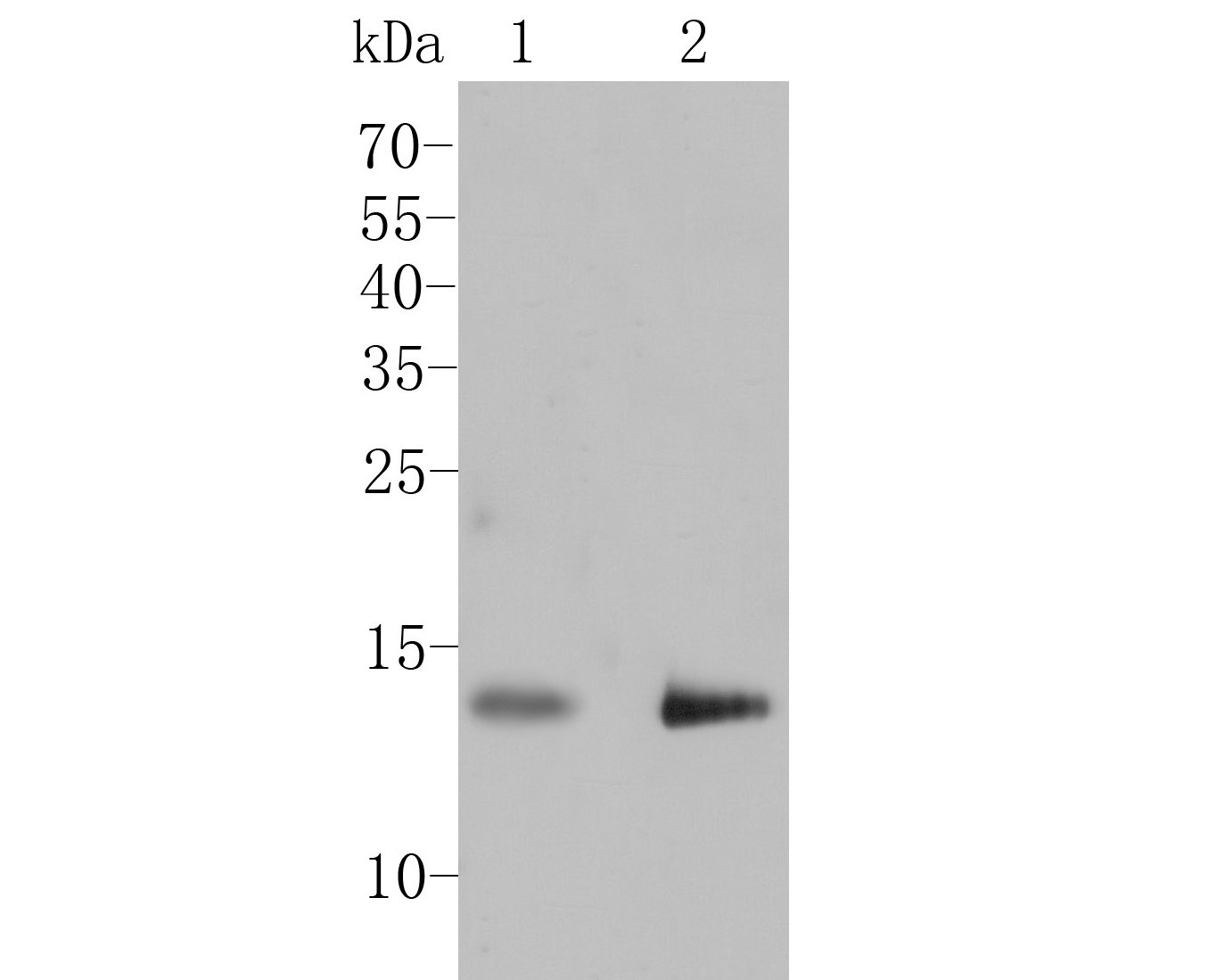
Fig1: Western blot analysis of GABARAPL2 on different lysates. Proteins were transferred to a PVDF membrane and blocked with 5% BSA in PBS for 1 hour at room temperature. The primary antibody (ER1901-73, 1/100) was used in 5% BSA at room temperature for 2 hours. Goat Anti-Rabbit IgG - HRP Secondary Antibody (HA1001) at 1:5,000 dilution was used for 1 hour at room temperature. Positive control: Lane 1: 293 cell lysate Lane 2: SHSY5Y cell lysateApplication
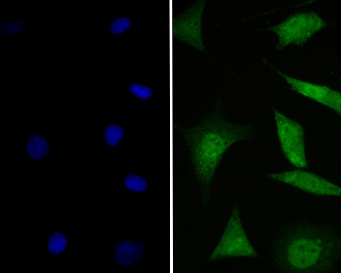
Fig2: ICC staining of GABARAPL2 in SHSY5Y cells (green). Formalin fixed cells were permeabilized with 0.1% Triton X-100 in TBS for 10 minutes at room temperature and blocked with 1% Blocker BSA for 15 minutes at room temperature. Cells were probed with the primary antibody (ER1901-73, 1/100) for 1 hour at room temperature, washed with PBS. Alexa Fluor®488 Goat anti-Rabbit IgG was used as the secondary antibody at 1/100 dilution. The nuclear counter stain is DAPI (blue).Application
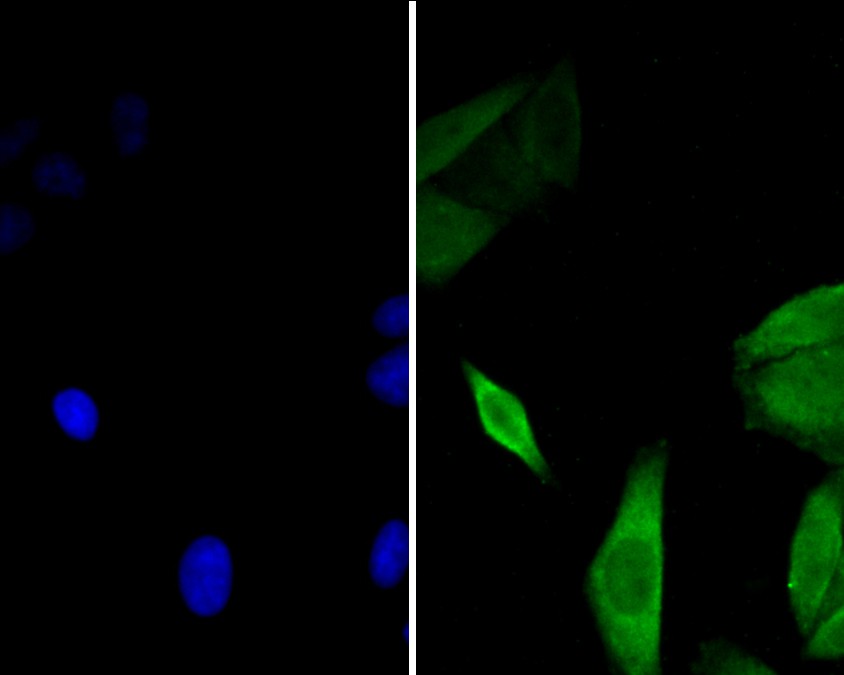
Fig3: ICC staining of GABARAPL2 in Siha cells (green). Formalin fixed cells were permeabilized with 0.1% Triton X-100 in TBS for 10 minutes at room temperature and blocked with 1% Blocker BSA for 15 minutes at room temperature. Cells were probed with the primary antibody (ER1901-73, 1/100) for 1 hour at room temperature, washed with PBS. Alexa Fluor®488 Goat anti-Rabbit IgG was used as the secondary antibody at 1/100 dilution. The nuclear counter stain is DAPI (blue).Application
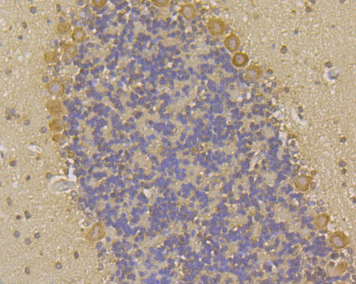
Fig4: Immunohistochemical analysis of paraffin-embedded Rat cerebellum tissue using anti-GABARAPL2 antibody. The section was pre-treated using heat mediated antigen retrieval with Tris-EDTA buffer (pH 8.0-8.4) for 20 minutes.The tissues were blocked in 5% BSA for 30 minutes at room temperature, washed with ddH2O and PBS, and then probed with the primary antibody (ER1901-73, 1/100) for 30 minutes at room temperature. The detection was performed using an HRP conjugated compact polymer system. DAB was used as the chromogen. Tissues were counterstained with hematoxylin and mounted with DPX.Application
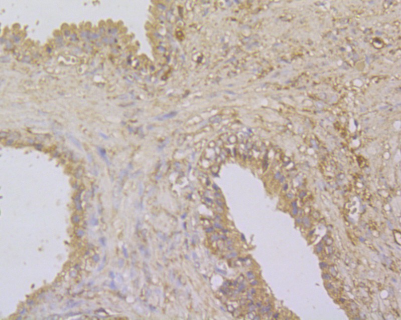
Fig5: Immunohistochemical analysis of paraffin-embedded Human prostate tissue using anti-GABARAPL2 antibody. The section was pre-treated using heat mediated antigen retrieval with Tris-EDTA buffer (pH 8.0-8.4) for 20 minutes.The tissues were blocked in 5% BSA for 30 minutes at room temperature, washed with ddH2O and PBS, and then probed with the primary antibody (ER1901-73, 1/100) for 30 minutes at room temperature. The detection was performed using an HRP conjugated compact polymer system. DAB was used as the chromogen. Tissues were counterstained with hematoxylin and mounted with DPX.Application
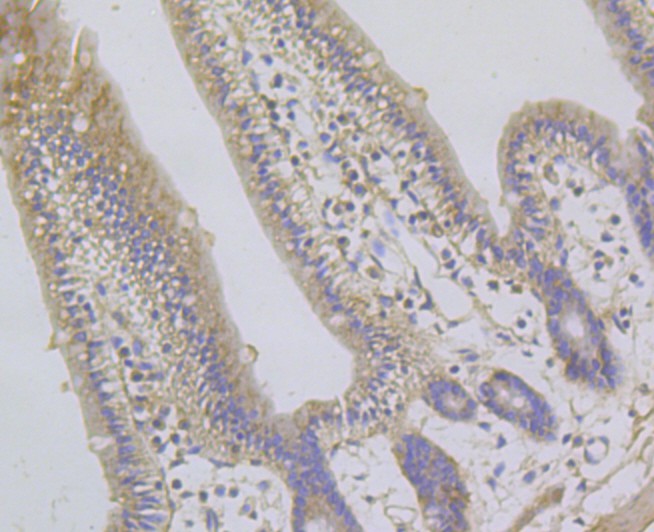
Fig6: Immunohistochemical analysis of paraffin-embedded Mouse small intestine tissue using anti-GABARAPL2 antibody. The section was pre-treated using heat mediated antigen retrieval with Tris-EDTA buffer (pH 8.0-8.4) for 20 minutes.The tissues were blocked in 5% BSA for 30 minutes at room temperature, washed with ddH2O and PBS, and then probed with the primary antibody (ER1901-73, 1/100) for 30 minutes at room temperature. The detection was performed using an HRP conjugated compact polymer system. DAB was used as the chromogen. Tissues were counterstained with hematoxylin and mounted with DPX.Application
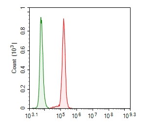
Fig7: Flow cytometric analysis of GABARAPL2 was done on SHSY5Y cells. The cells were fixed, permeabilized and stained with the primary antibody (ER1901-73, 1/100) (red). After incubation of the primary antibody at room temperature for an hour, the cells were stained with a Alexa Fluor 488-conjugated goat anti-rabbit IgG Secondary antibody at 1/500 dilution for 30 minutes.Unlabelled sample was used as a control (cells without incubation with primary antibody; green).| Product Name | GABARAPL2 |
|---|---|
| Antibody Type | Primary Antibodies |
| Antigen Alias | ATG8 antibody ATG8C antibody FLC3A antibody GABA(A) receptor-associated protein-like 2 antibody Gabarapl2 antibody Gamma-aminobutyric acid receptor-associated protein-like 2 antibody Ganglioside expression factor 2 antibody GATE-16 antibody GATE16 antibody GBRL2_HUMAN antibody GEF-2 antibody GEF2 antibody General protein transport factor p16 antibody Golgi-associated ATPase enhancer of 16 kDa antibody MAP1 light chain 3-related protein antibody |
| Product description | Ubiquitin-like modifier involved in intra-Golgi traffic. Modulates intra-Golgi transport through coupling between NSF activity and SNAREs activation. It first stimulates the ATPase activity of NSF which in turn stimulates the association with GOSR1 (By similarity). Involved in autophagy. Plays a role in mitophagy which contributes to regulate mitochondrial quantity and quality by eliminating the mitochondria to a basal level to fulfill cellular energy requirements and preventing excess ROS production. Whereas LC3s are involved in elongation of the phagophore membrane, the GABARAP/GATE-16 subfamily is essential for a later stage in autophagosome maturation. |
| Immunogen | Recombinant protein within human GABARAPL2 aa 1-117. |
| Clonality | Polyclonal |
|---|---|
| Isotype | IgG |
| Host Species | Rabbit |
| Tested Applications | WBIHCICCFC |
| WB:1:500-1:2000 IHC:1:50-1:200 ICC:1:50-1:200 FC:1:50-1:100 |
|
| Species Reactivity | HumanMouseRat |
| Concentration | 1mg/ml |
| Alternative Names | ATG8 antibody
ATG8C antibody
FLC3A antibody GABA(A) receptor-associated protein-like 2 antibody Gabarapl2 antibody Gamma-aminobutyric acid receptor-associated protein-like 2 antibody Ganglioside expression factor 2 antibody GATE-16 antibody GATE16 antibody GBRL2_HUMAN antibody GEF-2 antibody GEF2 antibody General protein transport factor p16 antibody Golgi-associated ATPase enhancer of 16 kDa antibody MAP1 light chain 3-related protein antibody |
|---|---|
| Molecular Weight(MW) | 14 kDa |
| Cellular Localization | Cytoplasmic vesicle. Golgi apparatus. |
Application
Fig1: Western blot analysis of GABARAPL2 on different lysates. Proteins were transferred to a PVDF membrane and blocked with 5% BSA in PBS for 1 hour at room temperature. The primary antibody (ER1901-73, 1/100) was used in 5% BSA at room temperature for 2 hours. Goat Anti-Rabbit IgG - HRP Secondary Antibody (HA1001) at 1:5,000 dilution was used for 1 hour at room temperature. Positive control: Lane 1: 293 cell lysate Lane 2: SHSY5Y cell lysate
Application
Fig2: ICC staining of GABARAPL2 in SHSY5Y cells (green). Formalin fixed cells were permeabilized with 0.1% Triton X-100 in TBS for 10 minutes at room temperature and blocked with 1% Blocker BSA for 15 minutes at room temperature. Cells were probed with the primary antibody (ER1901-73, 1/100) for 1 hour at room temperature, washed with PBS. Alexa Fluor®488 Goat anti-Rabbit IgG was used as the secondary antibody at 1/100 dilution. The nuclear counter stain is DAPI (blue).
Application
Fig3: ICC staining of GABARAPL2 in Siha cells (green). Formalin fixed cells were permeabilized with 0.1% Triton X-100 in TBS for 10 minutes at room temperature and blocked with 1% Blocker BSA for 15 minutes at room temperature. Cells were probed with the primary antibody (ER1901-73, 1/100) for 1 hour at room temperature, washed with PBS. Alexa Fluor®488 Goat anti-Rabbit IgG was used as the secondary antibody at 1/100 dilution. The nuclear counter stain is DAPI (blue).
Application
Fig4: Immunohistochemical analysis of paraffin-embedded Rat cerebellum tissue using anti-GABARAPL2 antibody. The section was pre-treated using heat mediated antigen retrieval with Tris-EDTA buffer (pH 8.0-8.4) for 20 minutes.The tissues were blocked in 5% BSA for 30 minutes at room temperature, washed with ddH2O and PBS, and then probed with the primary antibody (ER1901-73, 1/100) for 30 minutes at room temperature. The detection was performed using an HRP conjugated compact polymer system. DAB was used as the chromogen. Tissues were counterstained with hematoxylin and mounted with DPX.
Application
Fig5: Immunohistochemical analysis of paraffin-embedded Human prostate tissue using anti-GABARAPL2 antibody. The section was pre-treated using heat mediated antigen retrieval with Tris-EDTA buffer (pH 8.0-8.4) for 20 minutes.The tissues were blocked in 5% BSA for 30 minutes at room temperature, washed with ddH2O and PBS, and then probed with the primary antibody (ER1901-73, 1/100) for 30 minutes at room temperature. The detection was performed using an HRP conjugated compact polymer system. DAB was used as the chromogen. Tissues were counterstained with hematoxylin and mounted with DPX.
Application
Fig6: Immunohistochemical analysis of paraffin-embedded Mouse small intestine tissue using anti-GABARAPL2 antibody. The section was pre-treated using heat mediated antigen retrieval with Tris-EDTA buffer (pH 8.0-8.4) for 20 minutes.The tissues were blocked in 5% BSA for 30 minutes at room temperature, washed with ddH2O and PBS, and then probed with the primary antibody (ER1901-73, 1/100) for 30 minutes at room temperature. The detection was performed using an HRP conjugated compact polymer system. DAB was used as the chromogen. Tissues were counterstained with hematoxylin and mounted with DPX.
Application
Fig7: Flow cytometric analysis of GABARAPL2 was done on SHSY5Y cells. The cells were fixed, permeabilized and stained with the primary antibody (ER1901-73, 1/100) (red). After incubation of the primary antibody at room temperature for an hour, the cells were stained with a Alexa Fluor 488-conjugated goat anti-rabbit IgG Secondary antibody at 1/500 dilution for 30 minutes.Unlabelled sample was used as a control (cells without incubation with primary antibody; green).| Positive Control | 293 cell lysate, SHSY5Y cell lysate, SHSY5Y, Siha, Rat cerebellum tissue, Human prostate tissue, Mouse small intestine tissue. |
|---|---|
| Application Notes | WB:1:500-1:2000 IHC:1:50-1:200 ICC:1:50-1:200 FC:1:50-1:100 |
| Form | Liquid |
|---|---|
| Storage Instructions | Store at +4℃ after thawing. Aliquot store at -20℃. Avoid repeated freeze / thaw cycles. |
| Storage Buffer | 1*TBS (pH7.4), 1%BSA, 50%Glycerol. Preservative: 0.05% Sodium Azide. |


