Application
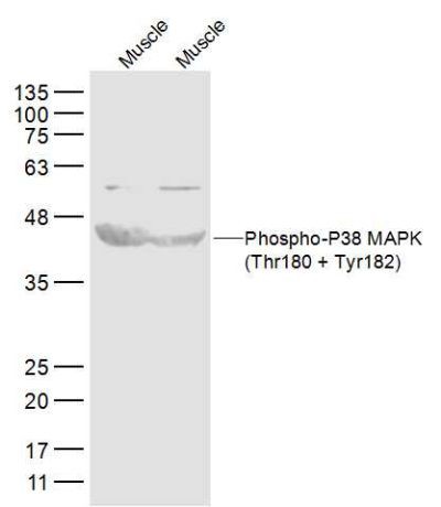
Fig1: Western blot analysis of Phospho-P38 MAPK (Thr180 + Tyr182) on different tissue lysates using anti-Phospho-P38 MAPK (Thr180 + Tyr182) at 1/300 dilution. Positive control: Line 1: Mouse muscle tissue Line 2: Rat muscle tissueApplication
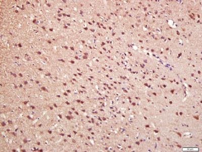
Fig2: Immunohistochemical analysis of paraffin-embedded rat brain tissue using anti-Phospho-P38 MAPK (Thr180 + Tyr182) antibody. Counter stained with hematoxylin.Application
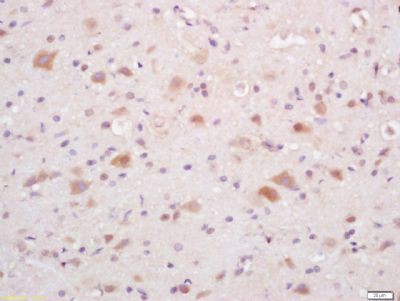
Fig3: Immunohistochemical analysis of paraffin-embedded mouse brain tissue using anti-Phospho-P38 MAPK (Thr180 + Tyr182) antibody. Counter stained with hematoxylin.Application
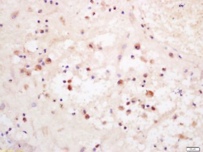
Fig4: Immunohistochemical analysis of paraffin-embedded human placenta tissue using anti-Phospho-P38 MAPK (Thr180 + Tyr182) antibody. Counter stained with hematoxylin.Application
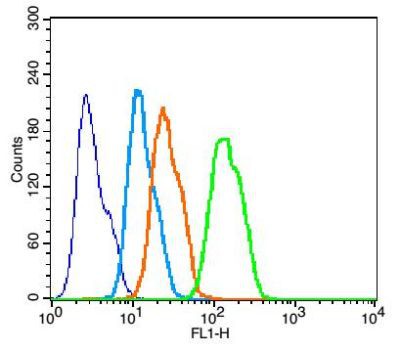
Fig5: Flow cytometric analysis of HepG2 cells with Phospho-P38 MAPK (Thr180 + Tyr182) antibody at 1/50 dilution (green) compared with blank control (blue) and isotype control (orange). Goat anti-rabbit IgG-FITC was used as the secondary antibody (white blue).| Product Name | Phospho-P38 MAPK (Thr180 + Tyr182) |
|---|---|
| Antibody Type | Primary Antibodies |
| Product description | The protein encoded by this gene is a member of the MAP kinase family. MAP kinases act as an integration point for multiple biochemical signals, and are involved in a wide variety of cellular processes such as proliferation, differentiation, transcription regulation and development. This kinase is activated by various environmental stresses and proinflammatory cytokines. The activation requires its phosphorylation by MAP kinase kinases(MKKs), or its autophosphorylation triggered by the interaction of MAP3K7IP1/TAB1 protein with this kinase. The substrates of this kinase include transcription regulator ATF2, MEF2C, and MAX, cell cycle regulator CDC25B, and tumor suppressor p53, which suggest the roles of this kinase in stress related transcription and cell cycle regulation, as well as in genotoxic stress response. Four alternatively spliced transcript variants of this gene encoding distinct isoforms have been reported. |
| Immunogen | KLH conjugated Synthesised phosphopeptide derived from human p38 MAPK around the phosphorylation site of Thr180/Tyr182: M(p-T)G(p-Y)VA. |
| Modification | p-Thr180 + Tyr182 |
| Clonality | Polyclonal |
|---|---|
| Isotype | IgG |
| Host Species | Rabbit |
| Tested Applications | WBIHCFC |
| ELISA:: WB:1:500-1:2,000 IP:: ICC:: IF:: IHC:1:400-1:800 FC:1:50 |
|
| Species Reactivity | HumanMouseRat |
| Concentration | 1mg/ml |
| Molecular Weight(MW) | 42 kDa |
|---|---|
| Cellular Localization | Cytoplasm. Nucleus. |
Application
Fig1: Western blot analysis of Phospho-P38 MAPK (Thr180 + Tyr182) on different tissue lysates using anti-Phospho-P38 MAPK (Thr180 + Tyr182) at 1/300 dilution. Positive control: Line 1: Mouse muscle tissue Line 2: Rat muscle tissue
Application
Fig2: Immunohistochemical analysis of paraffin-embedded rat brain tissue using anti-Phospho-P38 MAPK (Thr180 + Tyr182) antibody. Counter stained with hematoxylin.
Application
Fig3: Immunohistochemical analysis of paraffin-embedded mouse brain tissue using anti-Phospho-P38 MAPK (Thr180 + Tyr182) antibody. Counter stained with hematoxylin.
Application
Fig4: Immunohistochemical analysis of paraffin-embedded human placenta tissue using anti-Phospho-P38 MAPK (Thr180 + Tyr182) antibody. Counter stained with hematoxylin.
Application
Fig5: Flow cytometric analysis of HepG2 cells with Phospho-P38 MAPK (Thr180 + Tyr182) antibody at 1/50 dilution (green) compared with blank control (blue) and isotype control (orange). Goat anti-rabbit IgG-FITC was used as the secondary antibody (white blue).| Positive Control | Mouse muscle tissue, rat muscle tissue, rat brain tissue, mouse brain tissue, human placenta tissue, HepG2. |
|---|---|
| Application Notes | ELISA:: WB:1:500-1:2,000 IP:: ICC:: IF:: IHC:1:400-1:800 FC:1:50 |
| Form | Liquid |
|---|---|
| Storage Instructions | Store at -20 |
| Storage Buffer | 0.01M TBS(pH7.4) with 1% BSA, 0.03% Proclin300 and 50% Glycerol. |


