Application
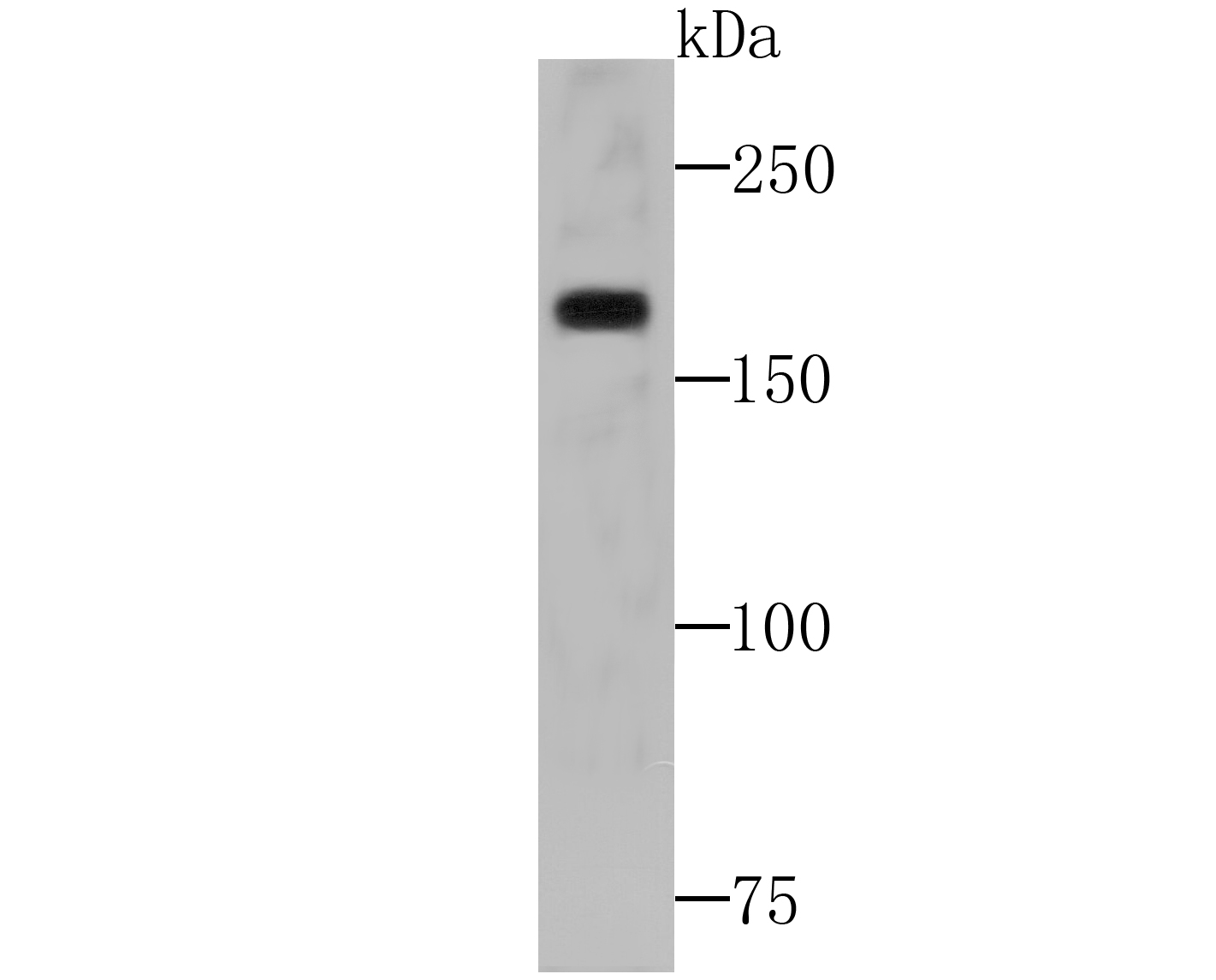
Fig1: Western blot analysis of WSTF on SiHa cell lysates using anti-WSTF at 1/500 dilution.Application
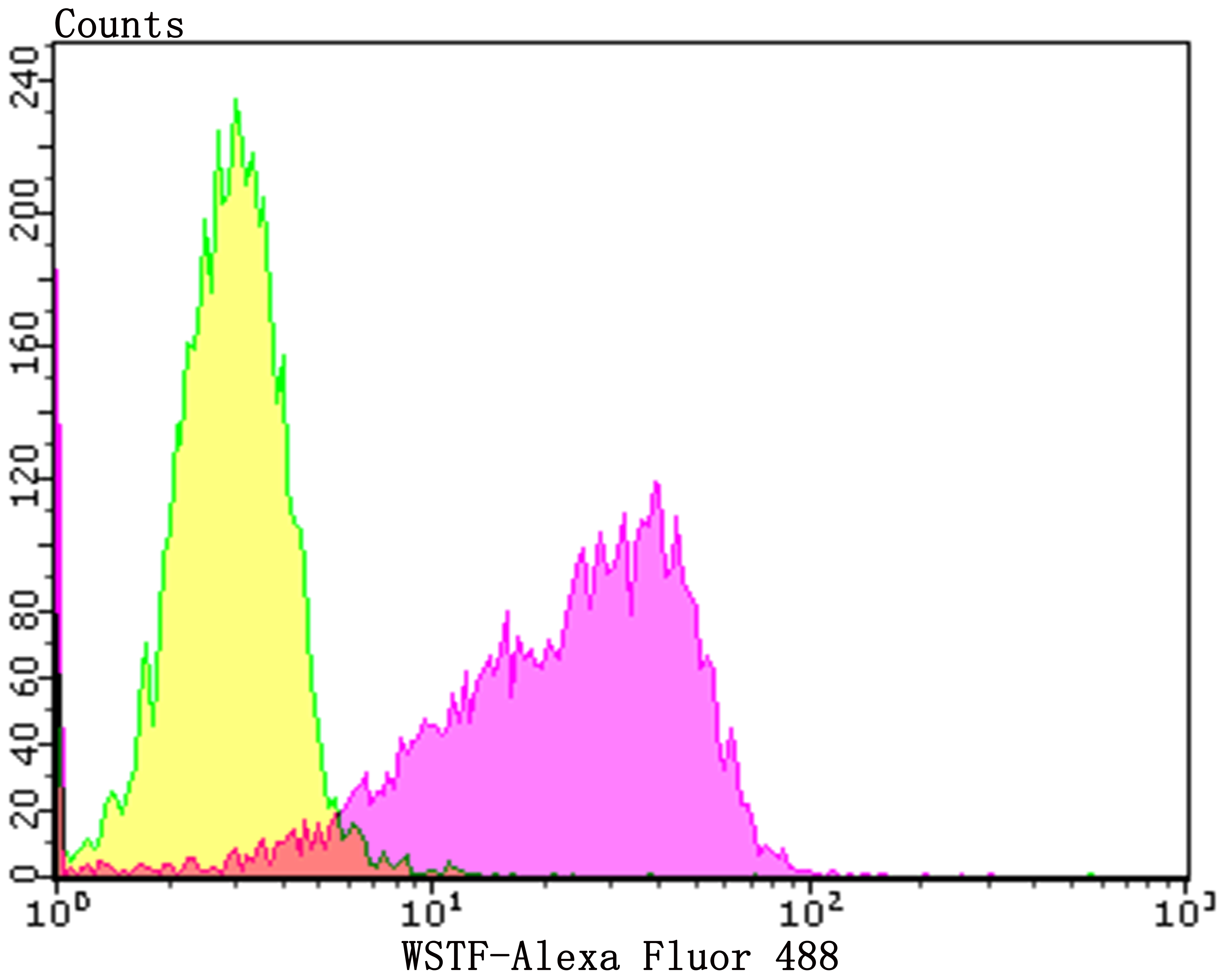
Fig2: Flow cytometric analysis of K562 cells with WSTF antibody at 1/50 dilution (Fuchsia) compared with an unlabelled control (cells without incubation with primary antibody; Yellow). Alexa Fluor 488-conjugated goat anti-rabbit IgG was used as the secondary antibody.| Product Name | WSTF [JG36-32] |
|---|---|
| Antibody Type | Primary Antibodies |
| Antigen Alias | baz1b antibody BAZ1B_HUMAN antibody Bromodomain adjacent to zinc finger domain protein 1B antibody hWALP 2 antibody hWALP-2 antibody hWALP2 antibody transcription factor WSTF antibody Tyrosine-protein kinase BAZ1B antibody WALP-2 antibody WALP2 antibody WBRS 9 antibody WBRS-9 antibody WBRS9 antibody WBSC 10 antibody WBSC-10 antibody WBSC10 antibody WBSCR10 antibody WBSCR9 antibody Williams Beuren syndrome chromosome region 9 protein antibody Williams syndrome transcription factor antibody Williams-Beuren syndrome chromosomal region 10 protein antibody Williams-Beuren syndrome chromosomal region 9 protein antibody WSTF antibody |
| Product description | Atypical tyrosine-protein kinase that plays a central role in chromatin remodeling and acts as a transcription regulator. Involved in DNA damage response by phosphorylating 'Tyr-142' of histone H2AX (H2AXY142ph). H2AXY142ph plays a central role in DNA repair and acts as a mark that distinguishes between apoptotic and repair responses to genotoxic stress. Essential component of the WICH complex, a chromatin remodeling complex that mobilizes nucleosomes and reconfigures irregular chromatin to a regular nucleosomal array structure. The WICH complex regulates the transcription of various genes, has a role in RNA polymerase I and RNA polymerase III transcription, mediates the histone H2AX phosphorylation at 'Tyr-142', and is involved in the maintenance of chromatin structures during DNA replication processes. In the complex, it mediates the recruitment of the WICH complex to replication foci during DNA replication. |
| Immunogen | Recombinant protein within C-terminal human WSTF. |
| Clonality | Monoclonal |
|---|---|
| Isotype | IgG |
| Host Species | Recombinant rabbit |
| Tested Applications | WBFC |
| WB:1:500-1:1,000 FC:1:50-1:100 |
|
| Species Reactivity | HumanMouseRat |
| Concentration | 1mg/ml |
| Alternative Names | baz1b antibody BAZ1B_HUMAN antibody Bromodomain adjacent to zinc finger domain protein 1B antibody hWALP 2 antibody hWALP-2 antibody hWALP2 antibody transcription factor WSTF antibody Tyrosine-protein kinase BAZ1B antibody WALP-2 antibody WALP2 antibody WBRS 9 antibody WBRS-9 antibody WBRS9 antibody WBSC 10 antibody WBSC-10 antibody WBSC10 antibody WBSCR10 antibody WBSCR9 antibody Williams Beuren syndrome chromosome region 9 protein antibody Williams syndrome transcription factor antibody Williams-Beuren syndrome chromosomal region 10 protein antibody Williams-Beuren syndrome chromosomal region 9 protein antibody WSTF antibody |
|---|---|
| Molecular Weight(MW) | 171 kDa |
| Cellular Localization | Nucleus. |
| SwissProt ID | Q9UIG0 |
|---|
Application
Fig1: Western blot analysis of WSTF on SiHa cell lysates using anti-WSTF at 1/500 dilution.
Application
Fig2: Flow cytometric analysis of K562 cells with WSTF antibody at 1/50 dilution (Fuchsia) compared with an unlabelled control (cells without incubation with primary antibody; Yellow). Alexa Fluor 488-conjugated goat anti-rabbit IgG was used as the secondary antibody.| Positive Control | SiHa, K562. |
|---|---|
| Application Notes | WB:1:500-1:1,000 FC:1:50-1:100 |
| Form | Liquid |
|---|---|
| Storage Instructions | Store at +4℃ after thawing. Aliquot store at -20℃. Avoid repeated freeze / thaw cycles. |
| Storage Buffer | 1*TBS (pH7.4), 1%BSA, 40%Glycerol. Preservative: 0.05% Sodium Azide. |


