Application
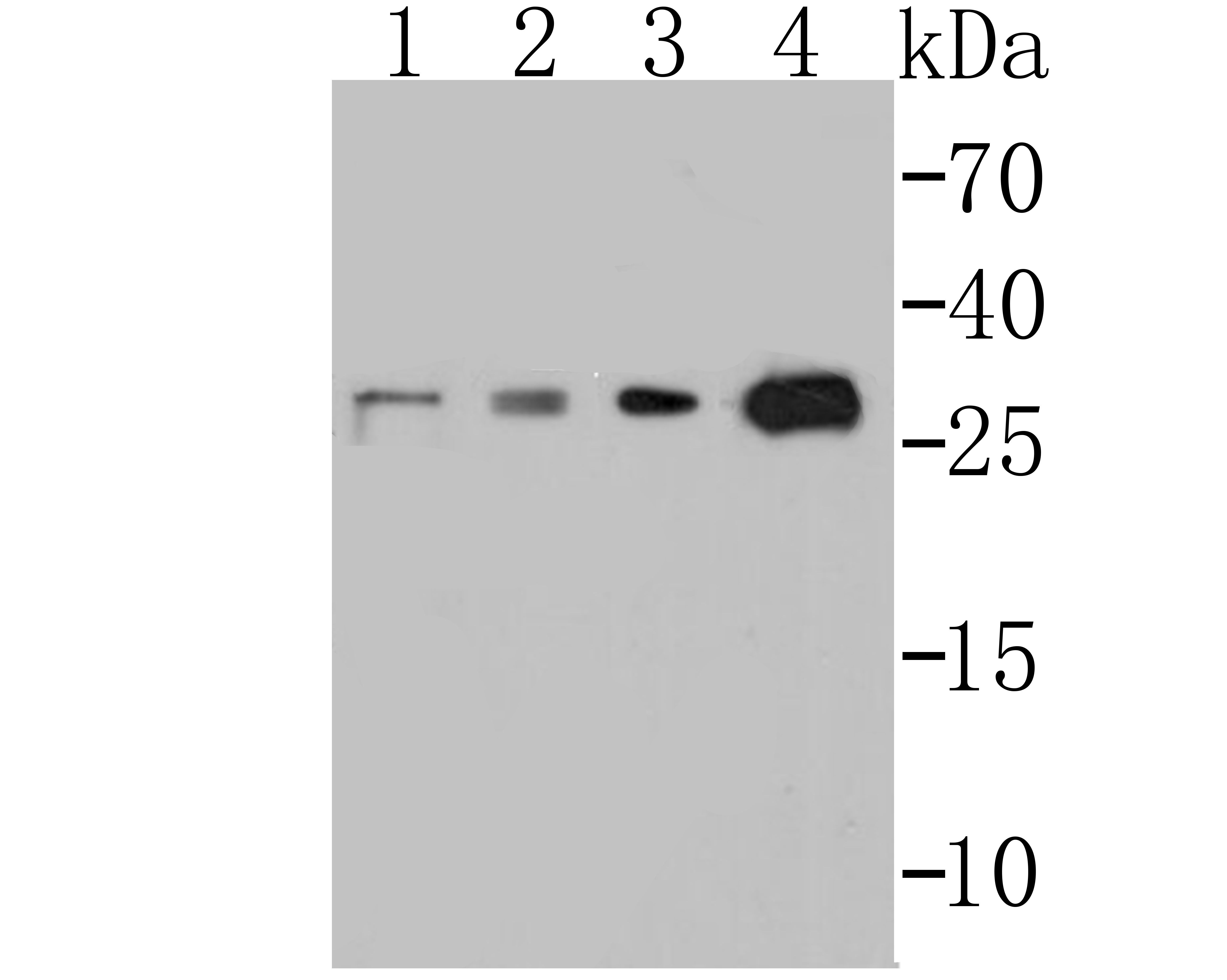
Fig1: Western blot analysis of p53DINP1 on different cell lysates using anti-p53DINP1 at 1/500 dilution. Positive control: Line 1: SH-SY-5Y Line 2: HepG2 Line 3: Mouse stomach Line 3: Human stomachApplication
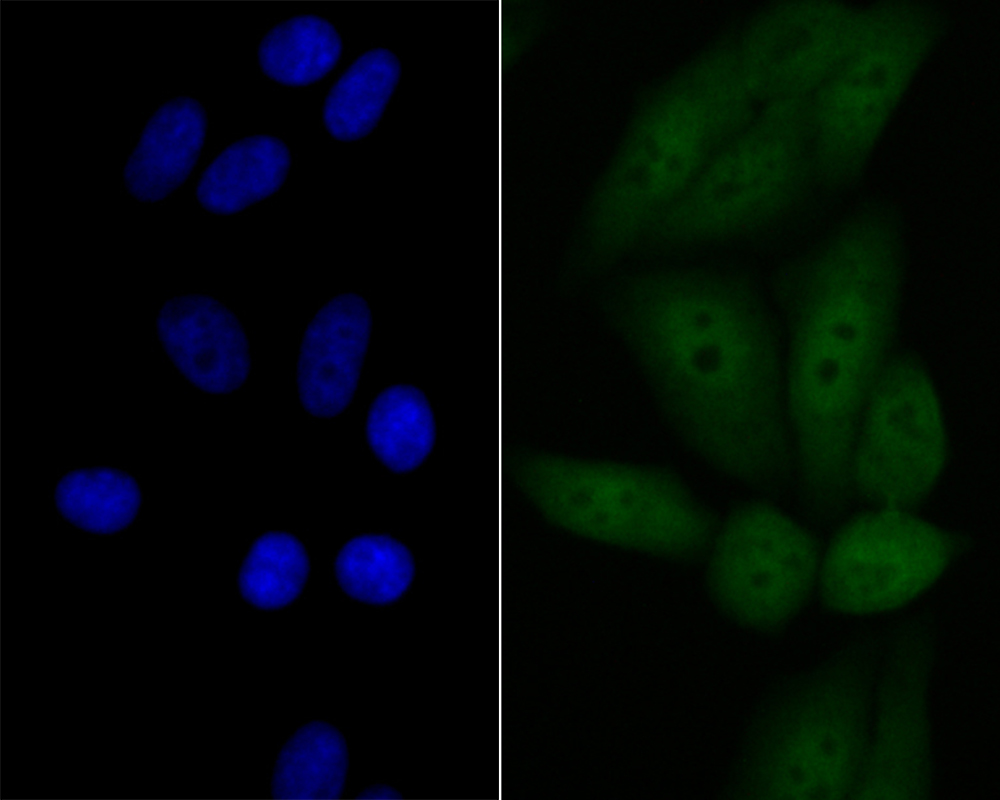
Fig2: ICC staining p53DINP1 in HepG2 cells (green). The nuclear counter stain is DAPI (blue). Cells were fixed in paraformaldehyde, permeabilised with 0.25% Triton X100/PBS.Application
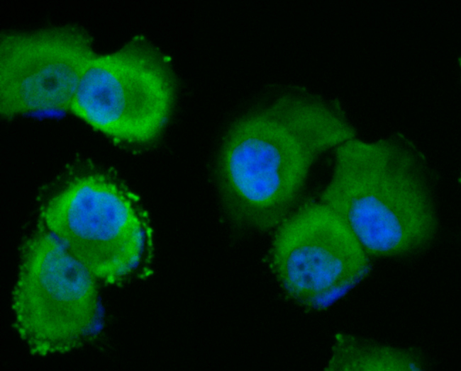
Fig3: ICC staining p53DINP1 in HUVEC cells (green). The nuclear counter stain is DAPI (blue). Cells were fixed in paraformaldehyde, permeabilised with 0.25% Triton X100/PBS.Application
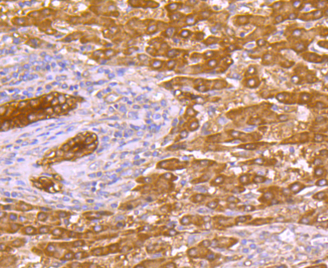
Fig4: Immunohistochemical analysis of paraffin-embedded huamn liver cancer tissue using anti-p53DINP1 antibody. Counter stained with hematoxylin.Application
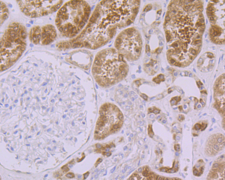
Fig5: Immunohistochemical analysis of paraffin-embedded human kidney tissue using anti-p53DINP1 antibody. Counter stained with hematoxylin.Application
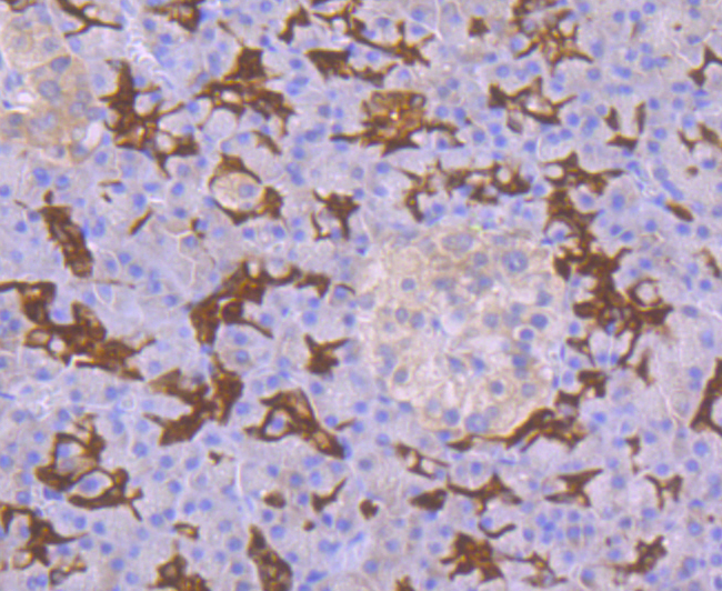
Fig6: Immunohistochemical analysis of paraffin-embedded human pancreas tissue using anti-p53DINP1 antibody. Counter stained with hematoxylin.| Product Name | p53 DINP1 [JG37-64] |
|---|---|
| Antibody Type | Primary Antibodies |
| Antigen Alias | DKFZp434M1317 antibody FLJ22139 antibody p53 damage inducible nuclear protein 1 antibody p53 dependent damage inducible nuclear protein 1 antibody p53 inducible nuclear protein 1 antibody p53 inducible p53DINP1 antibody p53-dependent damage-inducible nuclear protein 1 antibody p53DINP1 antibody SIP antibody Stress induced protein antibody Stress-induced protein antibody T53I1_HUMAN antibody Teap antibody TP53 DINP1 antibody TP53 INP1 antibody TP53DINP1 antibody TP53INP1 antibody TP53INP1A antibody TP53INP1B antibody Tumor protein p53 inducible nuclear protein 1 antibody Tumor protein p53-inducible nuclear protein 1 antibody |
| Product description | Antiproliferative and proapoptotic protein involved in cell stress response which acts as a dual regulator of transcription and autophagy. Acts as a positive regulator of autophagy. In response to cellular stress or activation of autophagy, relocates to autophagosomes where it interacts with autophagosome-associated proteins GABARAP, GABARAPL1/L2, MAP1LC3A/B/C and regulates autophagy. Acts as an antioxidant and plays a major role in p53/TP53-driven oxidative stress response. Possesses both a p53/TP53-independent intracellular reactive oxygen species (ROS) regulatory function and a p53/TP53-dependent transcription regulatory function. Positively regulates p53/TP53 and p73/TP73 and stimulates their capacity to induce apoptosis and regulate cell cycle. In response to double-strand DNA breaks, promotes p53/TP53 phosphorylation on 'Ser-46' and subsequent apoptosis. Acts as a tumor suppressor by inducing cell death by an autophagy and caspase-dependent mechanism. Can reduce cell migration by regulating the expression of SPARC. |
| Immunogen | Full length recombinant protein of human p53DINP1. |
| Clonality | Monoclonal |
|---|---|
| Isotype | IgG |
| Host Species | Recombinant rabbit |
| Tested Applications | WBICCIFIHC |
| WB:1:500-1:1,000 ICC:1:50-1:200 IHC:1:50-1:200 |
|
| Species Reactivity | HumanMouseRat |
| Concentration | 1mg/ml |
| Alternative Names | DKFZp434M1317 antibody FLJ22139 antibody p53 damage inducible nuclear protein 1 antibody p53 dependent damage inducible nuclear protein 1 antibody p53 inducible nuclear protein 1 antibody p53 inducible p53DINP1 antibody p53-dependent damage-inducible nuclear protein 1 antibody p53DINP1 antibody SIP antibody Stress induced protein antibody Stress-induced protein antibody T53I1_HUMAN antibody Teap antibody TP53 DINP1 antibody TP53 INP1 antibody TP53DINP1 antibody TP53INP1 antibody TP53INP1A antibody TP53INP1B antibody Tumor protein p53 inducible nuclear protein 1 antibody Tumor protein p53-inducible nuclear protein 1 antibody |
|---|---|
| Molecular Weight(MW) | 27 kDa |
| Cellular Localization | Cytosol. Nucleus. |
| SwissProt ID | Q96A56 |
|---|
Application
Fig1: Western blot analysis of p53DINP1 on different cell lysates using anti-p53DINP1 at 1/500 dilution. Positive control: Line 1: SH-SY-5Y Line 2: HepG2 Line 3: Mouse stomach Line 3: Human stomach
Application
Fig2: ICC staining p53DINP1 in HepG2 cells (green). The nuclear counter stain is DAPI (blue). Cells were fixed in paraformaldehyde, permeabilised with 0.25% Triton X100/PBS.
Application
Fig3: ICC staining p53DINP1 in HUVEC cells (green). The nuclear counter stain is DAPI (blue). Cells were fixed in paraformaldehyde, permeabilised with 0.25% Triton X100/PBS.
Application
Fig4: Immunohistochemical analysis of paraffin-embedded huamn liver cancer tissue using anti-p53DINP1 antibody. Counter stained with hematoxylin.
Application
Fig5: Immunohistochemical analysis of paraffin-embedded human kidney tissue using anti-p53DINP1 antibody. Counter stained with hematoxylin.
Application
Fig6: Immunohistochemical analysis of paraffin-embedded human pancreas tissue using anti-p53DINP1 antibody. Counter stained with hematoxylin.| Positive Control | SH-SY-5Y, HUVEC, HepG2, Mouse stomach tissue, Human stomach tissue, human liver cancer tissue, human kidney tissue, human pancreas tissue. |
|---|---|
| Application Notes | WB:1:500-1:1,000 ICC:1:50-1:200 IHC:1:50-1:200 |
| Form | Liquid |
|---|---|
| Storage Instructions | Store at +4℃ after thawing. Aliquot store at -20℃. Avoid repeated freeze / thaw cycles. |
| Storage Buffer | 1*TBS (pH7.4), 1%BSA, 40%Glycerol. Preservative: 0.05% Sodium Azide. |


