Application
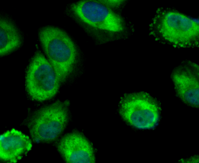
Fig1: ICC staining Mib1 in HUVEC cells (green). The nuclear counter stain is DAPI (blue). Cells were fixed in paraformaldehyde, permeabilised with 0.25% Triton X100/PBS.Application
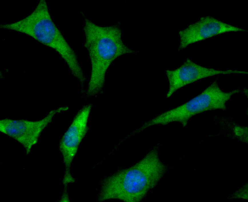
Fig2: ICC staining Mib1 in SH-SY-5Y cells (green). The nuclear counter stain is DAPI (blue). Cells were fixed in paraformaldehyde, permeabilised with 0.25% Triton X100/PBS.Application
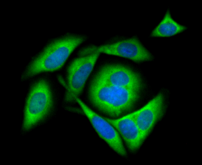
Fig3: ICC staining Mib1 in SiHa cells (green). The nuclear counter stain is DAPI (blue). Cells were fixed in paraformaldehyde, permeabilised with 0.25% Triton X100/PBS.Application
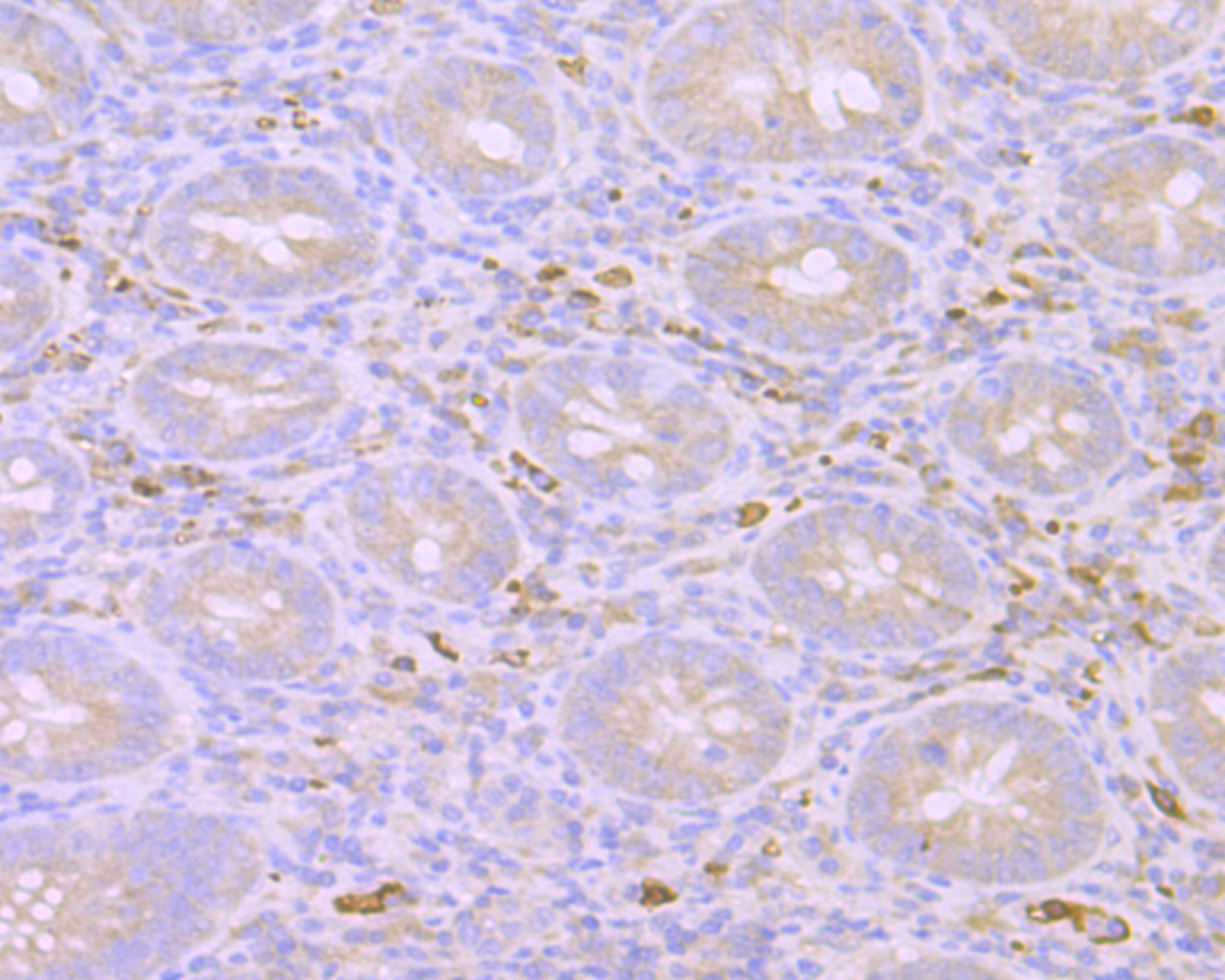
Fig4: Immunohistochemical analysis of paraffin-embedded human appendix tissue using anti-Mib1 antibody. Counter stained with hematoxylin.Application
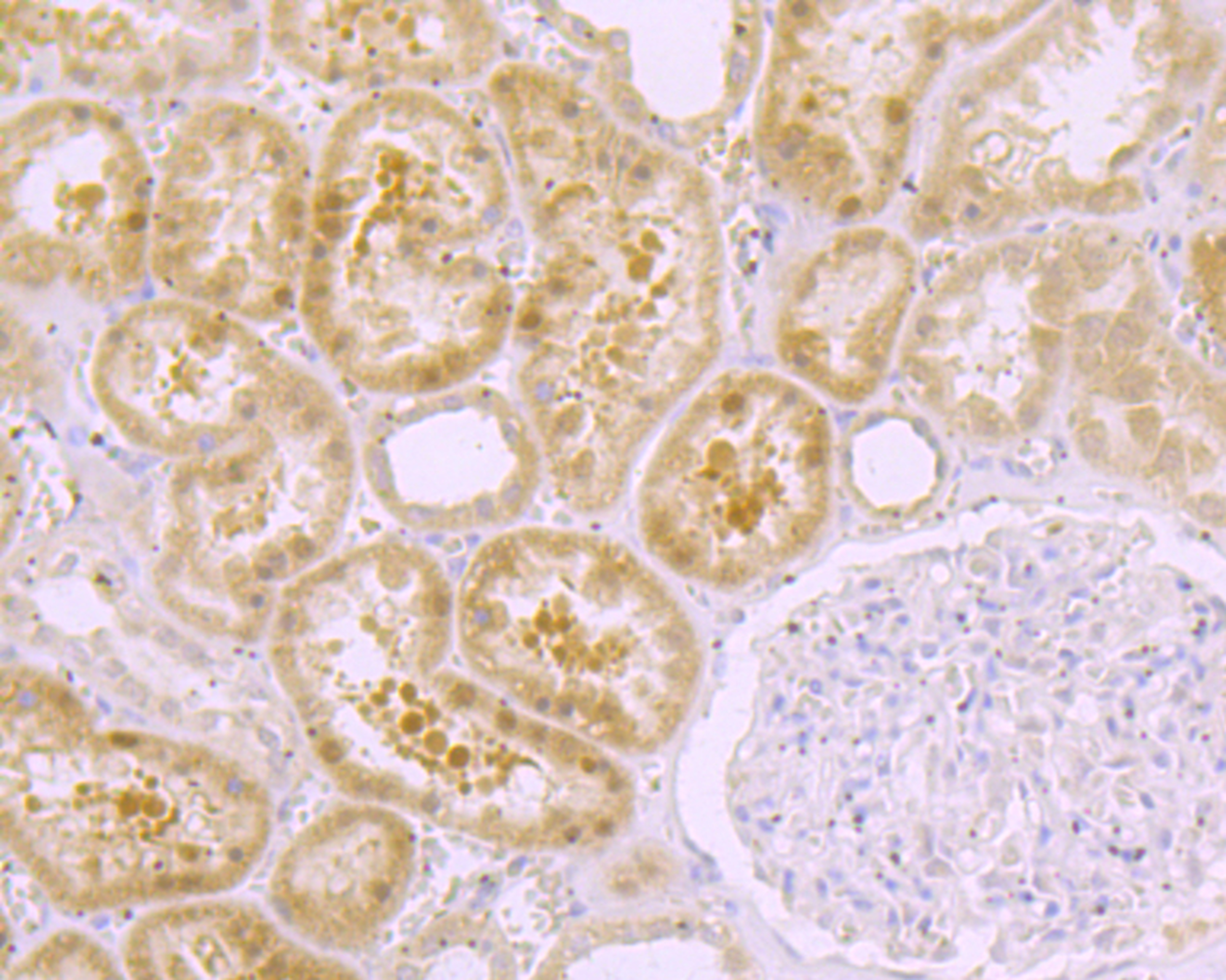
Fig5: Immunohistochemical analysis of paraffin-embedded human kidney tissue using anti-Mib1 antibody. Counter stained with hematoxylin.| Product Name | Mib1 [JG69-34] |
|---|---|
| Antibody Type | Primary Antibodies |
| Antigen Alias | DAPK-interacting protein 1 antibody Dip 1 antibody DIP-1 antibody Dip1 antibody E3 ubiquitin protein ligase MIB 1 antibody E3 ubiquitin protein ligase MIB1 antibody E3 ubiquitin-protein ligase mib1 antibody KIAA1323 antibody LVNC7 antibody MIB antibody mib1 antibody MIB1_HUMAN antibody Mind bomb homolog 1 antibody Mindbomb E3 ubiquitin protein ligase 1 antibody Ubiquitin ligase mind bomb antibody Zinc finger ZZ type with ankyrin repeat domain protein 2 antibody ZZANK2 antibody ZZZ6 antibody |
| Product description | E3 ubiquitin-protein ligase that mediates ubiquitination of Delta receptors, which act as ligands of Notch proteins. Positively regulates the Delta-mediated Notch signaling by ubiquitinating the intracellular domain of Delta, leading to endocytosis of Delta receptors. Probably mediates ubiquitination and subsequent proteasomal degradation of DAPK1, thereby antagonizing anti-apoptotic effects of DAPK1 to promote TNF-induced apoptosis (By similarity). Involved in ubiquitination of centriolar satellite CEP131, CEP290 and PCM1 proteins and hence inhibits primary cilium formation in proliferating cells. Mediates 'Lys-63'-linked polyubiquitination of TBK1, which probably participates in kinase activation. |
| Immunogen | Recombinant protein within N-terminal human Mib1. |
| Clonality | Monoclonal |
|---|---|
| Isotype | IgG |
| Host Species | Recombinant rabbit |
| Tested Applications | WBICCIHC |
| ICC:1:50-1:200 IHC:1:50-1:200 WB:1:500 |
|
| Species Reactivity | HumanMouse |
| Concentration | 1mg/ml |
| Alternative Names | DAPK-interacting protein 1 antibody Dip 1 antibody DIP-1 antibody Dip1 antibody E3 ubiquitin protein ligase MIB 1 antibody E3 ubiquitin protein ligase MIB1 antibody E3 ubiquitin-protein ligase mib1 antibody KIAA1323 antibody LVNC7 antibody MIB antibody mib1 antibody MIB1_HUMAN antibody Mind bomb homolog 1 antibody Mindbomb E3 ubiquitin protein ligase 1 antibody Ubiquitin ligase mind bomb antibody Zinc finger ZZ type with ankyrin repeat domain protein 2 antibody ZZANK2 antibody ZZZ6 antibody |
|---|---|
| Molecular Weight(MW) | 110 kDa |
| Cellular Localization | Plasma membrane. Cytoplasm. Cytoskeleton. |
| SwissProt ID | Q86YT6 |
|---|
Application
Fig1: ICC staining Mib1 in HUVEC cells (green). The nuclear counter stain is DAPI (blue). Cells were fixed in paraformaldehyde, permeabilised with 0.25% Triton X100/PBS.
Application
Fig2: ICC staining Mib1 in SH-SY-5Y cells (green). The nuclear counter stain is DAPI (blue). Cells were fixed in paraformaldehyde, permeabilised with 0.25% Triton X100/PBS.
Application
Fig3: ICC staining Mib1 in SiHa cells (green). The nuclear counter stain is DAPI (blue). Cells were fixed in paraformaldehyde, permeabilised with 0.25% Triton X100/PBS.
Application
Fig4: Immunohistochemical analysis of paraffin-embedded human appendix tissue using anti-Mib1 antibody. Counter stained with hematoxylin.
Application
Fig5: Immunohistochemical analysis of paraffin-embedded human kidney tissue using anti-Mib1 antibody. Counter stained with hematoxylin.| Positive Control | HUVEC, SH-SY-5Y, SiHa, human appendix tissue, human kidney tissue. |
|---|---|
| Application Notes | ICC:1:50-1:200 IHC:1:50-1:200 WB:1:500 |
| Form | Liquid |
|---|---|
| Storage Instructions | Store at +4℃ after thawing. Aliquot store at -20℃. Avoid repeated freeze / thaw cycles. |
| Storage Buffer | 1*TBS (pH7.4), 1%BSA, 40%Glycerol. Preservative: 0.05% Sodium Azide. |


