Application
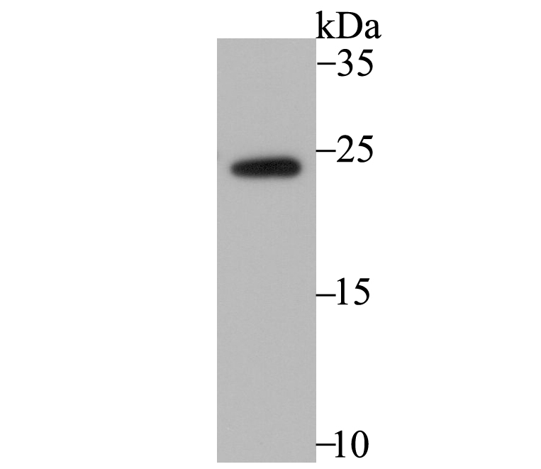
Fig1: Western blot analysis of RHEB on mouse placenta tissue lysate using anti-RHEB antibody at 1/1,000 dilution.Application
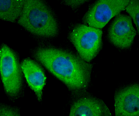
Fig2: ICC staining RHEB in A431 cells (green). The nuclear counter stain is DAPI (blue). Cells were fixed in paraformaldehyde, permeabilised with 0.25% Triton X100/PBS.Application
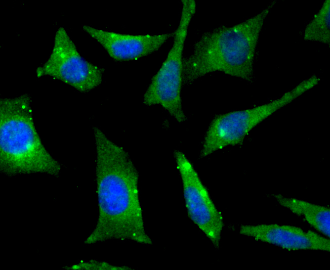
Fig3: ICC staining RHEB in SH-SY-5Y cells (green). The nuclear counter stain is DAPI (blue). Cells were fixed in paraformaldehyde, permeabilised with 0.25% Triton X100/PBS.Application
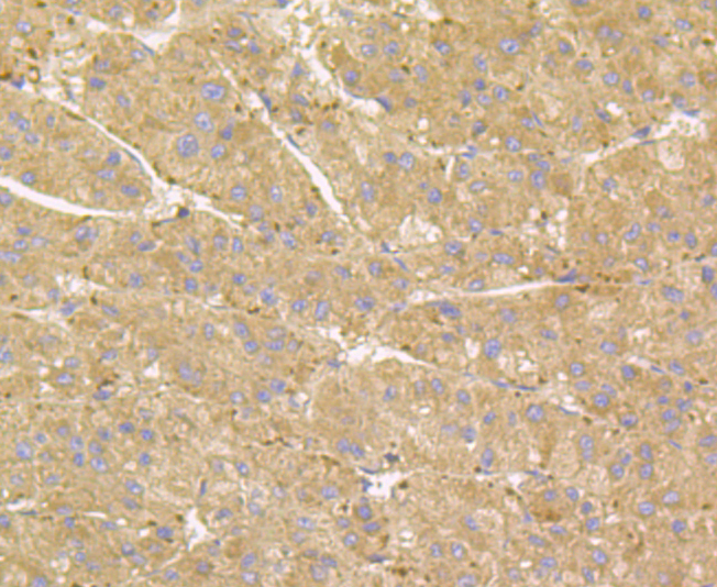
Fig4: Immunohistochemical analysis of paraffin-embedded human liver tissue using anti-RHEB antibody. Counter stained with hematoxylin.Application
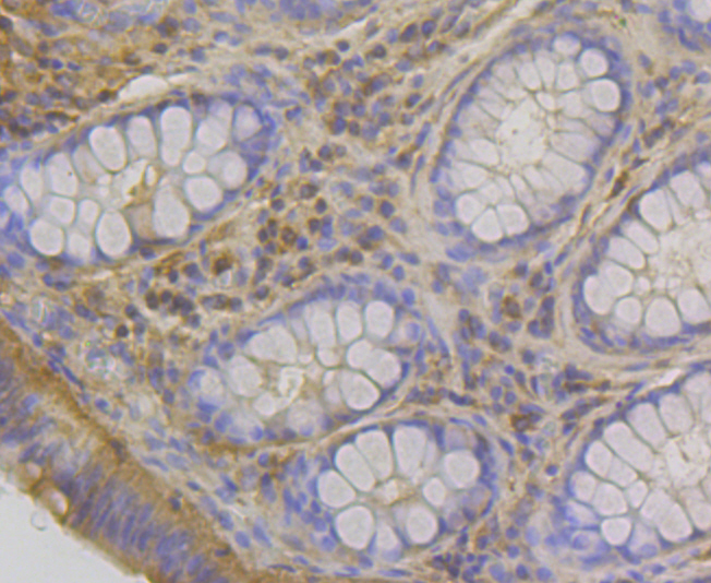
Fig5: Immunohistochemical analysis of paraffin-embedded human colon tissue using anti-RHEB antibody. Counter stained with hematoxylin.Application
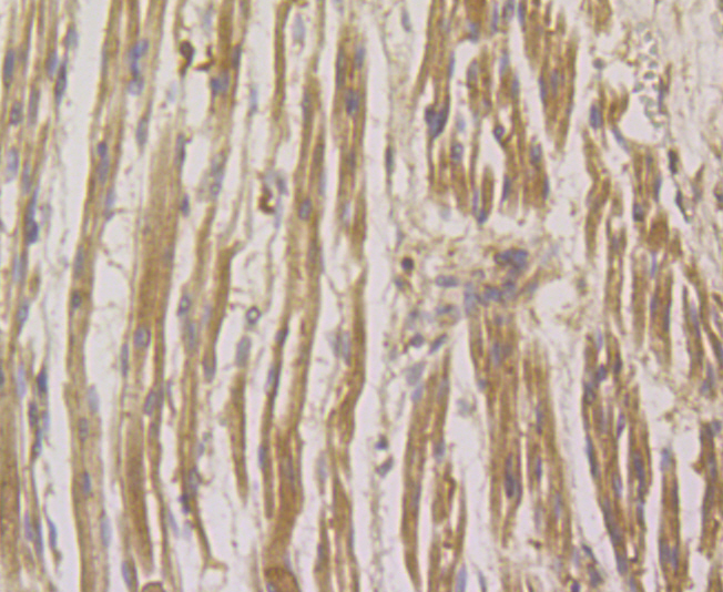
Fig6: Immunohistochemical analysis of paraffin-embedded human fetal skeletal muscle tissue using anti-RHEB antibody. Counter stained with hematoxylin.Application
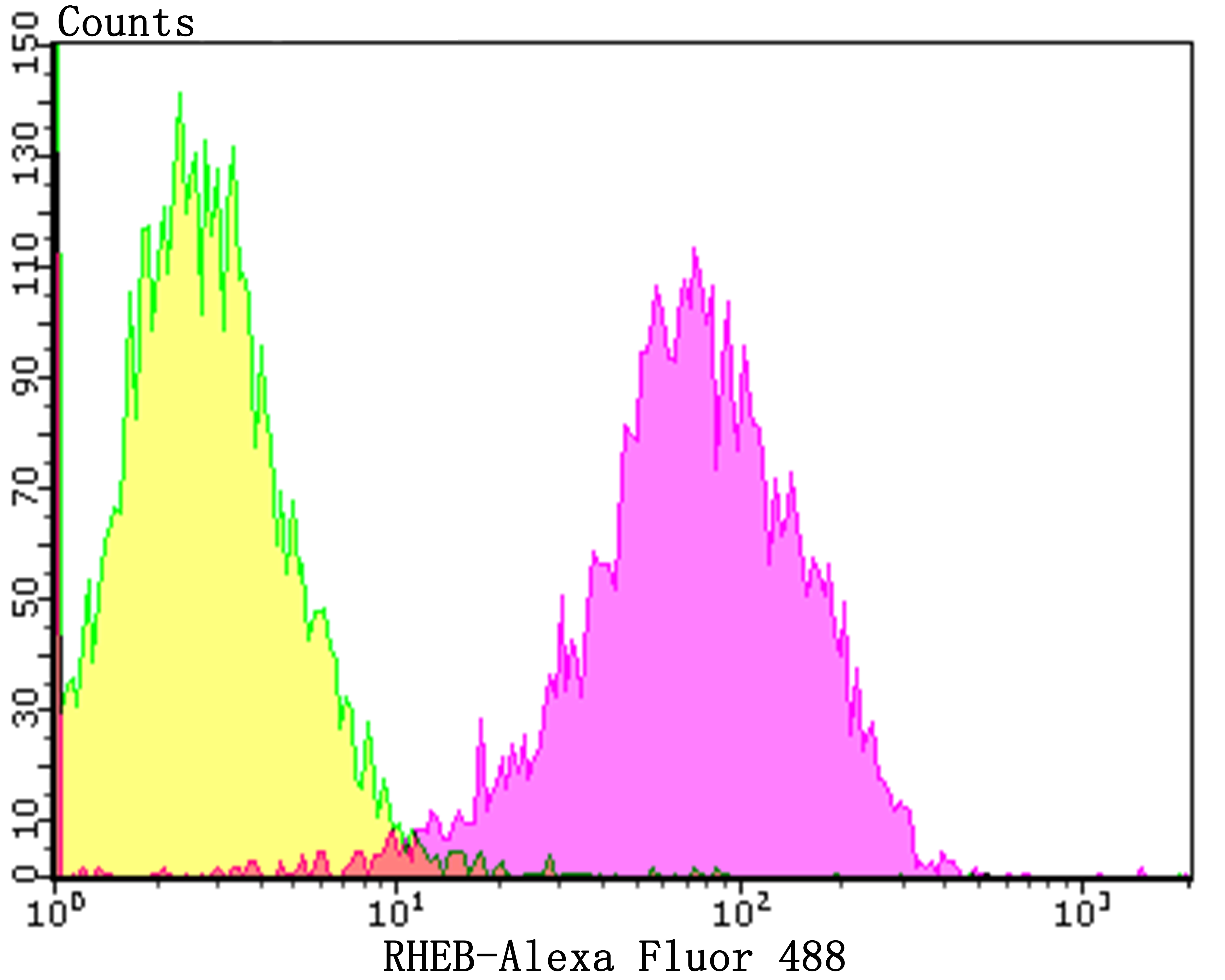
Fig7: Flow cytometric analysis of A431 cells with RHEB antibody at 1/100 dilution (yellow) compared with an unlabelled control (cells without incubation with primary antibody; purple).Alexa Fluor 488-conjugated goat anti-rabbit IgG was used as the secondary antibody.| Product Name | RHEB [JG37-12] |
|---|---|
| Antibody Type | Primary Antibodies |
| Antigen Alias | Ras homolog enriched in brain 2, formerly antibody GTP binding protein Rheb antibody GTP-binding protein Rheb antibody MGC111559 antibody Ras homolog enriched in brain 2 antibody Ras homolog enriched in brain antibody RHEB 2 antibody Rheb antibody RHEB_HUMAN antibody RHEB2 antibody RHEB2, formerly antibody |
| Product description | H-, K- and N-Ras represent the prototype members of a family of small G proteins which are frequently activated to an oncogenic state in a wide variety of human tumors. Activation is due to point mutations at position 12 or 61 within their coding sequence. Such mutations cause these proteins to be constitutively converted to their active GTP-bound rather than the inactive GDP-bound state. The related human R-Ras gene was initially cloned by low stringency hybridization methods. Position 38 or 87 mutants of R-Ras (analogous to positions 12 and 61 in H-Ras) have been shown to be capable of activating oncogenic function. Ras p21 in its active GTP binding state binds to Raf-1, resulting in activation of the MAP kinase signaling cascade. An additional member of the Ras family, Rheb (Ras-related GTP-binding protein), also interacts with Raf-1. This interaction is potentiated by growth factors and agents that increase cAMP levels. |
| Immunogen | Recombinant protein within human RHEB aa 1-250. |
| Clonality | Monoclonal |
|---|---|
| Isotype | IgG |
| Host Species | Recombinant rabbit |
| Tested Applications | WBICCIFIHCFC |
| WB:1:500-1:2,000 ICC:1:50-1:200 IHC:1:50-1:200 FC:1:50-1:100 |
|
| Species Reactivity | HumanMouse |
| Concentration | 1mg/ml |
| Alternative Names | Ras homolog enriched in brain 2 formerly antibody GTP binding protein Rheb antibody GTP-binding protein Rheb antibody MGC111559 antibody Ras homolog enriched in brain 2 antibody Ras homolog enriched in brain antibody RHEB 2 antibody Rheb antibody RHEB_HUMAN antibody RHEB2 antibody RHEB2 formerly antibody |
|---|---|
| Molecular Weight(MW) | 20 kDa |
| Cellular Localization | Cytoplasm. Golgi apparatus. Endoplasmic reticulum. |
Application
Fig1: Western blot analysis of RHEB on mouse placenta tissue lysate using anti-RHEB antibody at 1/1,000 dilution.
Application
Fig2: ICC staining RHEB in A431 cells (green). The nuclear counter stain is DAPI (blue). Cells were fixed in paraformaldehyde, permeabilised with 0.25% Triton X100/PBS.
Application
Fig3: ICC staining RHEB in SH-SY-5Y cells (green). The nuclear counter stain is DAPI (blue). Cells were fixed in paraformaldehyde, permeabilised with 0.25% Triton X100/PBS.
Application
Fig4: Immunohistochemical analysis of paraffin-embedded human liver tissue using anti-RHEB antibody. Counter stained with hematoxylin.
Application
Fig5: Immunohistochemical analysis of paraffin-embedded human colon tissue using anti-RHEB antibody. Counter stained with hematoxylin.
Application
Fig6: Immunohistochemical analysis of paraffin-embedded human fetal skeletal muscle tissue using anti-RHEB antibody. Counter stained with hematoxylin.
Application
Fig7: Flow cytometric analysis of A431 cells with RHEB antibody at 1/100 dilution (yellow) compared with an unlabelled control (cells without incubation with primary antibody; purple).Alexa Fluor 488-conjugated goat anti-rabbit IgG was used as the secondary antibody.| Positive Control | Mouse placenta tissue lysate, A431, SH-SY-5Y, human liver tissue, human colon tissue, human fetal skeletal muscle tissue. |
|---|---|
| Application Notes | WB:1:500-1:2,000 ICC:1:50-1:200 IHC:1:50-1:200 FC:1:50-1:100 |
| Form | Liquid |
|---|---|
| Storage Instructions | Store at +4℃ after thawing. Aliquot store at -20℃. Avoid repeated freeze / thaw cycles. |
| Storage Buffer | 1*TBS (pH7.4), 1%BSA, 40%Glycerol. Preservative: 0.05% Sodium Azide. |


