Application
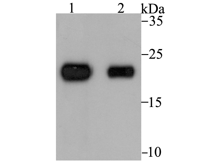
Fig1: Western blot analysis of Tetranectin on rat skin (1) and human skin (2) tissue lysates using anti-Tetranectin antibody at 1/1,000 dilution.Application
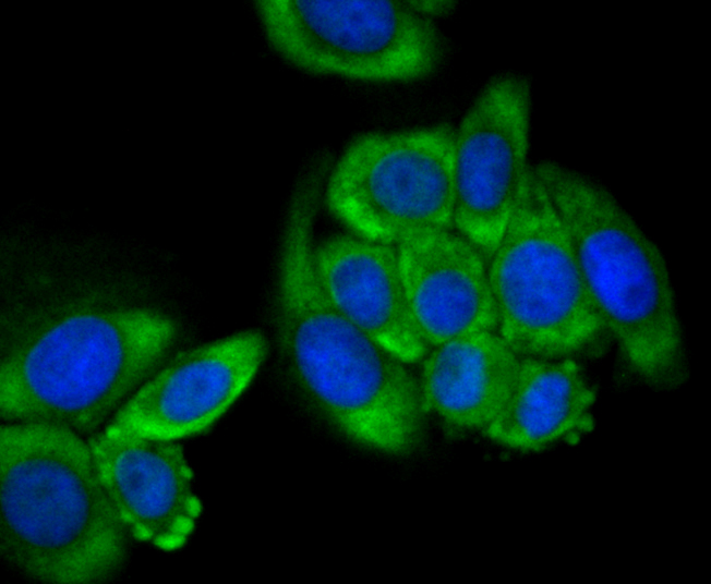
Fig2: ICC staining Tetranectin in HepG2 cells (green). The nuclear counter stain is DAPI (blue). Cells were fixed in paraformaldehyde, permeabilised with 0.25% Triton X100/PBS.Application
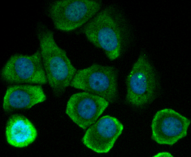
Fig3: ICC staining Tetranectin in MCF-7 cells (green). The nuclear counter stain is DAPI (blue). Cells were fixed in paraformaldehyde, permeabilised with 0.25% Triton X100/PBS.Application
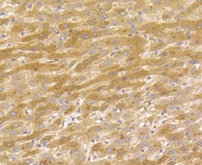
Fig4: Immunohistochemical analysis of paraffin-embedded human liver cancer tissue using anti-Tetranectin antibody. Counter stained with hematoxylin.Application
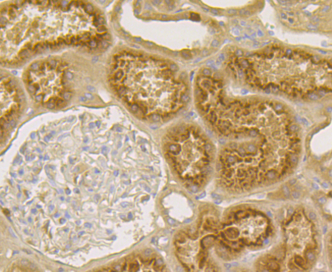
Fig5: Immunohistochemical analysis of paraffin-embedded human kidney tissue using anti-Tetranectin antibody. Counter stained with hematoxylin.| Product Name | Tetranectin [JG37-16] |
|---|---|
| Antibody Type | Primary Antibodies |
| Antigen Alias | C type lectin domain family 3 member B antibody C-type lectin domain family 3 member B antibody Clec3b antibody Plasminogen kringle 4 binding protein antibody Plasminogen kringle 4-binding protein antibody TETN_HUMAN antibody Tetranectin (plasminogen binding protein) antibody Tetranectin antibody TN antibody TNA antibody |
| Product description | Tetranectin is a homotrimeric glycoprotein present in plasma and various tissue locations that binds to calcium, heparin, and plasminogen kringle 4. Tetranectin may play a prominent role in tissue remodeling as well as in the regulation of proteolytic processes via its binding and indirect activation of plasminogen. Tetranectin is found in the extracellular matrix (ECM) of certain carcinomas, but is not present in the ECM of normal tissues. Extracellular proteolysis is an important factor in the ability of malignant cells to penetrate normal tissues and metastasize. Decreased plasma tetranectin or increased tetranectin in stroma of cancers correlates with cancer progression and a grim prognosis. Tetranectin may also influence cancer growth by altering activities of plasminogen or the plasminogen fragment, angiostatin which inhibits tumor cell proliferation. |
| Immunogen | Recombinant protein within human Tetranectin aa 100-250. |
| Clonality | Monoclonal |
|---|---|
| Isotype | IgG |
| Host Species | Recombinant rabbit |
| Tested Applications | WBICCIFIHCIP |
| WB:1:1,000-1:5,000 ICC:1:50-1:200 IHC:1:50-1:200 IP:1:10-1:50 |
|
| Species Reactivity | HumanMouseRat |
| Concentration | 1mg/ml |
| Alternative Names | C type lectin domain family 3 member B antibody C-type lectin domain family 3 member B antibody Clec3b antibody Plasminogen kringle 4 binding protein antibody Plasminogen kringle 4-binding protein antibody TETN_HUMAN antibody Tetranectin (plasminogen binding protein) antibody Tetranectin antibody TN antibody TNA antibody |
|---|---|
| Molecular Weight(MW) | 22 kDa |
| Cellular Localization | Secreted. |
Application
Fig1: Western blot analysis of Tetranectin on rat skin (1) and human skin (2) tissue lysates using anti-Tetranectin antibody at 1/1,000 dilution.
Application
Fig2: ICC staining Tetranectin in HepG2 cells (green). The nuclear counter stain is DAPI (blue). Cells were fixed in paraformaldehyde, permeabilised with 0.25% Triton X100/PBS.
Application
Fig3: ICC staining Tetranectin in MCF-7 cells (green). The nuclear counter stain is DAPI (blue). Cells were fixed in paraformaldehyde, permeabilised with 0.25% Triton X100/PBS.
Application
Fig4: Immunohistochemical analysis of paraffin-embedded human liver cancer tissue using anti-Tetranectin antibody. Counter stained with hematoxylin.
Application
Fig5: Immunohistochemical analysis of paraffin-embedded human kidney tissue using anti-Tetranectin antibody. Counter stained with hematoxylin.| Positive Control | Rat skin tissue lysate, human skin tissue lysate, HepG2, MCF-7, human liver cancer tissue, human kidney tissue. |
|---|---|
| Application Notes | WB:1:1,000-1:5,000 ICC:1:50-1:200 IHC:1:50-1:200 IP:1:10-1:50 |
| Form | Liquid |
|---|---|
| Storage Instructions | Store at +4℃ after thawing. Aliquot store at -20℃. Avoid repeated freeze / thaw cycles. |
| Storage Buffer | 1*TBS (pH7.4), 1%BSA, 40%Glycerol. Preservative: 0.05% Sodium Azide. |


