Application
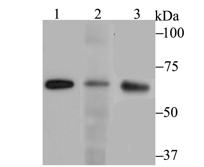
Fig1: Western blot analysis of RanGAP1 on different cell lysates using anti-RanGAP1 antibody at 1/1,000 dilution. Positive control: Lane 1: MCF-7 Lane 2: SiHa Lane 3: 293Application
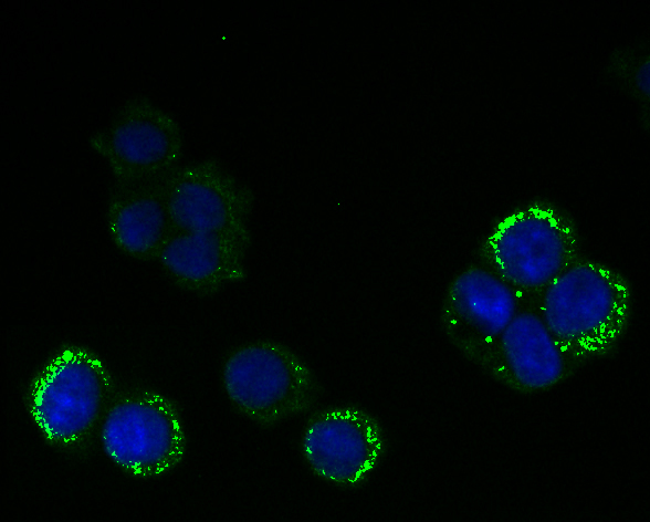
Fig2: ICC staining RanGAP1 (green) in 293T cells. The nuclear counter stain is DAPI (blue). Cells were fixed in paraformaldehyde, permeabilised with 0.25% Triton X100/PBS.Application
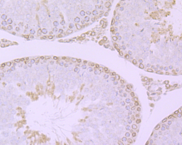
Fig3: Immunohistochemical analysis of paraffin-embedded rat testis tissue using anti-RanGAP1 antibody. Counter stained with hematoxylin.Application
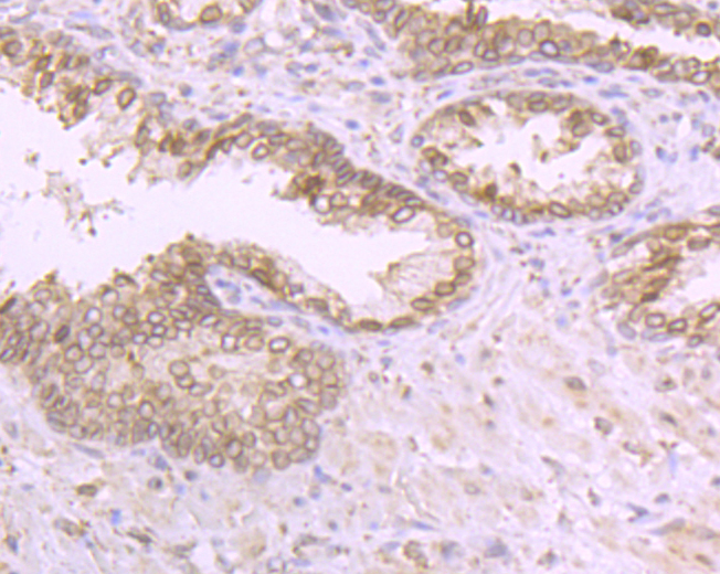
Fig4: Immunohistochemical analysis of paraffin-embedded human prostate tissue using anti-RanGAP1 antibody. Counter stained with hematoxylin.Application
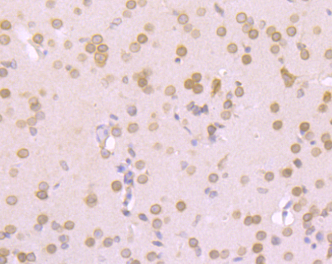
Fig5: Immunohistochemical analysis of paraffin-embedded mouse brain tissue using anti-RanGAP1 antibody. Counter stained with hematoxylin.Application
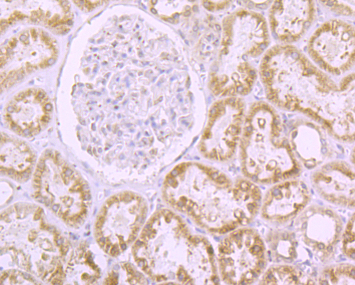
Fig6: Immunohistochemical analysis of paraffin-embedded human kidney tissue using anti-RanGAP1 antibody. Counter stained with hematoxylin.Application
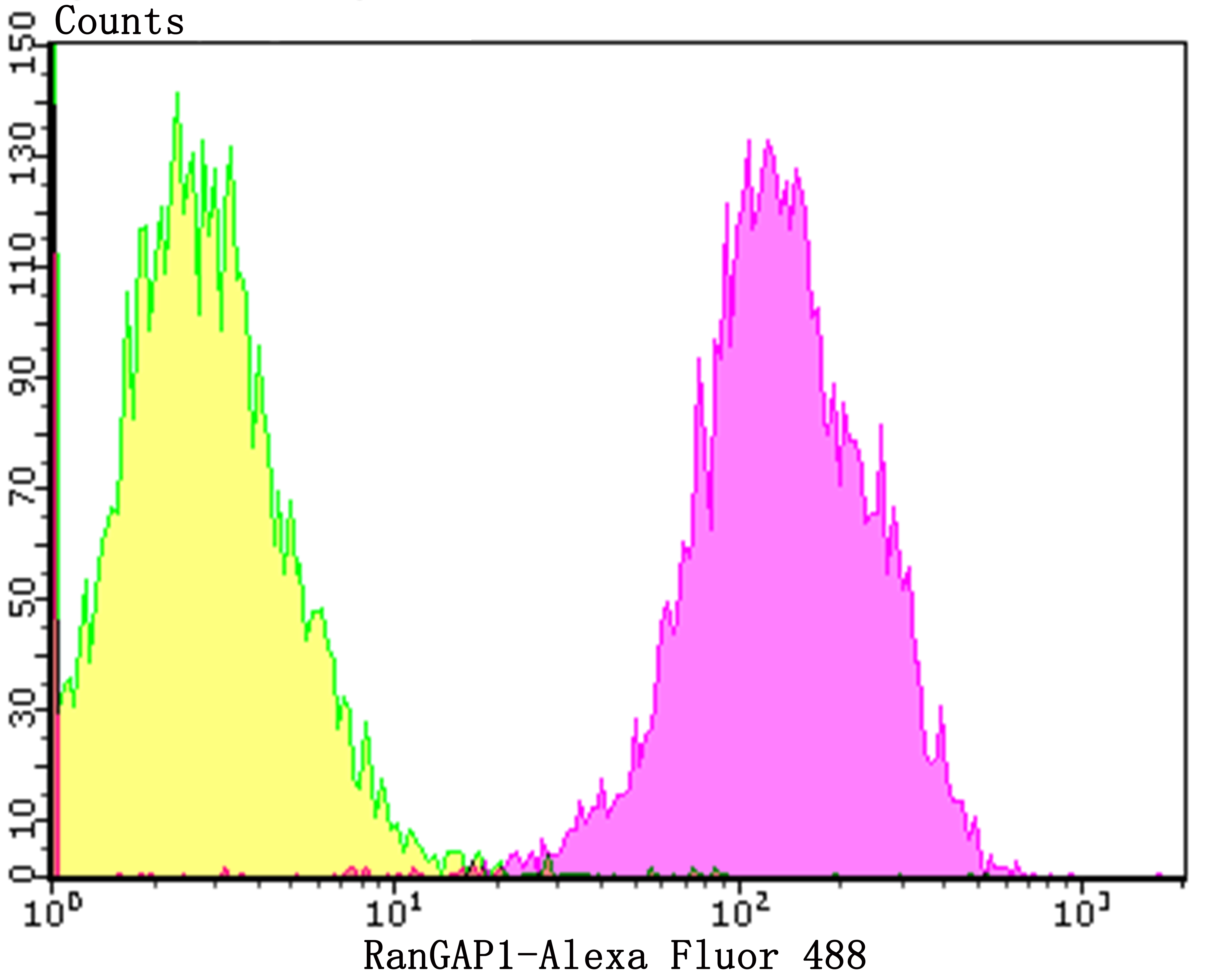
Fig7: Flow cytometric analysis of A431 cells with RanGAP1 antibody at 1/100 dilution (purple) compared with an unlabelled control (cells without incubation with primary antibody; yellow). Alexa Fluor 488-conjugated goat anti-rabbit IgG was used as the secondary antibody.| Product Name | RanGAP1 [JG37-41] |
|---|---|
| Antibody Type | Primary Antibodies |
| Antigen Alias | Fug 1 antibody Fug1 antibody GTPase-activating protein, RAN, 1 antibody KIAA1835 antibody MGC20266 antibody OTTHUMP00000028918 antibody OTTHUMP00000198755 antibody OTTHUMP00000198756 antibody OTTHUMP00000198757 antibody OTTHUMP00000198758 antibody RAGP1_HUMAN antibody Ran 1 antibody RAN GTPase activating protein 1 antibody Ran GTPase-activating protein 1 antibody Ran1 antibody RANGAP 1 antibody RANGAP antibody RanGAP1 antibody SD antibody Segregation distorter homolog antibody Segregation distortion antibody |
| Product description | The small Ras related protein Ran, also called TC4, is a nuclear localized GTPase implicated in a diverse array of cellular processes including DNA replication, entry into and exit from mitosis and the transport of RNA and proteins through the nuclear pore complex. Like Ras, active Ran GTP and inactive Ran GDP levels are tightly regulated by guanine nucleotide exchange factors (GEFs) and GTPase-activating proteins (GAPs). The abundant GEF RCC1 (regulator of chromosome condensation 1) increases the rate at which Ran exchanges GDP for GTP. Ran GAP1 opposes the effects of RCC1 by increasing the rate at which Ran hydrolyzes GTP to GDP. A protein designated Ran BP1 has no intrinsic GAP activity and functions as a GEF inhibitor deactivating RCC1 and thereby indirectly increasing the ratio of Ran GDP to Ran GTP. Ran BP2 has been proposed as the Ran GTP docking site at the periphery of the nuclear pore complex. |
| Immunogen | Recombinant protein within human RanGAP1 aa 1-200. |
| Clonality | Monoclonal |
|---|---|
| Isotype | IgG |
| Host Species | Recombinant rabbit |
| Tested Applications | WBICCIFIHCFCIP |
| WB:1:500-1:2,000 ICC/IF:1:100 IHC:1:50-1:200 FC:1:50-1:100 IP:1:10-1:50 |
|
| Species Reactivity | HumanMouseRat |
| Concentration | 1mg/ml |
| Alternative Names | Fug 1 antibody
Fug1 antibody
GTPase-activating protein RAN 1 antibody KIAA1835 antibody MGC20266 antibody OTTHUMP00000028918 antibody OTTHUMP00000198755 antibody OTTHUMP00000198756 antibody OTTHUMP00000198757 antibody OTTHUMP00000198758 antibody RAGP1_HUMAN antibody Ran 1 antibody RAN GTPase activating protein 1 antibody Ran GTPase-activating protein 1 antibody Ran1 antibody RANGAP 1 antibody RANGAP antibody RanGAP1 antibody SD antibody Segregation distorter homolog antibody Segregation distortion antibody |
|---|---|
| Molecular Weight(MW) | 64 kDa |
| Cellular Localization | Cytoplasm. Cytoskeleton. Nucleus. |
| SwissProt ID | P46060 |
|---|
Application
Fig1: Western blot analysis of RanGAP1 on different cell lysates using anti-RanGAP1 antibody at 1/1,000 dilution. Positive control: Lane 1: MCF-7 Lane 2: SiHa Lane 3: 293
Application
Fig2: ICC staining RanGAP1 (green) in 293T cells. The nuclear counter stain is DAPI (blue). Cells were fixed in paraformaldehyde, permeabilised with 0.25% Triton X100/PBS.
Application
Fig3: Immunohistochemical analysis of paraffin-embedded rat testis tissue using anti-RanGAP1 antibody. Counter stained with hematoxylin.
Application
Fig4: Immunohistochemical analysis of paraffin-embedded human prostate tissue using anti-RanGAP1 antibody. Counter stained with hematoxylin.
Application
Fig5: Immunohistochemical analysis of paraffin-embedded mouse brain tissue using anti-RanGAP1 antibody. Counter stained with hematoxylin.
Application
Fig6: Immunohistochemical analysis of paraffin-embedded human kidney tissue using anti-RanGAP1 antibody. Counter stained with hematoxylin.
Application
Fig7: Flow cytometric analysis of A431 cells with RanGAP1 antibody at 1/100 dilution (purple) compared with an unlabelled control (cells without incubation with primary antibody; yellow). Alexa Fluor 488-conjugated goat anti-rabbit IgG was used as the secondary antibody.| Positive Control | MCF-7, SiHa, 293, A431, 293T, rat testis tissue, human prostate tissue, mouse brain tissue, human kidney tissue. |
|---|---|
| Application Notes | WB:1:500-1:2,000 ICC/IF:1:100 IHC:1:50-1:200 FC:1:50-1:100 IP:1:10-1:50 |
| Form | Liquid |
|---|---|
| Storage Instructions | Store at +4℃ after thawing. Aliquot store at -20℃. Avoid repeated freeze / thaw cycles. |
| Storage Buffer | 1*TBS (pH7.4), 1%BSA, 40%Glycerol. Preservative: 0.05% Sodium Azide. |


