Application
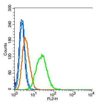
Fig1: Blank control: RSC 96 (blue). Primary Antibody:Rabbit Anti-AKAP12 antibody(ER1903-55), Dilution: 1μg in 100 μL 1X PBS containing 0.5% BSA; Isotype Control Antibody: Rabbit IgG(orange) ,used under the same conditions ); Secondary Antibody: Goat anti-rabbit IgG-PE(white blue), Dilution: 1:200 in 1 X PBS containing 0.5% BSA. Protocol The cells were fixed with 2% paraformaldehyde (10 min) , then permeabilized with 90% ice-cold methanol for 30 min on ice. Antibody (ER1903-55, 1μg /1x10^6 cells) were incubated for 30 min on the ice, followed by 1 X PBS containing 0.5% BSA + 1 0% goat serum (15 min) to block non-specific protein-protein interactions. Then the Goat Anti-rabbit IgG/PE antibody was added into the blocking buffer mentioned above to react with the primary antibody of ER1903-55 at 1/200 dilution for 30 min on ice. Acquisition of 20,000 events was performed.Application
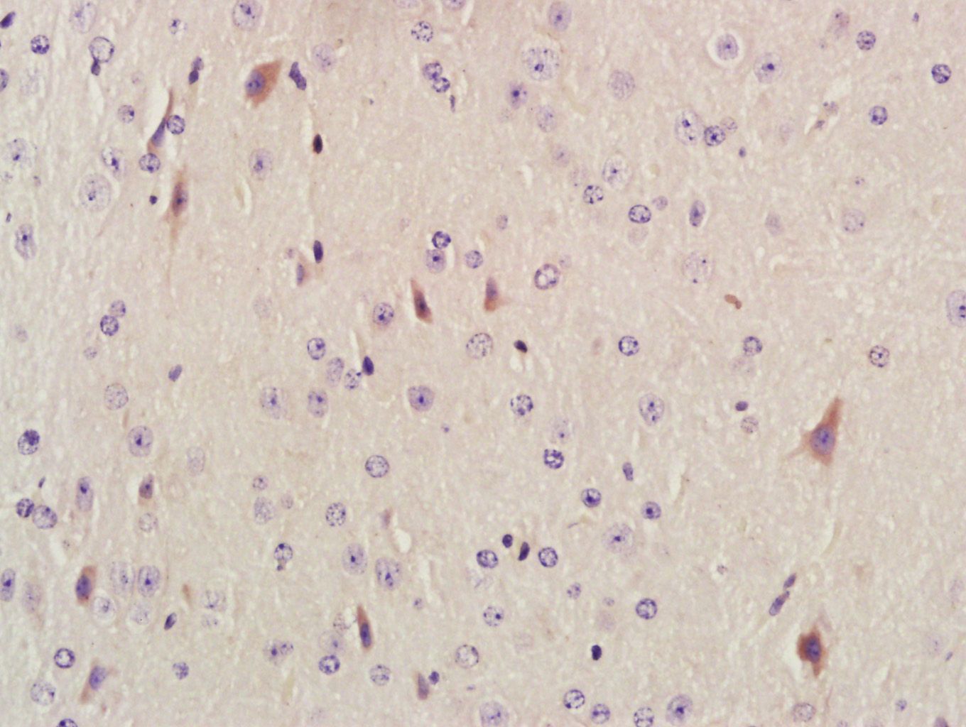
Fig2: Paraformaldehyde-fixed, paraffin embedded (Mouse brain); Antigen retrieval by boiling in sodium citrate buffer (pH6.0) for 15min; Block endogenous peroxidase by 3% hydrogen peroxide for 20 minutes; Blocking buffer (normal goat serum) at 37℃ for 30min; Antibody incubation with (AKAP12) Polyclonal Antibody, Unconjugated (ER1903-55) at 1:400 overnight at 4℃, followed by a conjugated secondary antibody (sp-0023) for 20 minutes and DAB staining.Application
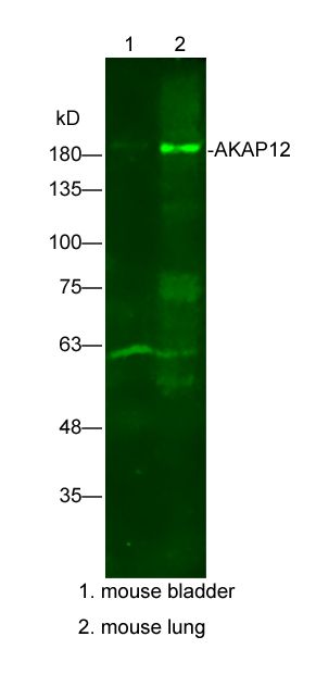
Fig3: Sample: Lane1: mouse bladder Lysate at 25 ug Lane2: mouse lung Lysate at 25 ug Primary: Anti-AKAP12(ER1903-55) at 1/300 dilution Secondary: IRDye800CW Goat Anti-Rabbit IgG at 1/20000 dilution Predicted band size: 191kD Observed band size: 191kD| Product Name | AKAP12 |
|---|---|
| Antibody Type | Primary Antibodies |
| Antigen Alias | A kinase (PRKA) anchor protein (gravin) 12 A kinase Anchor Protein 12 A kinase anchor protein 250kDa AKAP 12 AKAP 250 AKAP250 DKFZp686M0430 DKFZp686O0331 Gercelin Gravin Kinase scaffold protein gravin Myasthenia gravis autoantigen gravin Srcs5 SSeCKS Tsga12. |
| Product description | Anchoring protein that mediates the subcellular compartmentation of protein kinase A (PKA) and protein kinase C (PKC). |
| Immunogen | KLH conjugated synthetic peptide derived from human AKAP12 701-900/1684 |
| Clonality | Polyclonal |
|---|---|
| Isotype | IgG |
| Host Species | Rabbit |
| Tested Applications | WBIHC-PICC/IFFC |
| WB:1:500-2000: IHC-P:1:400-800: FC:1: |
|
| Species Reactivity | HumanMouseRatDog |
| Concentration | 1mg/ml |
| Alternative Names | A kinase (PRKA) anchor protein (gravin) 12 A kinase Anchor Protein 12 A kinase anchor protein 250kDa AKAP 12 AKAP 250 AKAP250 DKFZp686M0430 DKFZp686O0331 Gercelin Gravin Kinase scaffold protein gravin Myasthenia gravis autoantigen gravin Srcs5 SSeCKS Tsga12. |
|---|---|
| Molecular Weight(MW) | 191 KDa |
| Cellular Localization | Cytoplasm |
| SwissProt ID | Q02952 |
|---|
Application
Fig1: Blank control: RSC 96 (blue). Primary Antibody:Rabbit Anti-AKAP12 antibody(ER1903-55), Dilution: 1μg in 100 μL 1X PBS containing 0.5% BSA; Isotype Control Antibody: Rabbit IgG(orange) ,used under the same conditions ); Secondary Antibody: Goat anti-rabbit IgG-PE(white blue), Dilution: 1:200 in 1 X PBS containing 0.5% BSA. Protocol The cells were fixed with 2% paraformaldehyde (10 min) , then permeabilized with 90% ice-cold methanol for 30 min on ice. Antibody (ER1903-55, 1μg /1x10^6 cells) were incubated for 30 min on the ice, followed by 1 X PBS containing 0.5% BSA + 1 0% goat serum (15 min) to block non-specific protein-protein interactions. Then the Goat Anti-rabbit IgG/PE antibody was added into the blocking buffer mentioned above to react with the primary antibody of ER1903-55 at 1/200 dilution for 30 min on ice. Acquisition of 20,000 events was performed.
Application
Fig2: Paraformaldehyde-fixed, paraffin embedded (Mouse brain); Antigen retrieval by boiling in sodium citrate buffer (pH6.0) for 15min; Block endogenous peroxidase by 3% hydrogen peroxide for 20 minutes; Blocking buffer (normal goat serum) at 37℃ for 30min; Antibody incubation with (AKAP12) Polyclonal Antibody, Unconjugated (ER1903-55) at 1:400 overnight at 4℃, followed by a conjugated secondary antibody (sp-0023) for 20 minutes and DAB staining.
Application
Fig3: Sample: Lane1: mouse bladder Lysate at 25 ug Lane2: mouse lung Lysate at 25 ug Primary: Anti-AKAP12(ER1903-55) at 1/300 dilution Secondary: IRDye800CW Goat Anti-Rabbit IgG at 1/20000 dilution Predicted band size: 191kD Observed band size: 191kD| Positive Control | Expressed in endothelial cells, cultured fibroblasts and osteosarcoma, but not in platelets, leukocytes, monocytic cell lines or peripherical blood cells. |
|---|---|
| Application Notes | WB:1:500-2000: IHC-P:1:400-800: FC:1: |
| Form | Liquid |
|---|---|
| Storage Instructions | Store at -20℃ for one year. Avoid repeated freeze/thaw cycles. The lyophilized antibody is stable at room temperature for at least one month and for greater than a year when kept at -20℃. When reconstituted in sterile pH 7.4 0.01M PBS or diluent of antibody the antibody is stable for at least two weeks at 2-4℃. |
| Storage Buffer | 0.01M TBS(pH7.4) with 1% BSA, 0.03% Proclin300 and 50% Glycerol. |


