Application
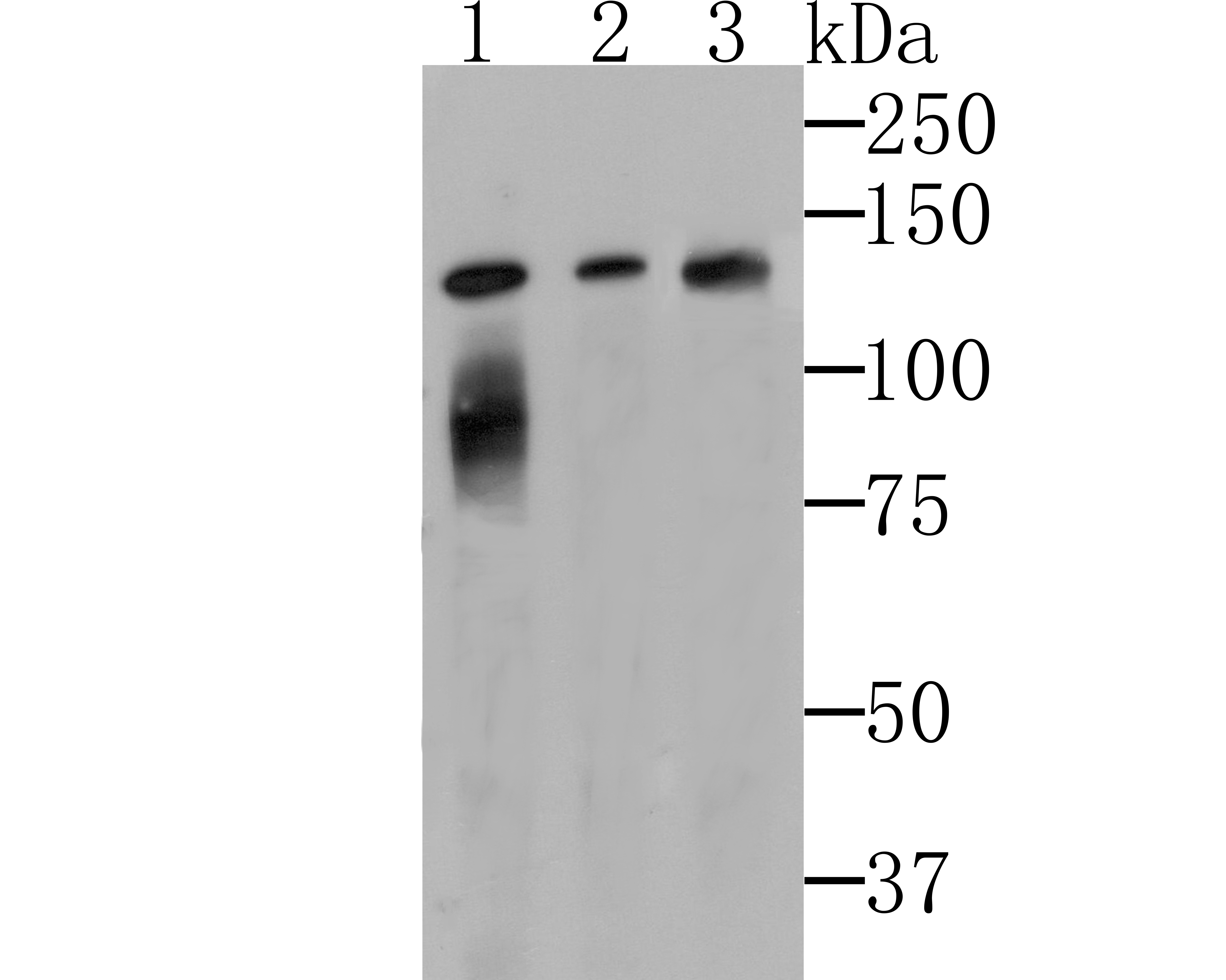
Fig1: Western blot analysis of Lipin 1 on different cell lysates using anti-Lipin 1 at 1/500 dilution. Positive control: Line 1: K562 Line 2: SH-SY-5Y Line 3: Mouse lungApplication
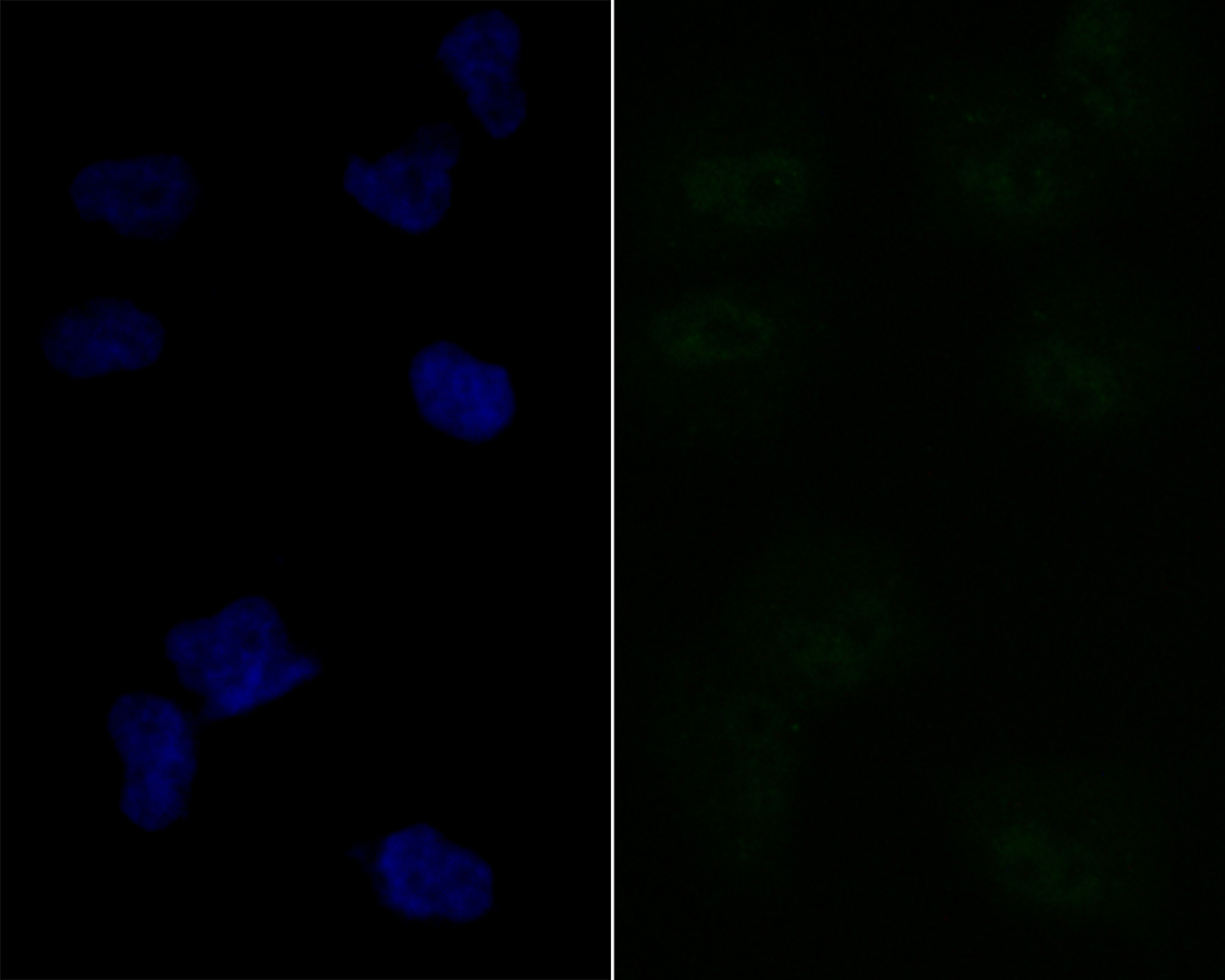
Fig2: ICC staining Lipin 1 in HUVEC cells (green). The nuclear counter stain is DAPI (blue). Cells were fixed in paraformaldehyde, permeabilised with 0.25% Triton X100/PBS.Application
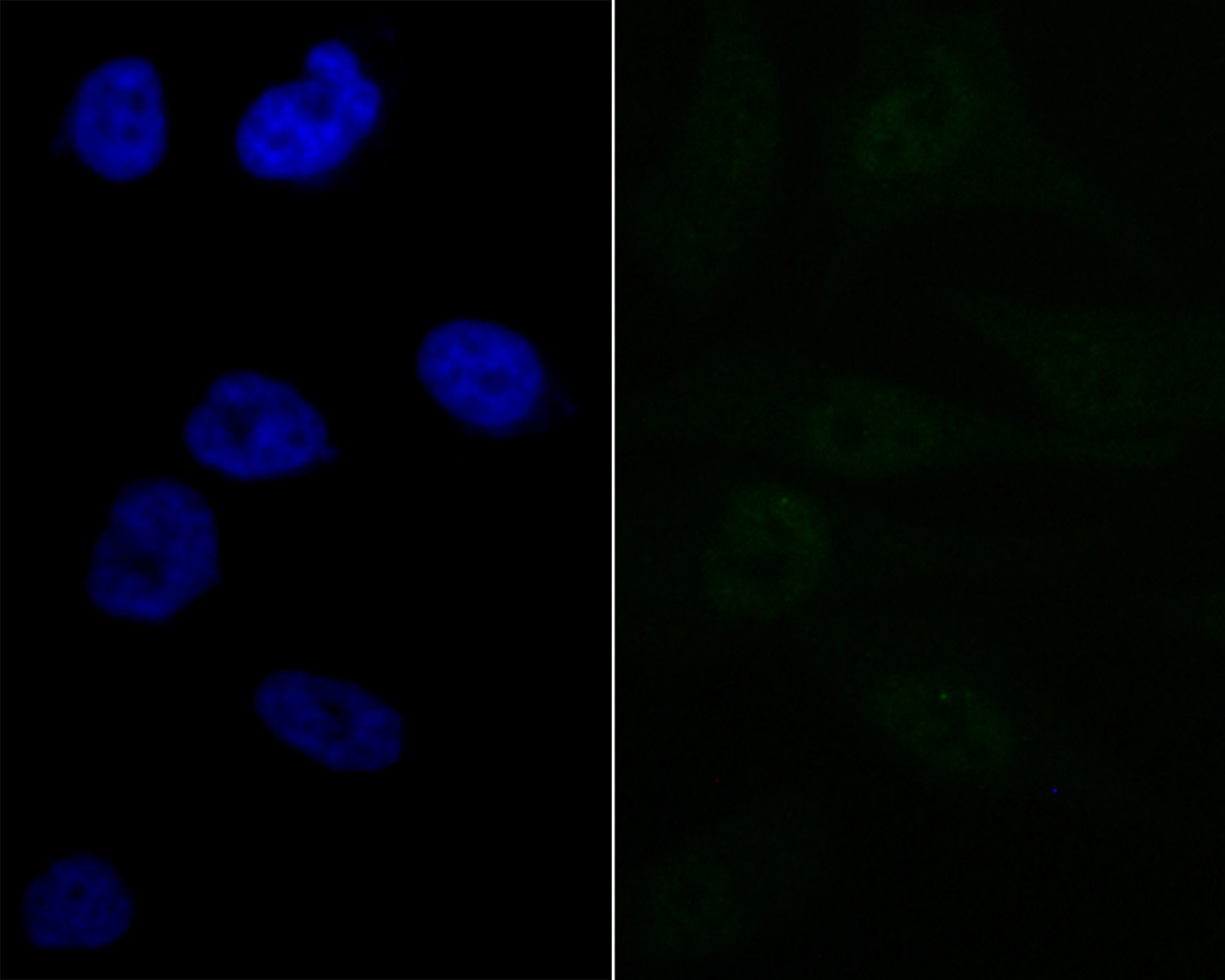
Fig3: ICC staining Lipin 1 in PC-3M cells (green). The nuclear counter stain is DAPI (blue). Cells were fixed in paraformaldehyde, permeabilised with 0.25% Triton X100/PBS.Application
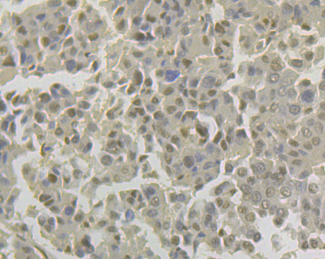
Fig4: Immunohistochemical analysis of paraffin-embedded human liver cancer tissue using anti-Lipin 1 antibody. Counter stained with hematoxylin.Application
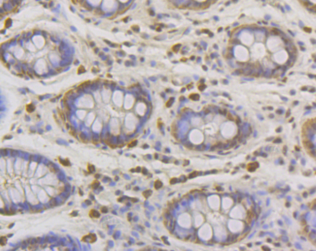
Fig5: Immunohistochemical analysis of paraffin-embedded human colon tissue using anti-Lipin 1 antibody. Counter stained with hematoxylin.Application
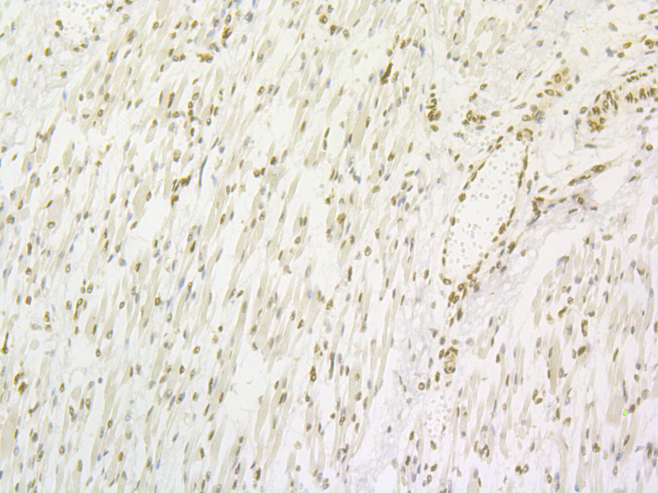
Fig6: Immunohistochemical analysis of paraffin-embedded human skeletal muscle tissue using anti-Lipin 1 antibody. Counter stained with hematoxylin.Application
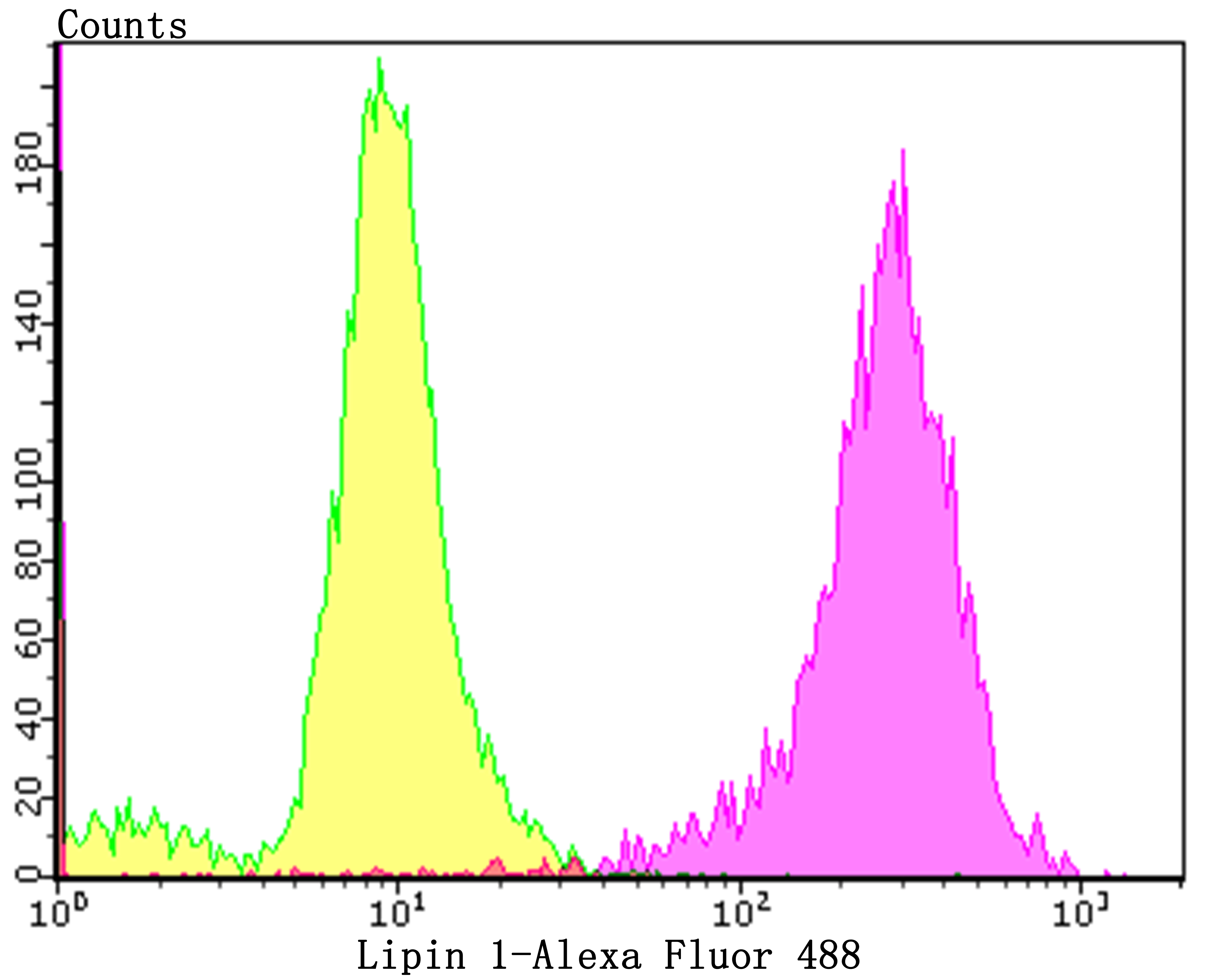
Fig7: Flow cytometric analysis of PC-3M cells with Lipin 1 antibody at 1/50 dilution (purple) compared with an unlabelled control (cells without incubation with primary antibody; yellow). Alexa Fluor 488-conjugated goat anti-rabbit IgG was used as the secondary antibody.| Product Name | Lipin 1 [JG37-69] |
|---|---|
| Antibody Type | Primary Antibodies |
| Antigen Alias | EC=3.1.3.4 antibody KIAA0188 antibody Lipin-1 antibody Lpin1 antibody LPIN1_HUMAN antibody PAP1 antibody Phosphatidate phosphatase LPIN1 antibody |
| Product description | Plays important roles in controlling the metabolism of fatty acids at different levels. Acts as a magnesium-dependent phosphatidate phosphatase enzyme which catalyzes the conversion of phosphatidic acid to diacylglycerol during triglyceride, phosphatidylcholine and phosphatidylethanolamine biosynthesis in the reticulum endoplasmic membrane. Acts also as a nuclear transcriptional coactivator for PPARGC1A/PPARA to modulate lipid metabolism gene expression (By similarity). Is involved in adipocyte differentiation. May also be involved in mitochondrial fission by converting phosphatidic acid to diacylglycerol (By similarity). |
| Immunogen | Recombinant protein within human Lipin 1 aa 50-250. |
| Clonality | Monoclonal |
|---|---|
| Isotype | IgG |
| Host Species | Recombinant rabbit |
| Tested Applications | WBICCIFIHCFCIP |
| WB:1:500-1:1,000 ICC:1:50-1:200 IHC:1:50-1:200 FC:1:50-1:100 |
|
| Species Reactivity | Human |
| Concentration | 1mg/ml |
| Alternative Names | EC=3.1.3.4 antibody KIAA0188 antibody Lipin-1 antibody Lpin1 antibody LPIN1_HUMAN antibody PAP1 antibody Phosphatidate phosphatase LPIN1 antibody |
|---|---|
| Molecular Weight(MW) | 98 kDa |
| Cellular Localization | Nucleus. Cytoplasm. Endoplasmic reticulum. |
| SwissProt ID | Q14693 |
|---|
Application
Fig1: Western blot analysis of Lipin 1 on different cell lysates using anti-Lipin 1 at 1/500 dilution. Positive control: Line 1: K562 Line 2: SH-SY-5Y Line 3: Mouse lung
Application
Fig2: ICC staining Lipin 1 in HUVEC cells (green). The nuclear counter stain is DAPI (blue). Cells were fixed in paraformaldehyde, permeabilised with 0.25% Triton X100/PBS.
Application
Fig3: ICC staining Lipin 1 in PC-3M cells (green). The nuclear counter stain is DAPI (blue). Cells were fixed in paraformaldehyde, permeabilised with 0.25% Triton X100/PBS.
Application
Fig4: Immunohistochemical analysis of paraffin-embedded human liver cancer tissue using anti-Lipin 1 antibody. Counter stained with hematoxylin.
Application
Fig5: Immunohistochemical analysis of paraffin-embedded human colon tissue using anti-Lipin 1 antibody. Counter stained with hematoxylin.
Application
Fig6: Immunohistochemical analysis of paraffin-embedded human skeletal muscle tissue using anti-Lipin 1 antibody. Counter stained with hematoxylin.
Application
Fig7: Flow cytometric analysis of PC-3M cells with Lipin 1 antibody at 1/50 dilution (purple) compared with an unlabelled control (cells without incubation with primary antibody; yellow). Alexa Fluor 488-conjugated goat anti-rabbit IgG was used as the secondary antibody.| Positive Control | K562, SH-SY-5Y, HUVEC, PC-3M, human liver cancer tissue, human colon tissue, human skeletal muscle tissue. |
|---|---|
| Application Notes | WB:1:500-1:1,000 ICC:1:50-1:200 IHC:1:50-1:200 FC:1:50-1:100 |
| Form | Liquid |
|---|---|
| Storage Instructions | Store at +4℃ after thawing. Aliquot store at -20℃. Avoid repeated freeze / thaw cycles. |
| Storage Buffer | 1*TBS (pH7.4), 1%BSA, 40%Glycerol. Preservative: 0.05% Sodium Azide. |


