Application
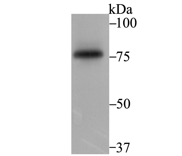
Fig1: Western blot analysis of SENP1 on K562 cell using anti-SENP1 antibody at 1/1,000 dilution.Application
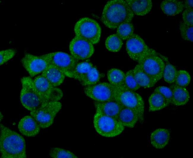
Fig2: ICC staining SENP1 in LOVO cells (green). The nuclear counter stain is DAPI (blue). Cells were fixed in paraformaldehyde, permeabilised with 0.25% Triton X100/PBS.Application
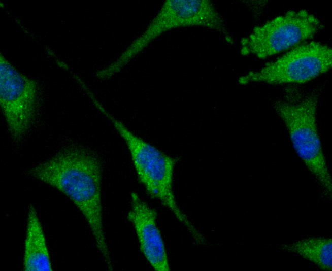
Fig3: ICC staining SENP1 in SH-SY-5Y cells (green). The nuclear counter stain is DAPI (blue). Cells were fixed in paraformaldehyde, permeabilised with 0.25% Triton X100/PBS.Application
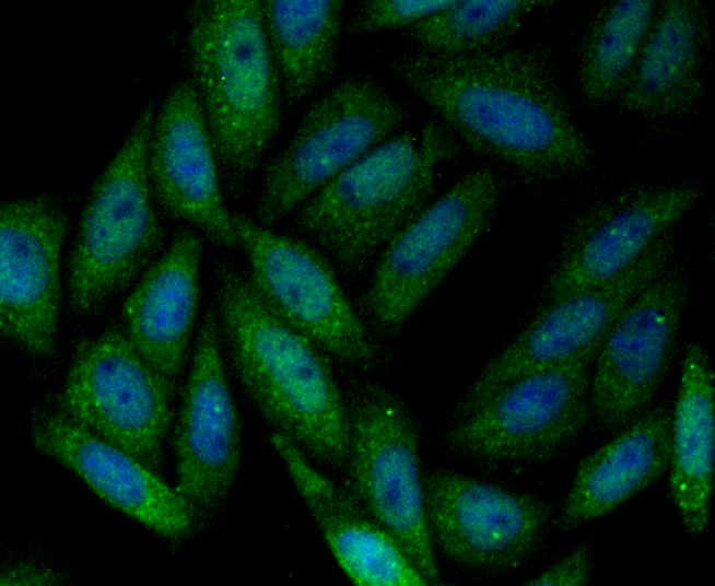
Fig4: ICC staining SENP1 in SiHa cells (green). The nuclear counter stain is DAPI (blue). Cells were fixed in paraformaldehyde, permeabilised with 0.25% Triton X100/PBS.Application
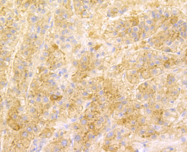
Fig5: Immunohistochemical analysis of paraffin-embedded human liver tissue using anti-SENP1 antibody. Counter stained with hematoxylin.Application
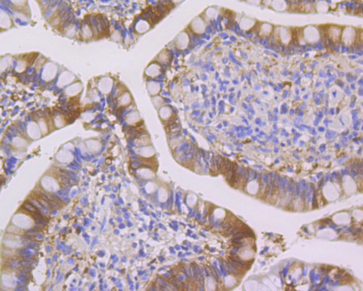
Fig6: Immunohistochemical analysis of paraffin-embedded human small Intestine tissue using anti-SENP1 antibody. Counter stained with hematoxylin.Application
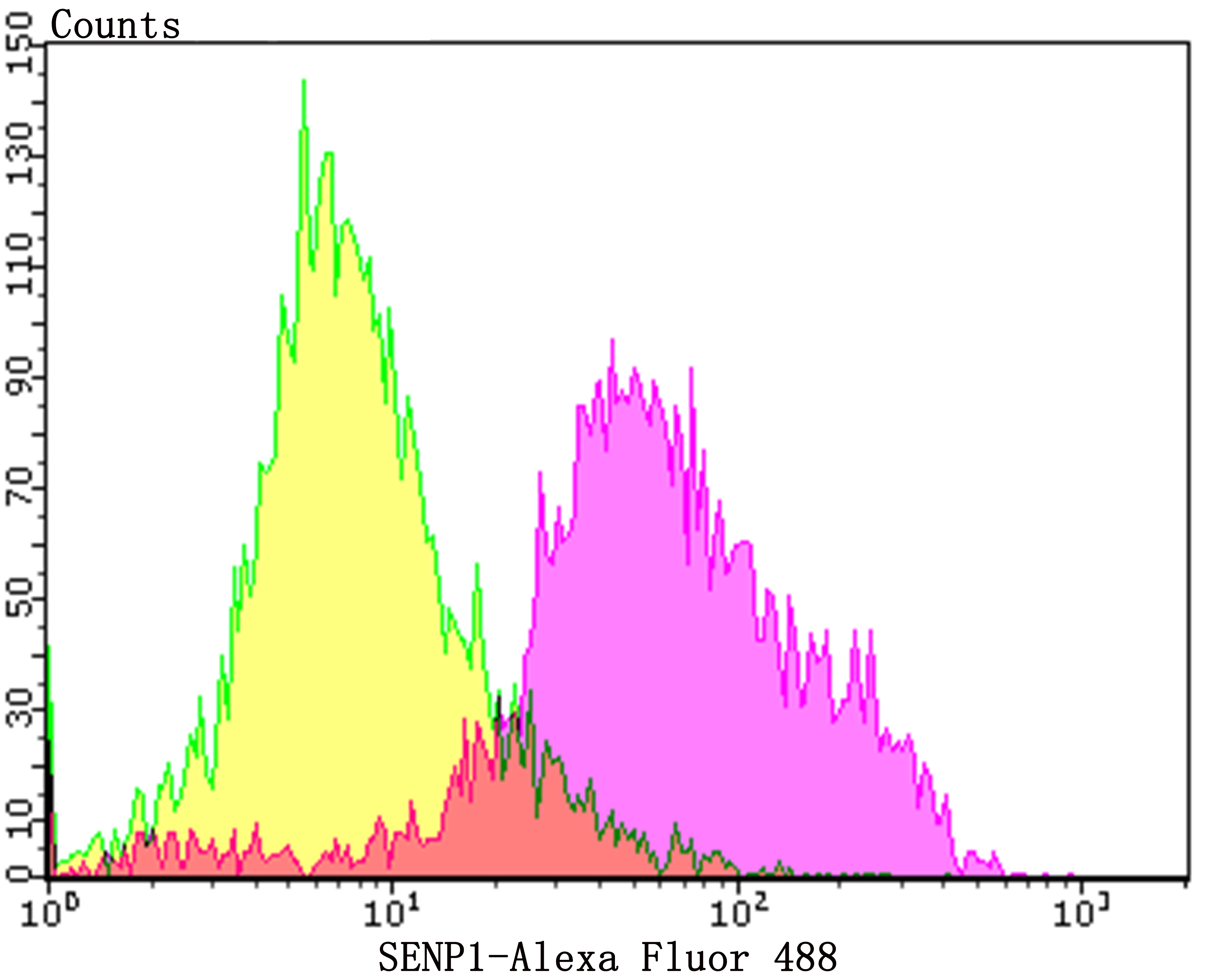
Fig7: Flow cytometric analysis of Daudi cells with SENP1 antibody at 1/100 dilution (purple) compared with an unlabelled control (cells without incubation with primary antibody; yellow). Alexa Fluor 488-conjugated goat anti-rabbit IgG was used as the secondary antibody.| Product Name | SENP1 [JG37-79] |
|---|---|
| Antibody Type | Primary Antibodies |
| Antigen Alias | SENP 1 antibody SENP1 antibody SENP1_HUMAN antibody Sentrin specific protease 1 antibody Sentrin-specific protease 1 antibody Sentrin/SUMO specific protease 1 antibody Sentrin/SUMO specific protease antibody Sentrin/SUMO specific protease SENP 1 antibody Sentrin/SUMO specific protease SENP1 antibody Sentrin/SUMO-specific protease SENP1 antibody SUMO1/sentrin specific peptidase 1 antibody SUMO1/sentrin specific protease 1 antibody SuPr 2 antibody SuPr2 antibody |
| Product description | Protease that catalyzes two essential functions in the SUMO pathway. The first is the hydrolysis of an alpha-linked peptide bond at the C-terminal end of the small ubiquitin-like modifier (SUMO) propeptides, SUMO1, SUMO2 and SUMO3 leading to the mature form of the proteins. The second is the deconjugation of SUMO1, SUMO2 and SUMO3 from targeted proteins, by cleaving an epsilon-linked peptide bond between the C-terminal glycine of the mature SUMO and the lysine epsilon-amino group of the target protein. Deconjugates SUMO1 from HIPK2. Deconjugates SUMO1 from HDAC1 and BHLHE40/DEC1, which decreases its transcriptional repression activity. Deconjugates SUMO1 from CLOCK, which decreases its transcriptional activation activity. Deconjugates SUMO2 from MTA1. Desumoylates CCAR2 which decreases its interaction with SIRT1. Deconjugates SUMO1 from GPS2. |
| Immunogen | Recombinant protein within human SENP1 aa 300-500. |
| Clonality | Monoclonal |
|---|---|
| Isotype | IgG |
| Host Species | Recombinant rabbit |
| Tested Applications | WBICCIFIHCFC |
| WB:1:500-1:2,000 ICC:1:50-1:200 IHC:1:50-1:200 FC:1:50-1:100 |
|
| Species Reactivity | Human |
| Concentration | 1mg/ml |
| Alternative Names | SENP 1 antibody SENP1 antibody SENP1_HUMAN antibody Sentrin specific protease 1 antibody Sentrin-specific protease 1 antibody Sentrin/SUMO specific protease 1 antibody Sentrin/SUMO specific protease antibody Sentrin/SUMO specific protease SENP 1 antibody Sentrin/SUMO specific protease SENP1 antibody Sentrin/SUMO-specific protease SENP1 antibody SUMO1/sentrin specific peptidase 1 antibody SUMO1/sentrin specific protease 1 antibody SuPr 2 antibody SuPr2 antibody |
|---|---|
| Molecular Weight(MW) | 73 kDa |
| Cellular Localization | Cytoplasm. Nucleus. |
| SwissProt ID | Q9P0U3 |
|---|
Application
Fig1: Western blot analysis of SENP1 on K562 cell using anti-SENP1 antibody at 1/1,000 dilution.
Application
Fig2: ICC staining SENP1 in LOVO cells (green). The nuclear counter stain is DAPI (blue). Cells were fixed in paraformaldehyde, permeabilised with 0.25% Triton X100/PBS.
Application
Fig3: ICC staining SENP1 in SH-SY-5Y cells (green). The nuclear counter stain is DAPI (blue). Cells were fixed in paraformaldehyde, permeabilised with 0.25% Triton X100/PBS.
Application
Fig4: ICC staining SENP1 in SiHa cells (green). The nuclear counter stain is DAPI (blue). Cells were fixed in paraformaldehyde, permeabilised with 0.25% Triton X100/PBS.
Application
Fig5: Immunohistochemical analysis of paraffin-embedded human liver tissue using anti-SENP1 antibody. Counter stained with hematoxylin.
Application
Fig6: Immunohistochemical analysis of paraffin-embedded human small Intestine tissue using anti-SENP1 antibody. Counter stained with hematoxylin.
Application
Fig7: Flow cytometric analysis of Daudi cells with SENP1 antibody at 1/100 dilution (purple) compared with an unlabelled control (cells without incubation with primary antibody; yellow). Alexa Fluor 488-conjugated goat anti-rabbit IgG was used as the secondary antibody.| Positive Control | K562, LOVO, SH-SY-5Y, SiHa, Daudi, human liver tissue, human small Intestine tissue. |
|---|---|
| Application Notes | WB:1:500-1:2,000 ICC:1:50-1:200 IHC:1:50-1:200 FC:1:50-1:100 |
| Form | Liquid |
|---|---|
| Storage Instructions | Store at +4℃ after thawing. Aliquot store at -20℃. Avoid repeated freeze / thaw cycles. |
| Storage Buffer | 1*TBS (pH7.4), 1%BSA, 40%Glycerol. Preservative: 0.05% Sodium Azide. |


