Application
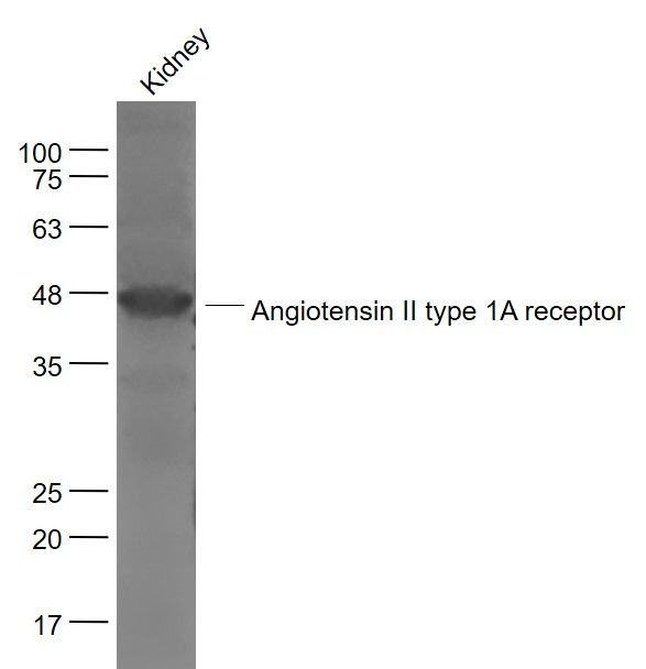
Fig1: Sample: Kidney (Mouse) Lysate at 40 ug Primary: Anti- Angiotensin II type 1A receptor (ER1903-69) at 1/1000 dilution Secondary: IRDye800CW Goat Anti-Rabbit IgG at 1/20000 dilution Predicted band size: 41 kD Observed band size: 42 kDApplication
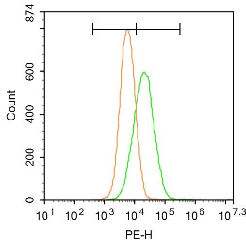
Fig2: Blank control: U87MG. Primary Antibody (green line): Rabbit Anti-Angiotensin II type 1A receptor antibody (ER1903-69) Dilution: 1μg /10^6 cells; Isotype Control Antibody (orange line): Rabbit IgG . Secondary Antibody : Goat anti-rabbit IgG-PE Dilution: 1μg /test. The cells were incubated in 5%BSA to block non-specific protein-protein interactions for 30 min at at room temperature .Cells stained with Primary Antibody for 30 min at room temperature. The secondary antibody used for 40 min at room temperature. Acquisition of 20,000 events was performed.Application
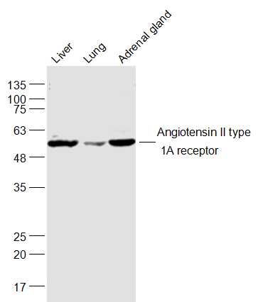
Fig3: Sample: Liver (Mouse) Lysate at 40 ug Lung (Mouse) Lysate at 40 ug Adrenal gland (Rat) Lysate at 40 ug Primary: Anti-Angiotensin II type 1A receptor (ER1903-69) at 1/1000 dilution Secondary: IRDye800CW Goat Anti-Rabbit IgG at 1/20000 dilution Predicted band size: 41 kD Observed band size: 56 kDApplication
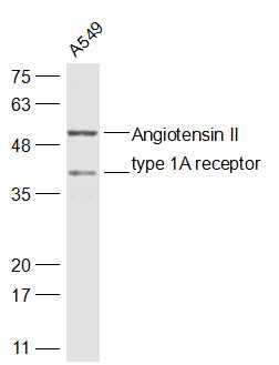
Fig4: Sample: A549(Human) Cell Lysate at 430 ug Primary: Anti-Angiotensin II type 1A receptor (ER1903-69) at 1/500 dilution Secondary: IRDye800CW Goat Anti-Rabbit IgG at 1/20000 dilution Predicted band size: 41 kD Observed band size: 41/51 kDApplication
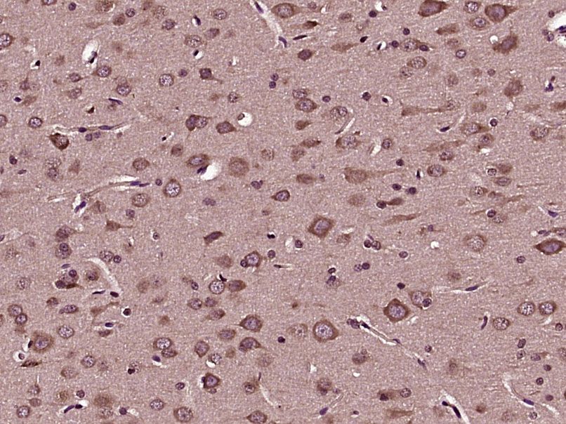
Fig5: Paraformaldehyde-fixed, paraffin embedded (Rat brain); Antigen retrieval by boiling in sodium citrate buffer (pH6.0) for 15min; Block endogenous peroxidase by 3% hydrogen peroxide for 20 minutes; Blocking buffer (normal goat serum) at 37℃ for 30min; Antibody incubation with (Angiotensin II type 1A receptor) Polyclonal Antibody, Unconjugated (ER1903-69) at 1:400 overnight at 4℃, followed by operating according to SP Kit(Rabbit) (sp-0023) instructionsand DAB staining.| Product Name | Angiotensin II type 1A receptor |
|---|---|
| Antibody Type | Primary Antibodies |
| Antigen Alias | AGTR1 Agtr1a AT1 AT1A AT1AR Type 1 angiotensin II receptor AGTR1_HUMAN AGTR1B AT2R1 AT2R1B. |
| Product description | Angiotensin II receptor type 1 or AT1 receptor is the best characterized angiotensin receptor. It has vasopressor effects and regulates aldosterone secretion. It is an important effector controlling blood pressure and volume in the cardiovascular system. Angiotensin II receptor antagonists are drugs indicated for hypertension, diabetic nephropathy and congestive heart failure. The AT1 receptor mediates the major cardiovascular effects of angiotensin II. Effects include vasoconstriction, aldosterone synthesis and secretion, increased vasopressin secretion, cardiac hypertrophy, augmentation of peripheral noradrenergic activity, vascular smooth muscle cells proliferation, decreased renal blood flow, renal renin inhibition, renal tubular sodium reuptake, modulation of central sympathetic nervous system activity, cardiac contractility, central osmocontrol and extracellular matrix formation. Angiotensin II receptor type 1 has been shown to interact with Zinc finger and BTB domain-containing protein 16. The protein's mRNA has been reported to interact with Mir-132 microRNA as part of an RNA silencing mechanism that reduces receptor expression. |
| Immunogen | KLH conjugated synthetic peptide derived from the middle of human Angiotensin II type 1A receptor 101-200/359 |
| Clonality | Polyclonal |
|---|---|
| Isotype | IgG |
| Host Species | Rabbit |
| Tested Applications | WBIHC-PFC |
| WB:1:500-2000: IHC-P:1:400-800: FC:3ug/Test: |
|
| Species Reactivity | HumanMouseRat |
| Concentration | 1mg/ml |
| Alternative Names | AGTR1
Agtr1a
AT1 AT1A AT1AR Type 1 angiotensin II receptor AGTR1_HUMAN AGTR1B AT2R1 AT2R1B. |
|---|---|
| Molecular Weight(MW) | 41 KDa |
| Cellular Localization | Cell membrane; Multi-pass membrane protein. |
| SwissProt ID | P30556 |
|---|
Application
Fig1: Sample: Kidney (Mouse) Lysate at 40 ug Primary: Anti- Angiotensin II type 1A receptor (ER1903-69) at 1/1000 dilution Secondary: IRDye800CW Goat Anti-Rabbit IgG at 1/20000 dilution Predicted band size: 41 kD Observed band size: 42 kD
Application
Fig2: Blank control: U87MG. Primary Antibody (green line): Rabbit Anti-Angiotensin II type 1A receptor antibody (ER1903-69) Dilution: 1μg /10^6 cells; Isotype Control Antibody (orange line): Rabbit IgG . Secondary Antibody : Goat anti-rabbit IgG-PE Dilution: 1μg /test. The cells were incubated in 5%BSA to block non-specific protein-protein interactions for 30 min at at room temperature .Cells stained with Primary Antibody for 30 min at room temperature. The secondary antibody used for 40 min at room temperature. Acquisition of 20,000 events was performed.
Application
Fig3: Sample: Liver (Mouse) Lysate at 40 ug Lung (Mouse) Lysate at 40 ug Adrenal gland (Rat) Lysate at 40 ug Primary: Anti-Angiotensin II type 1A receptor (ER1903-69) at 1/1000 dilution Secondary: IRDye800CW Goat Anti-Rabbit IgG at 1/20000 dilution Predicted band size: 41 kD Observed band size: 56 kD
Application
Fig4: Sample: A549(Human) Cell Lysate at 430 ug Primary: Anti-Angiotensin II type 1A receptor (ER1903-69) at 1/500 dilution Secondary: IRDye800CW Goat Anti-Rabbit IgG at 1/20000 dilution Predicted band size: 41 kD Observed band size: 41/51 kD
Application
Fig5: Paraformaldehyde-fixed, paraffin embedded (Rat brain); Antigen retrieval by boiling in sodium citrate buffer (pH6.0) for 15min; Block endogenous peroxidase by 3% hydrogen peroxide for 20 minutes; Blocking buffer (normal goat serum) at 37℃ for 30min; Antibody incubation with (Angiotensin II type 1A receptor) Polyclonal Antibody, Unconjugated (ER1903-69) at 1:400 overnight at 4℃, followed by operating according to SP Kit(Rabbit) (sp-0023) instructionsand DAB staining.| Positive Control | Liver, lung, adrenal and adrenocortical adenomas. |
|---|---|
| Application Notes | WB:1:500-2000: IHC-P:1:400-800: FC:3ug/Test: |
| Form | Liquid |
|---|---|
| Storage Instructions | Store at -20℃ for one year. Avoid repeated freeze/thaw cycles. |
| Storage Buffer | 0.01M TBS(pH7.4) with 1% BSA, 0.03% Proclin300 and 50% Glycerol. |


