Application
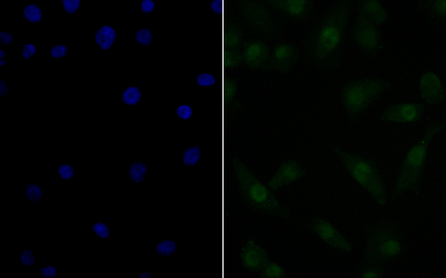
Fig1: ICC staining Nucleostemin in A549 cells (green). The nuclear counter stain is DAPI (blue). Cells were fixed in paraformaldehyde, permeabilised with 0.25% Triton X100/PBS.Application
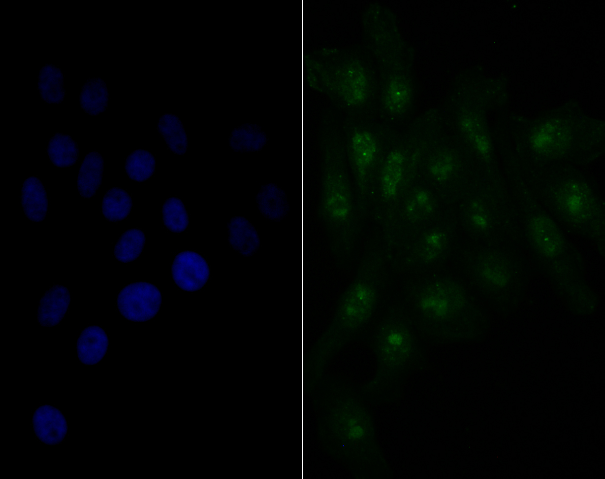
Fig2: ICC staining Nucleostemin in HepG2 cells (green). The nuclear counter stain is DAPI (blue). Cells were fixed in paraformaldehyde, permeabilised with 0.25% Triton X100/PBS.Application
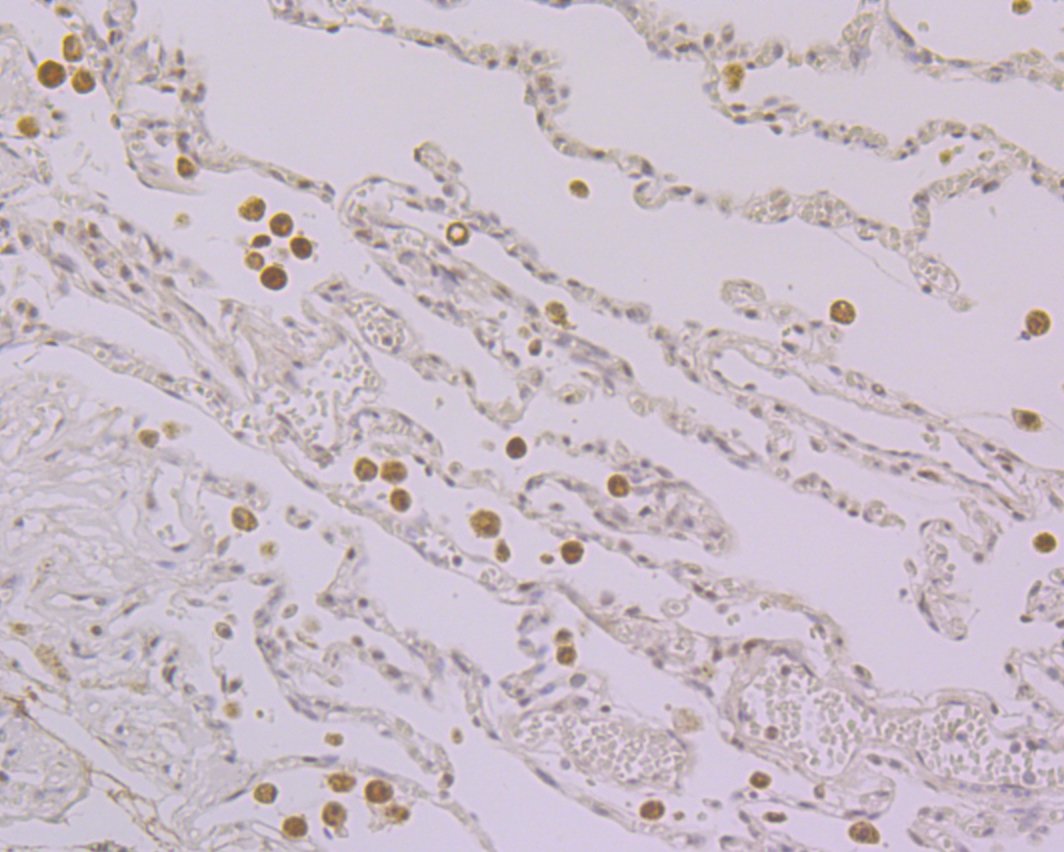
Fig3: Immunohistochemical analysis of paraffin-embedded human lung cancer tissue using anti-Nucleostemin antibody. Counter stained with hematoxylin.Application
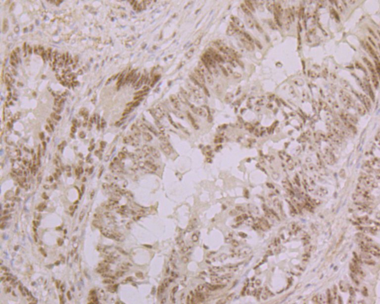
Fig4: Immunohistochemical analysis of paraffin-embedded human breast cancer tissue using anti-Nucleostemin antibody. Counter stained with hematoxylin.Application
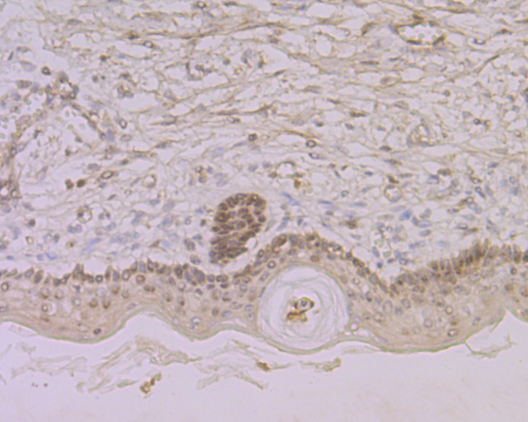
Fig5: Immunohistochemical analysis of paraffin-embedded human skin tissue using anti-Nucleostemin antibody. Counter stained with hematoxylin.Application
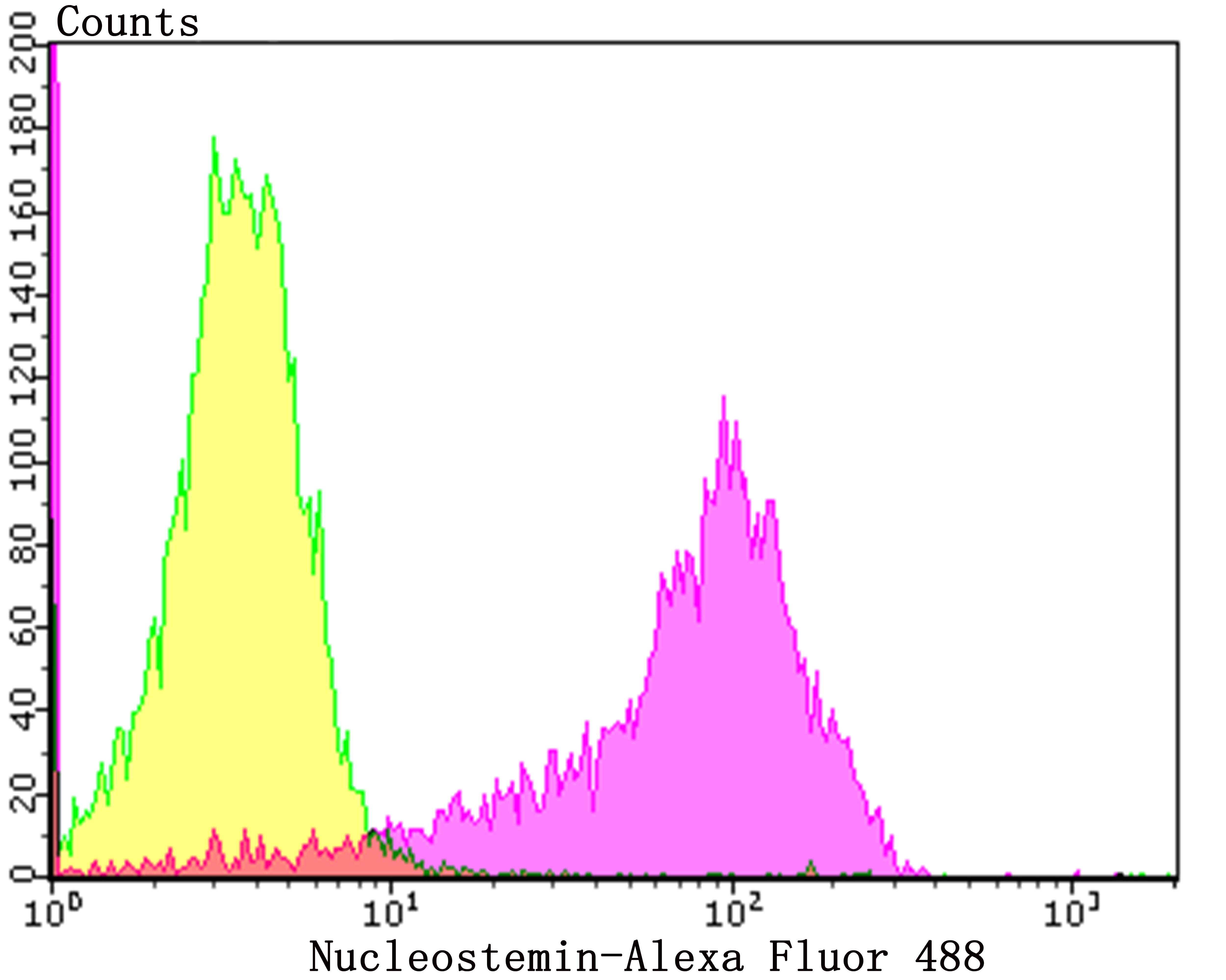
Fig6: Flow cytometric analysis of A549 cells with Nucleostemin antibody at 1/50 dilution (Fuchsia) compared with an unlabelled control (cells without incubation with primary antibody; Yellow). Alexa Fluor 488-conjugated goat anti-rabbit IgG was used as the secondary antibody.| Product Name | Nucleostemin [JG37-82] |
|---|---|
| Antibody Type | Primary Antibodies |
| Antigen Alias | C77032 antibody E2 induced gene 3 protein antibody E2-induced gene 3 protein antibody E2IG3 antibody Estradiol induced nucleotide binding protein antibody GNL3 antibody GNL3_HUMAN antibody Guanine nucleotide binding protein like 3 (nucleolar) antibody Guanine nucleotide binding protein like 3 antibody Guanine nucleotide-binding protein-like 3 antibody MGC800 antibody NNP47 antibody Novel nucleolar protein 47 antibody NS antibody Nucleolar GTP binding protein 3 antibody Nucleolar GTP-binding protein 3 antibody Nucleostemin antibody Putative nucleotide binding protein estradiol induced antibody |
| Product description | The protein encoded by this gene may interact with p53 and may be involved in tumorigenesis. The encoded protein also appears to be important for stem cell proliferation. Stabilizes MDM2 by preventing its ubiquitination, and hence proteasomal degradation. This protein is found in both the nucleus and nucleolus. Three transcript variants encoding two different isoforms have been found for this gene. |
| Immunogen | Recombinant protein corresponding to the C-terminus of human Nucleostemin. |
| Clonality | Monoclonal |
|---|---|
| Isotype | IgG |
| Host Species | Recombinant rabbit |
| Tested Applications | WBICCIHCFC |
| ICC:1:50-1:200 IHC:1:50-1:200 FC:1:50-1:100 WB:1:500 |
|
| Species Reactivity | Human |
| Concentration | 1mg/ml |
| Alternative Names | C77032 antibody E2 induced gene 3 protein antibody E2-induced gene 3 protein antibody E2IG3 antibody Estradiol induced nucleotide binding protein antibody GNL3 antibody GNL3_HUMAN antibody Guanine nucleotide binding protein like 3 (nucleolar) antibody Guanine nucleotide binding protein like 3 antibody Guanine nucleotide-binding protein-like 3 antibody MGC800 antibody NNP47 antibody Novel nucleolar protein 47 antibody NS antibody Nucleolar GTP binding protein 3 antibody Nucleolar GTP-binding protein 3 antibody Nucleostemin antibody Putative nucleotide binding protein estradiol induced antibody |
|---|---|
| Molecular Weight(MW) | 62 kDa |
| Cellular Localization | Nucleus. |
| SwissProt ID | Q9BVP2 |
|---|
Application
Fig1: ICC staining Nucleostemin in A549 cells (green). The nuclear counter stain is DAPI (blue). Cells were fixed in paraformaldehyde, permeabilised with 0.25% Triton X100/PBS.
Application
Fig2: ICC staining Nucleostemin in HepG2 cells (green). The nuclear counter stain is DAPI (blue). Cells were fixed in paraformaldehyde, permeabilised with 0.25% Triton X100/PBS.
Application
Fig3: Immunohistochemical analysis of paraffin-embedded human lung cancer tissue using anti-Nucleostemin antibody. Counter stained with hematoxylin.
Application
Fig4: Immunohistochemical analysis of paraffin-embedded human breast cancer tissue using anti-Nucleostemin antibody. Counter stained with hematoxylin.
Application
Fig5: Immunohistochemical analysis of paraffin-embedded human skin tissue using anti-Nucleostemin antibody. Counter stained with hematoxylin.
Application
Fig6: Flow cytometric analysis of A549 cells with Nucleostemin antibody at 1/50 dilution (Fuchsia) compared with an unlabelled control (cells without incubation with primary antibody; Yellow). Alexa Fluor 488-conjugated goat anti-rabbit IgG was used as the secondary antibody.| Positive Control | A549, HepG2, human lung cancer tissue, human breast cancer tissue, human skin tissue. |
|---|---|
| Application Notes | ICC:1:50-1:200 IHC:1:50-1:200 FC:1:50-1:100 WB:1:500 |
| Form | Liquid |
|---|---|
| Storage Instructions | Store at +4℃ after thawing. Aliquot store at -20℃. Avoid repeated freeze / thaw cycles. |
| Storage Buffer | 1*TBS (pH7.4), 1%BSA, 40%Glycerol. Preservative: 0.05% Sodium Azide. |


