Application
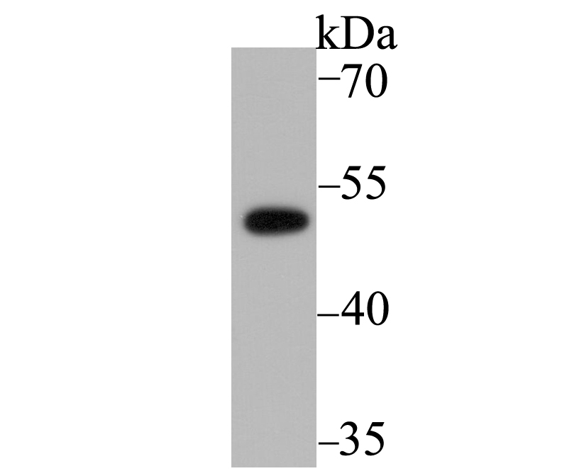
Fig1: Western blot analysis of PICK1 on SK-Br-3 cell lysate using anti-PICK1 antibody at 1/5,000 dilution.Application
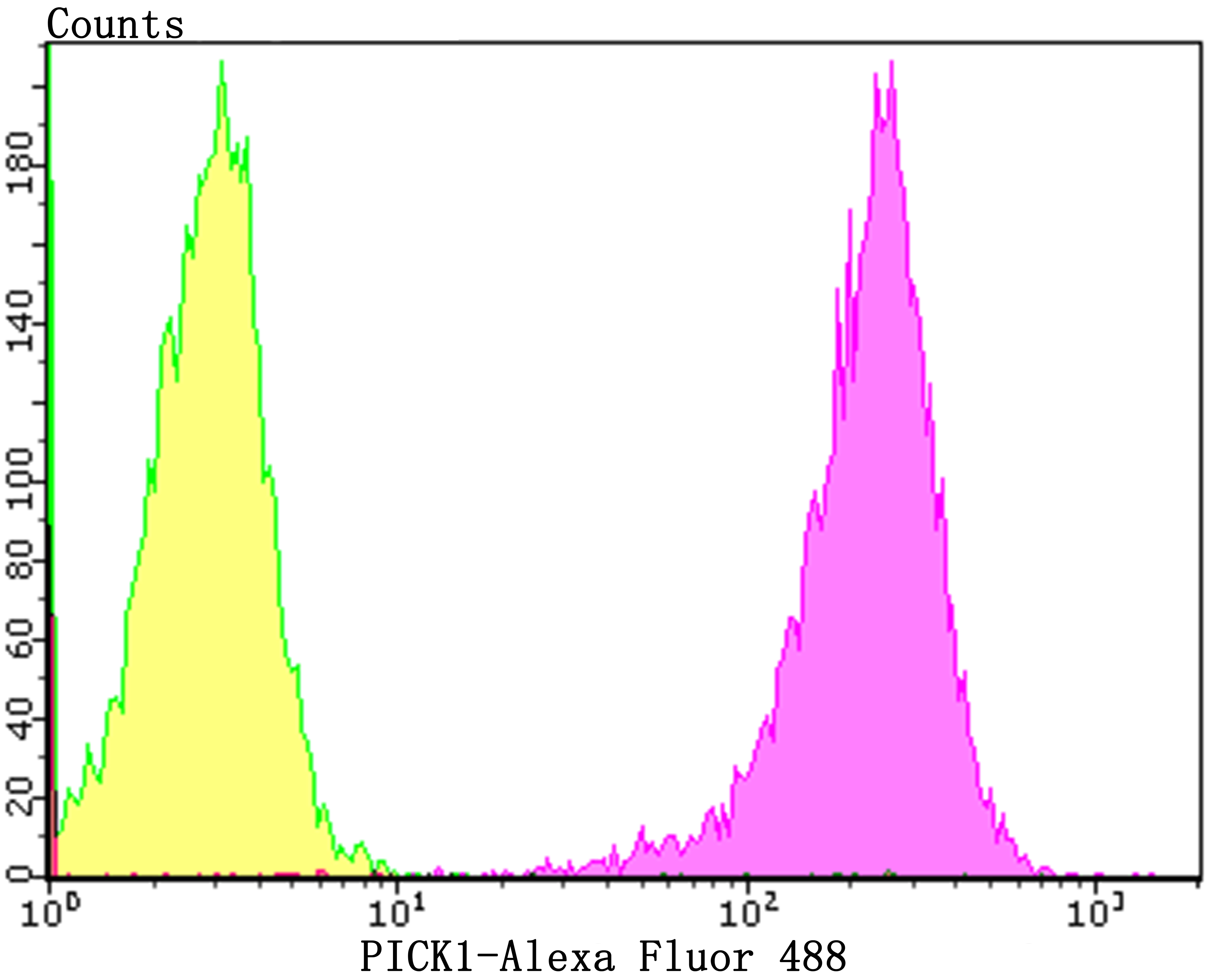
Fig2: Flow cytometric analysis of 293T cells with PICK1 antibody at 1/100 dilution (purple) compared with an unlabelled control (cells without incubation with primary antibody; yellow). Alexa Fluor 488-conjugated goat anti-rabbit IgG was used as the secondary antibody.| Product Name | PICK1 [JG30-80] |
|---|---|
| Antibody Type | Primary Antibodies |
| Antigen Alias | dJ1039K5 antibody MGC15204 antibody OTTHUMP00000028509 antibody PICK 1 antibody Pick1 antibody PICK1_HUMAN antibody PRKCA binding protein antibody PRKCA-binding protein antibody PRKCABP antibody Protein interacting with C kinase 1 antibody Protein interacting with PRKCA antibody Protein interacting with PRKCA 1 antibody Protein kinase C alpha binding protein antibody Protein kinase C-alpha-binding protein antibody Protein that interacts with C kinase 1 antibody |
| Product description | Protein interacting with C kinase 1 (PICK1) is a PDZ-domain containing protein that is located in the perinuclear region and is phosphorylated in response to PKCα activation. PKCα, which is essential for the regulation of proliferation and differentiation in numerous cell types, contains within its catalytic region a PDZ-binding domain that is absent from other PKC isoforms. Mutation of the PICK1 PDZ domain inhibits the binding of PICK1 to PKCα. PICK1 also interacts with the carboxy terminus of a-amino-3-hydroxy-5-methyl-isoxazole-4-propionic acid (AMPA) receptor, a neurotransmitter receptor located at excitatory synapses, suggesting that PICK1 plays a role in the modulation of synaptic transmission by targeting and anchoring AMPA to specific synapses. |
| Immunogen | Recombinant protein within N-terminal human PICK1. |
| Clonality | Monoclonal |
|---|---|
| Isotype | IgG |
| Host Species | Recombinant rabbit |
| Tested Applications | WBIPFC |
| WB:1:2,000-1:5,000 IP:1:10-1:50 FC:1:50-1:100 |
|
| Species Reactivity | HumanMouseRat |
| Concentration | 1mg/ml |
| Alternative Names | dJ1039K5 antibody MGC15204 antibody OTTHUMP00000028509 antibody PICK 1 antibody Pick1 antibody PICK1_HUMAN antibody PRKCA binding protein antibody PRKCA-binding protein antibody PRKCABP antibody Protein interacting with C kinase 1 antibody Protein interacting with PRKCA antibody Protein interacting with PRKCA 1 antibody Protein kinase C alpha binding protein antibody Protein kinase C-alpha-binding protein antibody Protein that interacts with C kinase 1 antibody |
|---|---|
| Molecular Weight(MW) | 47 kDa |
| Cellular Localization | Plasma membrane. Cytoskeleton. |
| SwissProt ID | Q9NRD5 |
|---|
Application
Fig1: Western blot analysis of PICK1 on SK-Br-3 cell lysate using anti-PICK1 antibody at 1/5,000 dilution.
Application
Fig2: Flow cytometric analysis of 293T cells with PICK1 antibody at 1/100 dilution (purple) compared with an unlabelled control (cells without incubation with primary antibody; yellow). Alexa Fluor 488-conjugated goat anti-rabbit IgG was used as the secondary antibody.| Positive Control | SK-Br-3, 293T. |
|---|---|
| Application Notes | WB:1:2,000-1:5,000 IP:1:10-1:50 FC:1:50-1:100 |
| Form | Liquid |
|---|---|
| Storage Instructions | Store at +4℃ after thawing. Aliquot store at -20℃. Avoid repeated freeze / thaw cycles. |
| Storage Buffer | 1*TBS (pH7.4), 1%BSA, 40%Glycerol. Preservative: 0.05% Sodium Azide. |


