Application
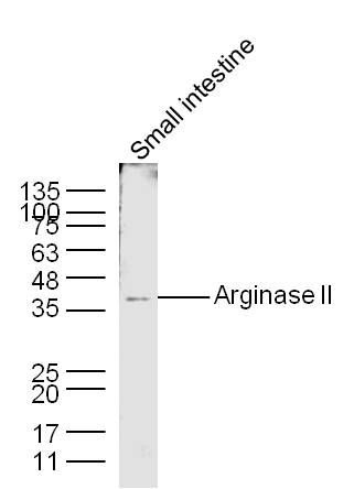
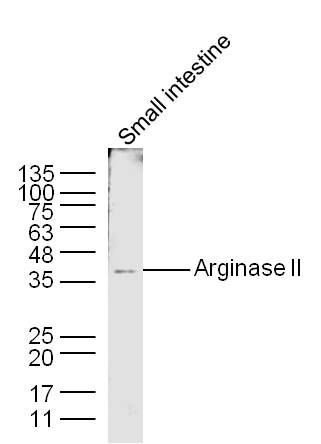
Fig1: Sample: Small intestine (Mouse) Lysate at 40 ug Primary: Anti-Arginase II (Bs- 11397R) at 1/300 dilution Secondary: IRDye800CW Goat Anti-Rabbit IgG at 1/20000 dilution Predicted band size: 36 kD Observed band size: 36 kDApplication
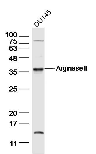
Fig2: Sample: DU145(human)cell Lysate at 30 ug Primary: Anti-Arginase II (ER1903-91) at 1/300 dilution Secondary: IRDye800CW Goat Anti-Rabbit IgG at 1/20000 dilution Predicted band size: 36kD Observed band size: 36 kDApplication
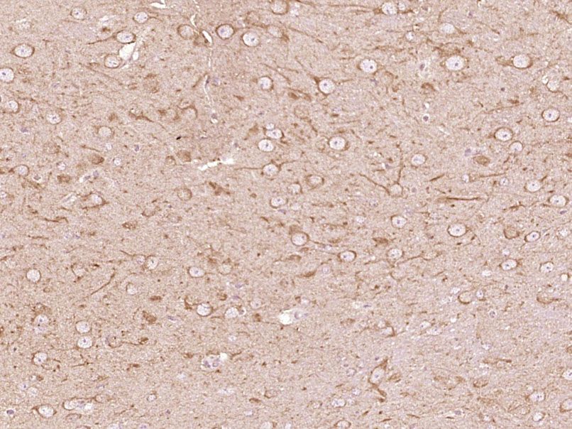
Fig3: Paraformaldehyde-fixed, paraffin embedded (Mouse brain); Antigen retrieval by boiling in sodium citrate buffer (pH6.0) for 15min; Block endogenous peroxidase by 3% hydrogen peroxide for 20 minutes; Blocking buffer (normal goat serum) at 37℃ for 30min; Antibody incubation with (Arginase II) Polyclonal Antibody, Unconjugated (ER1903-91) at 1:400 overnight at 4℃, followed by operating according to SP Kit(Rabbit) (sp-0023) instructionsand DAB staining.Application
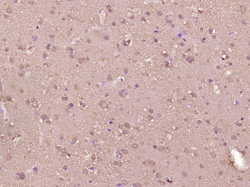
Fig4: Paraformaldehyde-fixed, paraffin embedded (Human brain glioma); Antigen retrieval by boiling in sodium citrate buffer (pH6.0) for 15min; Block endogenous peroxidase by 3% hydrogen peroxide for 20 minutes; Blocking buffer (normal goat serum) at 37℃ for 30min; Antibody incubation with (Arginase II) Polyclonal Antibody, Unconjugated (ER1903-91) at 1:400 overnight at 4℃, followed by operating according to SP Kit(Rabbit) (sp-0023) instructionsand DAB staining.Application
Fig5: Sample: Small intestine (Mouse) Lysate at 40 ug Primary: Anti-Arginase II (ER1903-91) at 1/300 dilution Secondary: IRDye800CW Goat Anti-Rabbit IgG at 1/20000 dilution Predicted band size: 36 kD Observed band size: 36 kD| Product Name | Arginase II |
|---|---|
| Antibody Type | Primary Antibodies |
| Antigen Alias | ARG 2 ARG2 ARGI2_HUMAN Arginase-2 Arginase 2Arginase2 Arginase liver Arginase type II Arginase II ArginaseII Arginase-II Arginase1 Arginase-2, mitochondrial Kidney-type arginase Non-hepatic arginase Type II arginase . |
| Product description | May play a role in the regulation of extra-urea cycle arginine metabolism and also in down-regulation of nitric oxide synthesis. Extrahepatic arginase functions to regulate L-arginine bioavailability to NO synthase. Since NO synthase is found in the penile corpus cavernosum smooth muscle, the clitoral corpus cavernosum and the vagina, arginase II plays a role in both male and female sexual arousal. It is therefore a potential target for the treatment of male and female sexual arousal disorders. |
| Immunogen | KLH conjugated synthetic peptide derived from human Arginase II 181-290/354 |
| Clonality | Polyclonal |
|---|---|
| Isotype | IgG |
| Host Species | Rabbit |
| Tested Applications | WBIHC-PICC/IF |
| WB:1:500-2000: IHC-P:1:400-800: IF:1:100-500: ICC:1:100-500: |
|
| Species Reactivity | HumanMouseRatChickenDogCowRabbit |
| Concentration | 1mg/ml |
| Alternative Names | ARG 2
ARG2
ARGI2_HUMAN
Arginase-2
Arginase 2Arginase2
Arginase liver
Arginase type II
Arginase II
ArginaseII
Arginase-II
Arginase1
Arginase-2 mitochondrial Kidney-type arginase Non-hepatic arginase Type II arginase . |
|---|---|
| Molecular Weight(MW) | 36 KDa |
| Cellular Localization | Cytoplasm. |
| SwissProt ID | P78540 |
|---|
Application
Fig1: Sample: Small intestine (Mouse) Lysate at 40 ug Primary: Anti-Arginase II (Bs- 11397R) at 1/300 dilution Secondary: IRDye800CW Goat Anti-Rabbit IgG at 1/20000 dilution Predicted band size: 36 kD Observed band size: 36 kD
Application
Fig2: Sample: DU145(human)cell Lysate at 30 ug Primary: Anti-Arginase II (ER1903-91) at 1/300 dilution Secondary: IRDye800CW Goat Anti-Rabbit IgG at 1/20000 dilution Predicted band size: 36kD Observed band size: 36 kD
Application
Fig3: Paraformaldehyde-fixed, paraffin embedded (Mouse brain); Antigen retrieval by boiling in sodium citrate buffer (pH6.0) for 15min; Block endogenous peroxidase by 3% hydrogen peroxide for 20 minutes; Blocking buffer (normal goat serum) at 37℃ for 30min; Antibody incubation with (Arginase II) Polyclonal Antibody, Unconjugated (ER1903-91) at 1:400 overnight at 4℃, followed by operating according to SP Kit(Rabbit) (sp-0023) instructionsand DAB staining.
Application
Fig4: Paraformaldehyde-fixed, paraffin embedded (Human brain glioma); Antigen retrieval by boiling in sodium citrate buffer (pH6.0) for 15min; Block endogenous peroxidase by 3% hydrogen peroxide for 20 minutes; Blocking buffer (normal goat serum) at 37℃ for 30min; Antibody incubation with (Arginase II) Polyclonal Antibody, Unconjugated (ER1903-91) at 1:400 overnight at 4℃, followed by operating according to SP Kit(Rabbit) (sp-0023) instructionsand DAB staining.
Application
Fig5: Sample: Small intestine (Mouse) Lysate at 40 ug Primary: Anti-Arginase II (ER1903-91) at 1/300 dilution Secondary: IRDye800CW Goat Anti-Rabbit IgG at 1/20000 dilution Predicted band size: 36 kD Observed band size: 36 kD| Positive Control | Expressed most strongly in kidney and prostate, much less strongly in the brain, skeletal muscle, placenta, lung, mammary gland, macrophage, uterus, testis and gut, but apparently not in the liver, heart and pancreas. |
|---|---|
| Application Notes | WB:1:500-2000: IHC-P:1:400-800: IF:1:100-500: ICC:1:100-500: |
| Form | Liquid |
|---|---|
| Storage Instructions | Store at -20℃ for one year. Avoid repeated freeze/thaw cycles. The lyophilized antibody is stable at room temperature for at least one month and for greater than a year when kept at -20℃. When reconstituted in sterile pH 7.4 0.01M PBS or diluent of antibody the antibody is stable for at least two weeks at 2-4℃. |
| Storage Buffer | 0.01M TBS(pH7.4) with 1% BSA, 0.03% Proclin300 and 50% Glycerol. |


